Loading video…


 15 Concepts
 10.4K views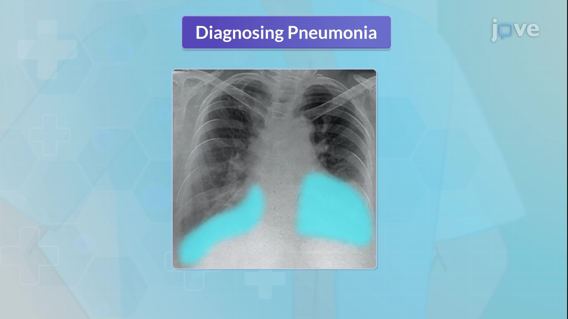

Video Summary: Pneumonia Iii Complications and Assessment Explained
Pneumonia complications can turn a treatable respiratory infection into a life-threatening emergency within hours. Understanding pneumonia III complications and assessment is crucial for recognizing when patients at Houston Methodist Hospital might develop dangerous conditions like respiratory failure or pleural effusion. This comprehensive tutorial covers the critical diagnostic tools healthcare providers use, from chest X-rays to arterial blood gas analysis, plus essential prevention strategies including proper vaccination schedules. Watch the full video on JoVE Coach to master this concept with expert-led visuals and step-by-step explanations.
Pneumonia III complications and assessment represents an advanced understanding of how respiratory infections can rapidly progress to life-threatening conditions. Unlike simple pneumonia cases, this level requires healthcare providers to recognize subtle warning signs and implement comprehensive diagnostic protocols that can mean the difference between recovery and critical care admission.
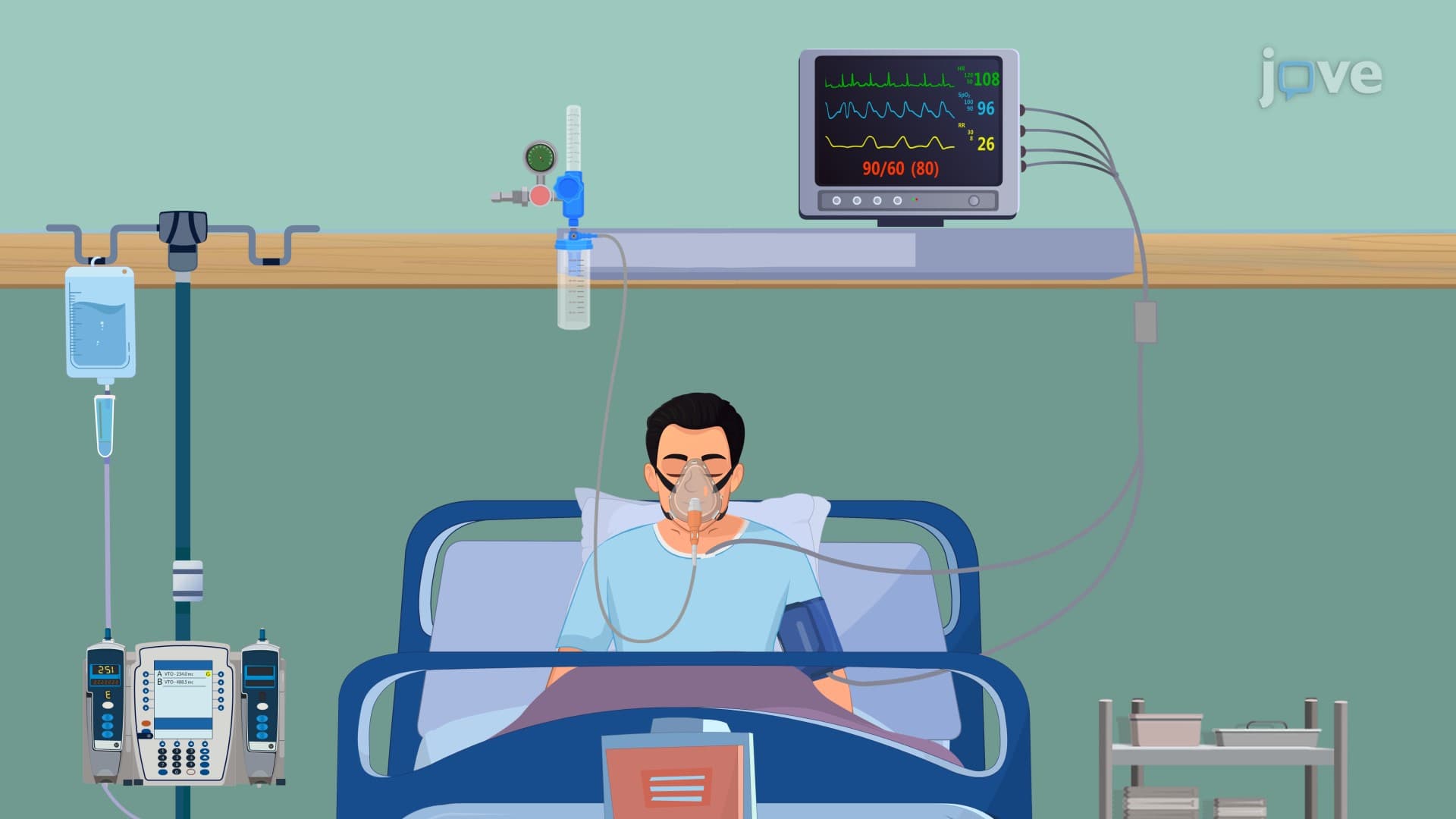
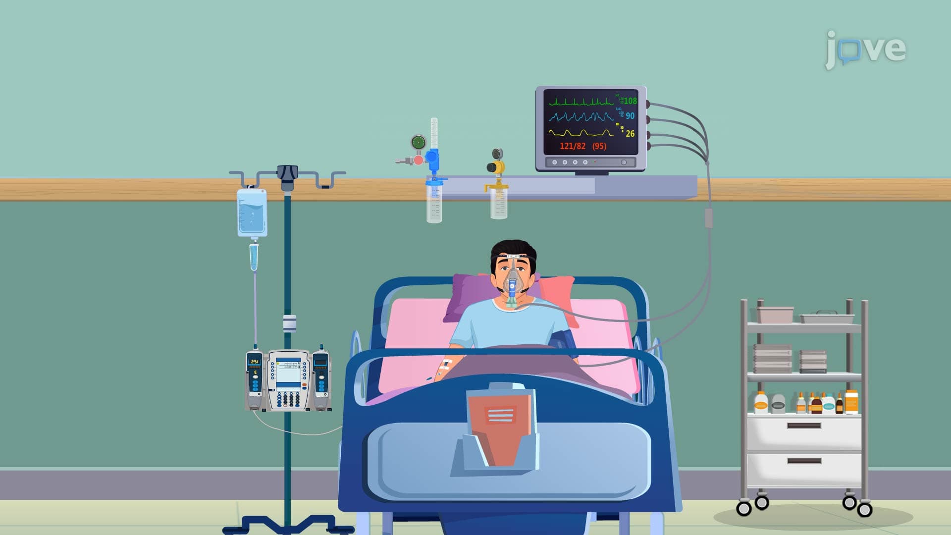
The most dangerous pneumonia complications develop when infection spreads beyond the lungs or overwhelms the body's compensatory mechanisms. Hypotension occurs when bacterial toxins trigger systemic vasodilation, potentially leading to septic shock. At major medical centers like Mayo Clinic Rochester, emergency protocols immediately activate when pneumonia patients present with systolic blood pressure below 90 mmHg.
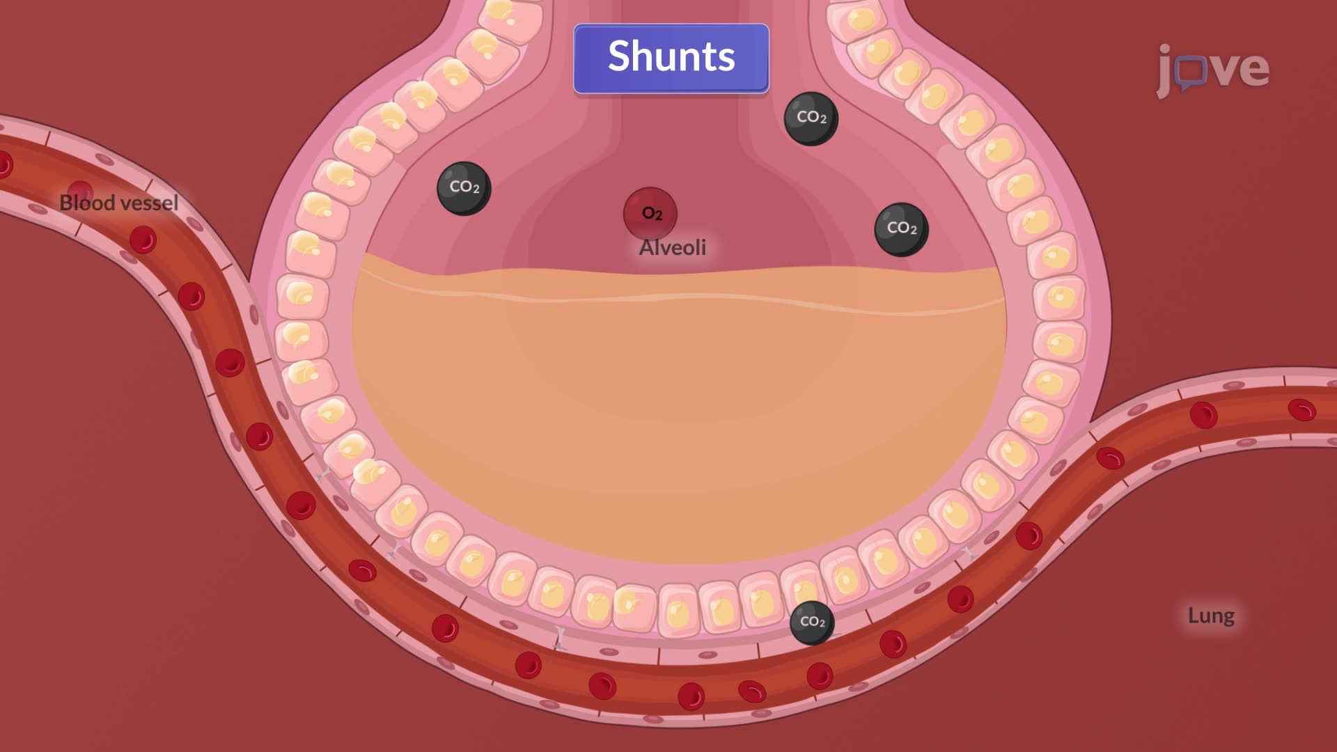
Pleural effusion represents fluid accumulation in the pleural space, often indicating severe inflammation or bacterial invasion. This complication appears in approximately 40% of hospitalized pneumonia patients and requires immediate thoracentesis for both diagnostic and therapeutic purposes. Atelectasis, or lung collapse, frequently accompanies severe pneumonia when airways become blocked by thick secretions or when patients cannot take deep breaths due to chest pain.
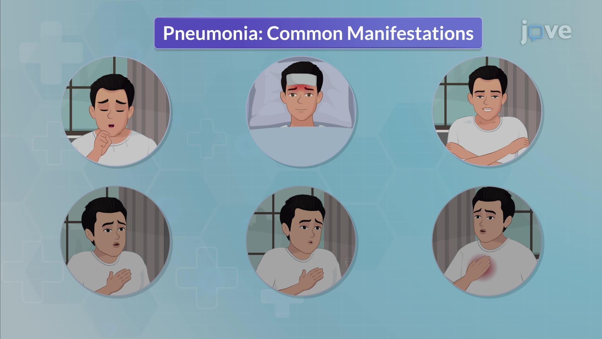
Modern pneumonia assessment extends far beyond the traditional chest X-ray. Healthcare providers now utilize a systematic approach combining clinical examination, laboratory analysis, and imaging studies. The physical examination focuses on identifying adventitious lung sounds-crackles indicate fluid in alveoli, while wheezes suggest airway narrowing from inflammation.
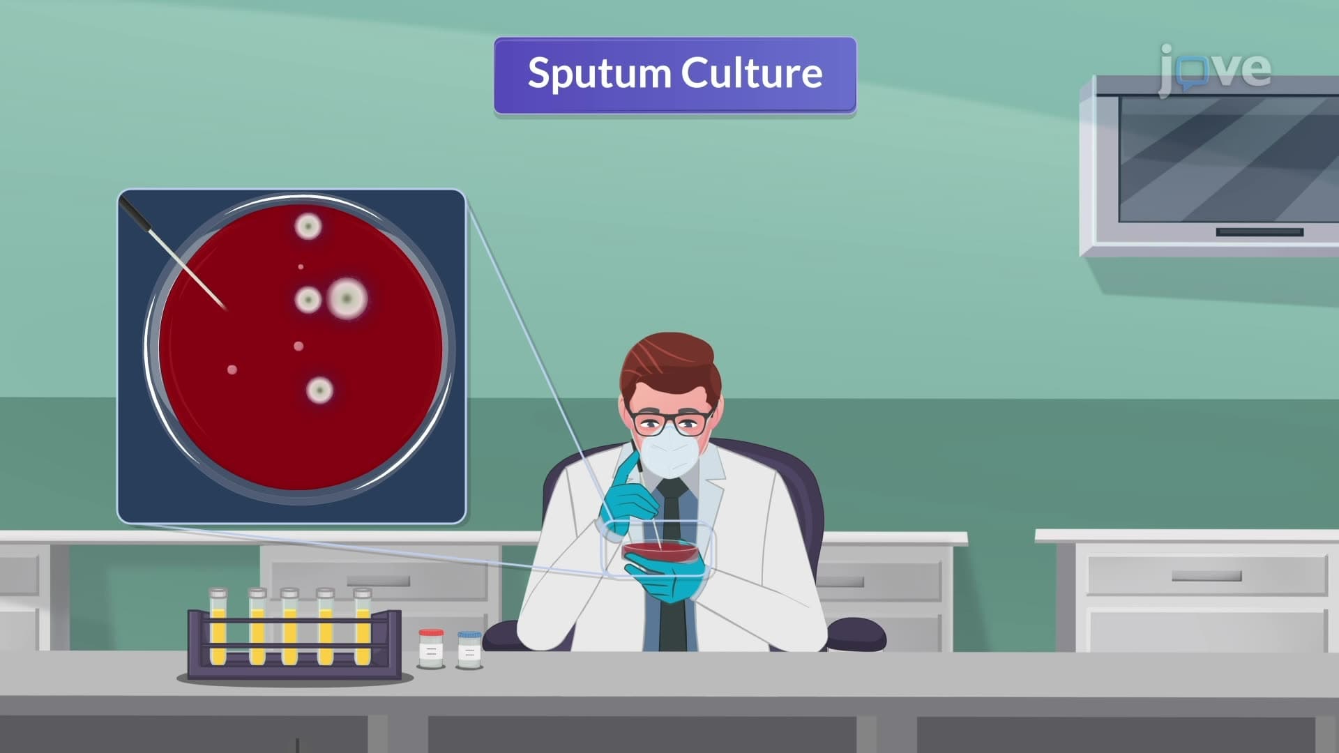
Laboratory diagnostics have revolutionized pneumonia assessment accuracy. Procalcitonin levels help distinguish bacterial from viral infections, with values above 0.5 ng/mL strongly suggesting bacterial etiology. C-reactive protein provides inflammation markers, while arterial blood gas analysis reveals the extent of respiratory compromise. Blood cultures, though positive in only 20% of cases, remain crucial for identifying causative organisms and guiding antibiotic therapy.
Effective pneumonia prevention combines individual health behaviors with population-level vaccination strategies. The CDC recommends PCV13 for all adults over 65, followed by PPSV23 one year later. This dual vaccination approach provides broader protection against pneumococcal strains responsible for the most severe complications.
Understanding these concepts proves essential for students preparing for health science careers and standardized exams like the MCAT or nursing entrance tests like HESI A2, where pneumonia assessment questions frequently appear in clinical reasoning sections.
Related Micro-courses