11 Concepts
5 Concepts
15 Concepts
11 Concepts
7 Concepts
10 Concepts
22 Concepts
11 Concepts
10 Concepts
8 Concepts
14 Concepts
17 Concepts
14 Concepts
19 Concepts
15 Concepts
16 Concepts
15 Concepts
7 Concepts
6 Concepts
7 Concepts
7 Concepts
27 Concepts
8 Concepts
9 Concepts
6 Concepts
10 Concepts
12 Concepts
10 Concepts
Diagnostic studies of respiratory system encompass essential tests including pulmonary function tests, imaging studies, blood gas analysis, and endoscopic procedures used to evaluate lung health and diagnose respiratory conditions. This comprehensive course through JoVE Coach covers diagnostic tests for respiratory conditions from sputum analysis and chest X-rays to advanced techniques like bronchoscopy and CT pulmonary imaging, providing healthcare students with critical skills for respiratory assessment in US clinical settings.
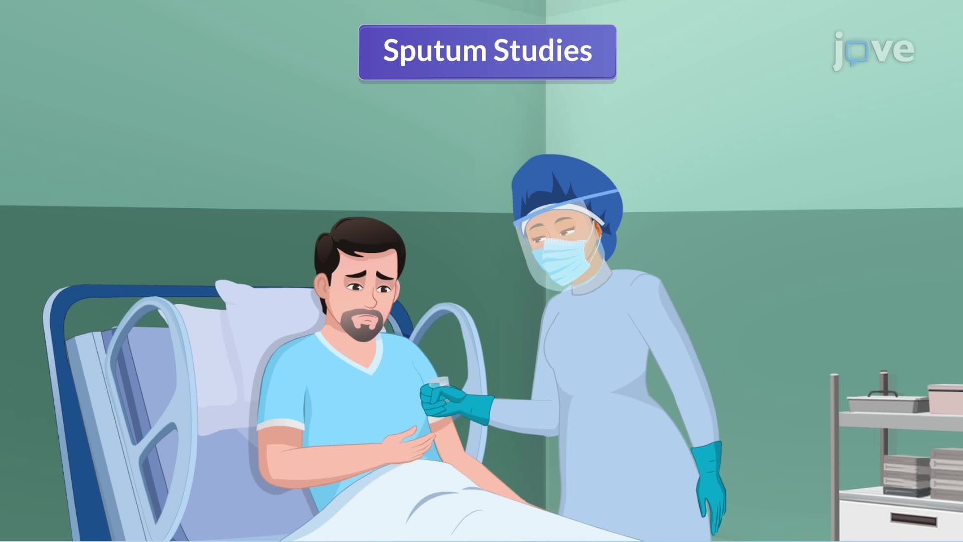
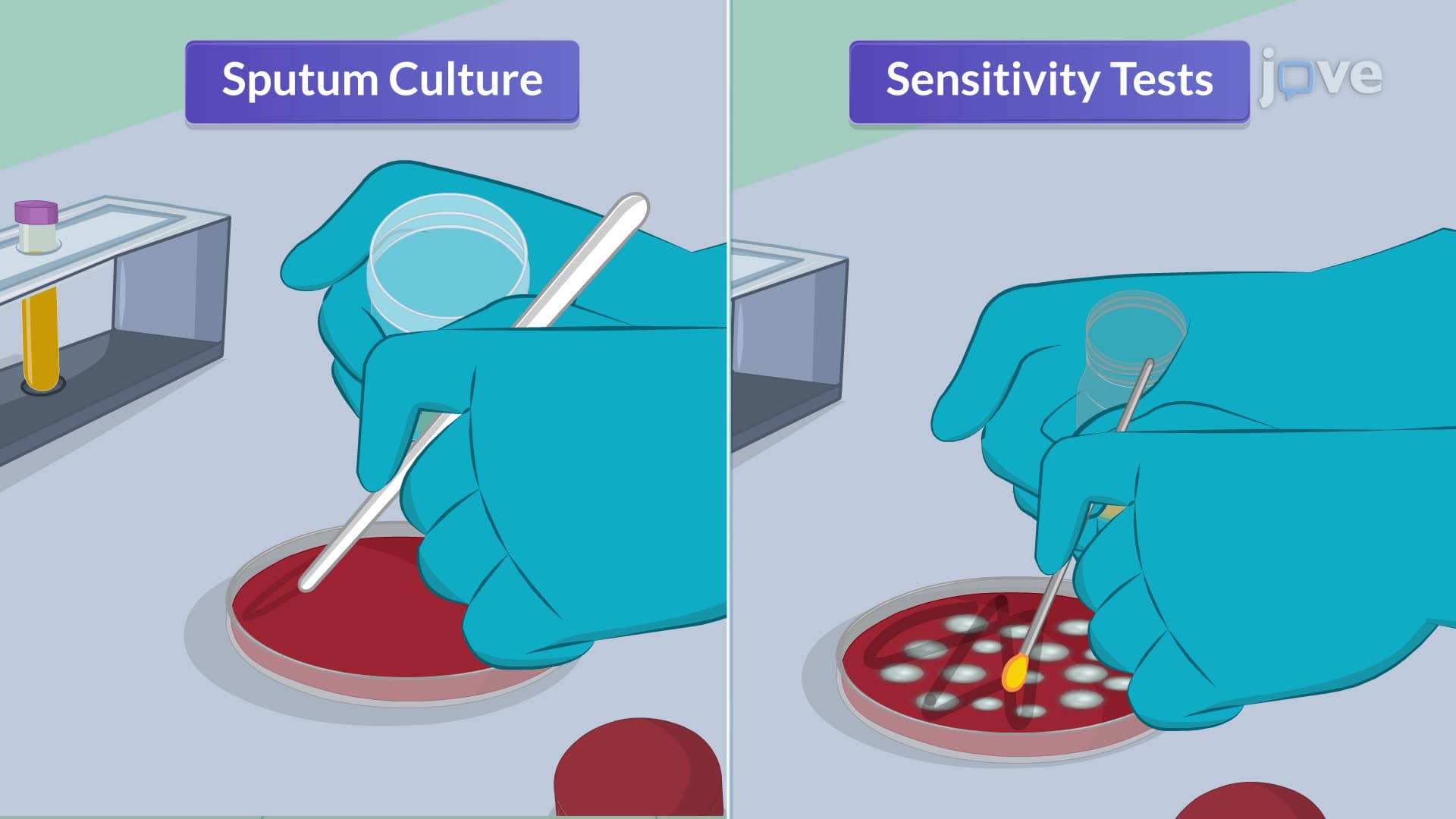
1. Sputum Analysis and Culture Studies Sputum studies form the foundation of respiratory diagnostics, utilizing three primary techniques: Gram stain classification of bacteria, cytology for cancer cell detection, and acid-fast smear for tuberculosis identification. Collection methods include expectoration, tracheal suction for tracheostomy patients, and bronchoscopy when standard collection fails. Proper technique requires early morning samples after mouth cleaning, sterile containers, and immediate laboratory transport. Culture and sensitivity testing identifies specific pathogens like Streptococcus pneumoniae, with preliminary results in 24 hours and complete antibiotic sensitivity in 48-72 hours, crucial for treating conditions like pneumonia in US hospitals.
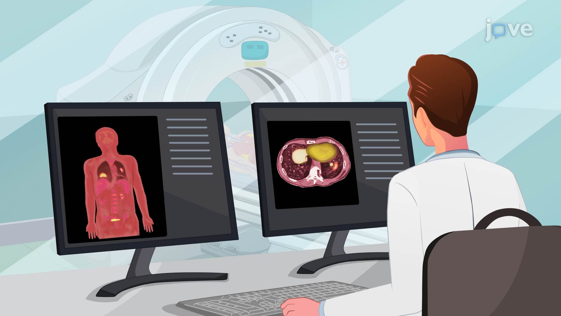
2. Radiological Imaging Techniques Chest X-rays provide initial respiratory screening, detecting fluid accumulation, tumors, and foreign bodies in emergency departments across US healthcare systems. CT pulmonary scans offer detailed cross-sectional views for identifying small nodules, mediastinal abnormalities, and hilar adenopathy that chest X-rays might miss. MRI uses magnetic fields for detailed lung tissue imaging, particularly valuable for bronchogenic carcinoma diagnosis. Ventilation-perfusion (V/Q) scans combine radioactive tracers to assess both airflow and blood flow, essential for diagnosing pulmonary embolism. Nursing responsibilities include patient preparation, contrast allergy screening, and post-procedure monitoring following protocols established by organizations like the American College of Radiology.
3. Advanced Imaging and Angiography Pulmonary angiograms represent invasive diagnostic procedures requiring catheter insertion through femoral veins into pulmonary arteries, using contrast medium to visualize blood vessel blockages. PET scans utilize radioactive glucose tracers to identify high-metabolic areas indicating cancer, infection, or inflammation, with 30-60 minute uptake periods before scanning. These procedures require comprehensive pre-procedure assessment including coagulation studies, allergy screening, and NPO status verification. Post-procedure care involves vital sign monitoring, hydration encouragement for tracer elimination, and observation for complications. These advanced techniques are commonly used in major US medical centers for complex respiratory diagnoses and cancer staging.
4. Endoscopic Respiratory Procedures Bronchoscopy enables direct visualization of airways from larynx to bronchi, serving both diagnostic and therapeutic purposes in US pulmonology practices. Diagnostic applications include tissue biopsy, secretion analysis, and airway assessment, while therapeutic uses encompass obstruction removal, bleeding control, and brachytherapy administration. Thoracoscopy examines pleural cavity through small incisions, evaluating effusions and collecting tissue samples. Pre-procedure requirements include 6-hour NPO status and informed consent, while post-procedure care focuses on vital sign monitoring and cough reflex assessment. These procedures are standard in US hospitals for lung cancer diagnosis, pleural disease evaluation, and therapeutic interventions.
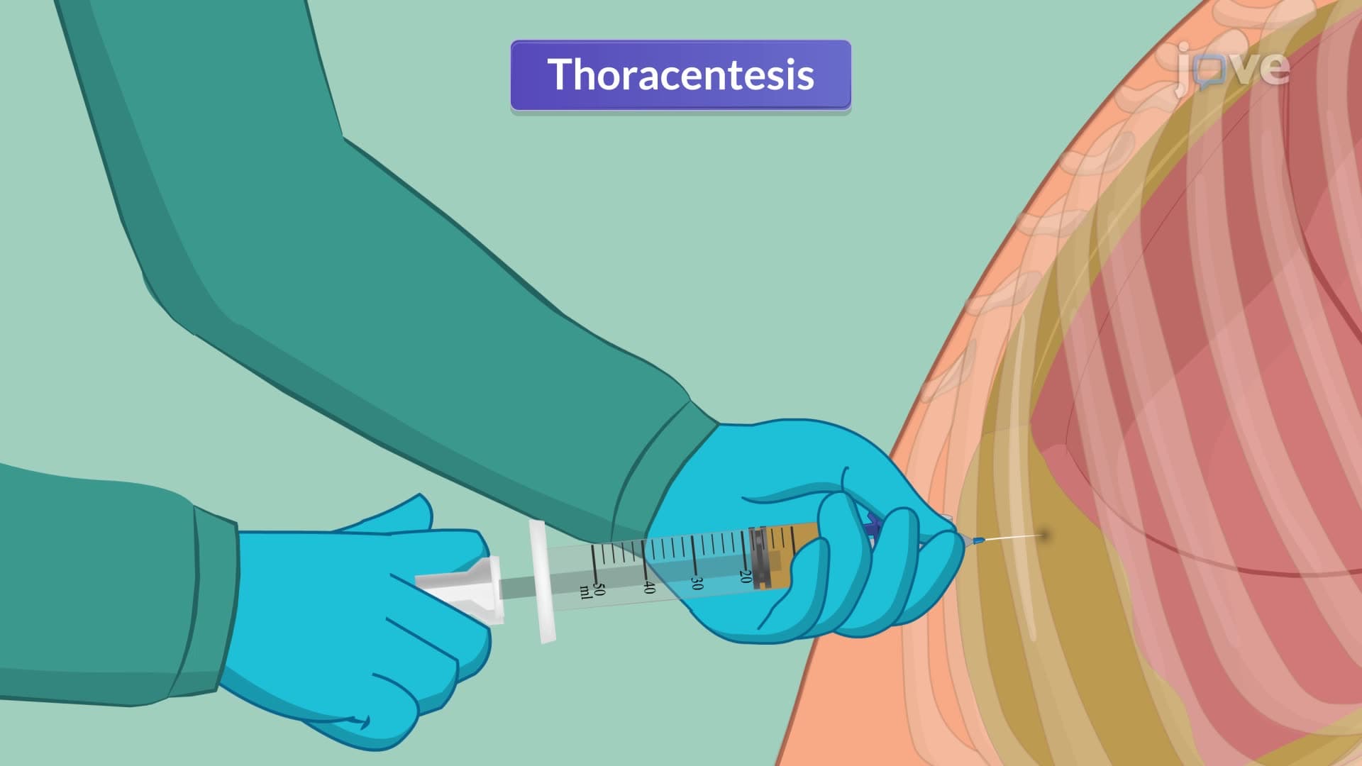
5. Thoracentesis and Pleural Procedures Thoracentesis involves large-bore needle insertion into pleural space under local anesthesia for both diagnostic fluid analysis and therapeutic fluid removal. Patient positioning requires upright sitting with arms supported on overbed tables, facilitating optimal needle insertion sites. The procedure addresses conditions like pleural effusion, pneumothorax, and empyema commonly seen in US intensive care units. Diagnostic analysis identifies infection, trauma, or malignancy through pleural fluid characteristics and laboratory studies. Therapeutic applications relieve respiratory distress from fluid accumulation. Nursing responsibilities encompass sterile technique maintenance, vital sign monitoring during fluid removal, and post-procedure chest X-ray verification following established US clinical protocols.
6. Blood Gas Analysis and Interpretation Arterial blood gas (ABG) analysis evaluates respiratory function through oxygen delivery and carbon dioxide removal assessment, while also indicating metabolic status via bicarbonate levels. Sample collection from radial, brachial, or femoral arteries requires heparinized syringes and immediate ice storage to prevent clotting. Venous blood gas (VBG) studies assess tissue oxygen consumption and delivery balance using pulmonary artery or central venous catheters. Critical pre-collection protocols include avoiding oxygen therapy changes and suctioning for 20 minutes before sampling. These studies are essential in US emergency departments and ICUs for managing respiratory failure, metabolic acidosis, and ventilator settings, with normal values and interpretation following American Association for Respiratory Care guidelines.
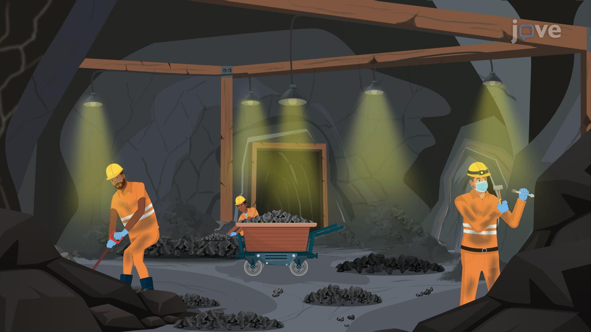
7. Pulmonary Function Testing and Spirometry Pulmonary function tests utilize spirometry to measure lung volumes and airflow rates, providing quantitative assessment of respiratory capacity and diagnosing conditions like asthma and COPD. The procedure involves patients breathing into spirometer mouthpieces while wearing nasal clips, performing various breathing maneuvers to measure forced vital capacity and expiratory volumes. Applications include chronic disease diagnosis, bronchodilator effectiveness evaluation, occupational screening in industries like coal mining, and pre-operative assessment for thoracic surgeries. Testing protocols require scheduling away from meals and withholding bronchodilators for 6 hours. Results interpretation follows American Thoracic Society standards, with normal values adjusted for age, gender, height, and ethnicity in diverse US patient populations.
8. Pulse Oximetry and Continuous Monitoring Pulse oximetry provides non-invasive continuous oxygen saturation monitoring using light absorption principles through fingertip, earlobe, or forehead sensors. Normal SpO2 values exceed 95%, with readings below 90% indicating tissue hypoxia requiring immediate intervention. The technology proves invaluable in US perioperative settings, critical care units, and emergency departments where anesthesia or decreased consciousness may mask hypoxia signs. Accuracy factors include patient motion, anemia, cold extremities, fluorescent lighting, nail polish, and skin pigmentation affecting light transmission. When accuracy is questioned, arterial blood gas analysis provides definitive oxygen saturation verification, following protocols established by organizations like the American Association for Respiratory Care and Joint Commission safety standards.