11 Concepts
5 Concepts
15 Concepts
11 Concepts
7 Concepts
10 Concepts
22 Concepts
11 Concepts
10 Concepts
8 Concepts
14 Concepts
17 Concepts
14 Concepts
19 Concepts
15 Concepts
16 Concepts
15 Concepts
7 Concepts
6 Concepts
7 Concepts
7 Concepts
27 Concepts
8 Concepts
9 Concepts
6 Concepts
10 Concepts
12 Concepts
10 Concepts
Cardiovascular diagnostic studies and procedures are essential tools used in US healthcare settings to assess heart function, detect cardiac abnormalities, and guide treatment decisions. This comprehensive JoVE Coach course covers blood-based cardiac diagnostic tests including troponin and lipid panels, electrocardiogram interpretation, advanced imaging techniques like echocardiography and cardiac CT, stress testing protocols, and invasive procedures such as cardiac catheterization used in American hospitals and clinics.
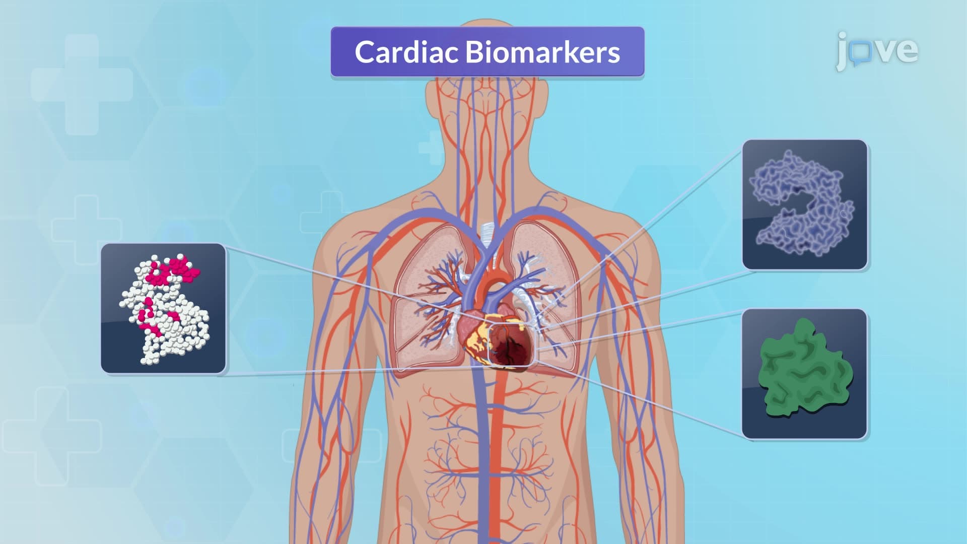
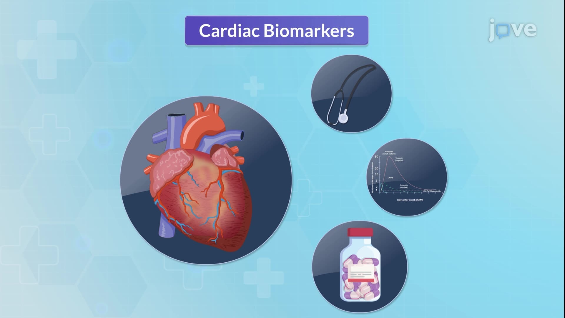
1. Cardiac Biomarker Analysis and Clinical Applications Understanding cardiac biomarkers is crucial for diagnosing acute coronary syndromes in US emergency departments. Troponins I and T are the gold standard markers, detectable within 4-6 hours post-myocardial injury and remaining elevated for 10-14 days, making them essential for both diagnosis and prognosis. High-sensitivity troponin assays used in American hospitals can detect cardiac injury as early as 90 minutes. Myoglobin serves as an early marker, rising within 1-3 hours but normalizing quickly. CK-MB is particularly valuable for detecting reinfarction due to its rapid normalization within 48 hours. Additional markers like BNP help distinguish cardiac from respiratory causes of dyspnea, while C-reactive protein indicates cardiovascular inflammation risk.
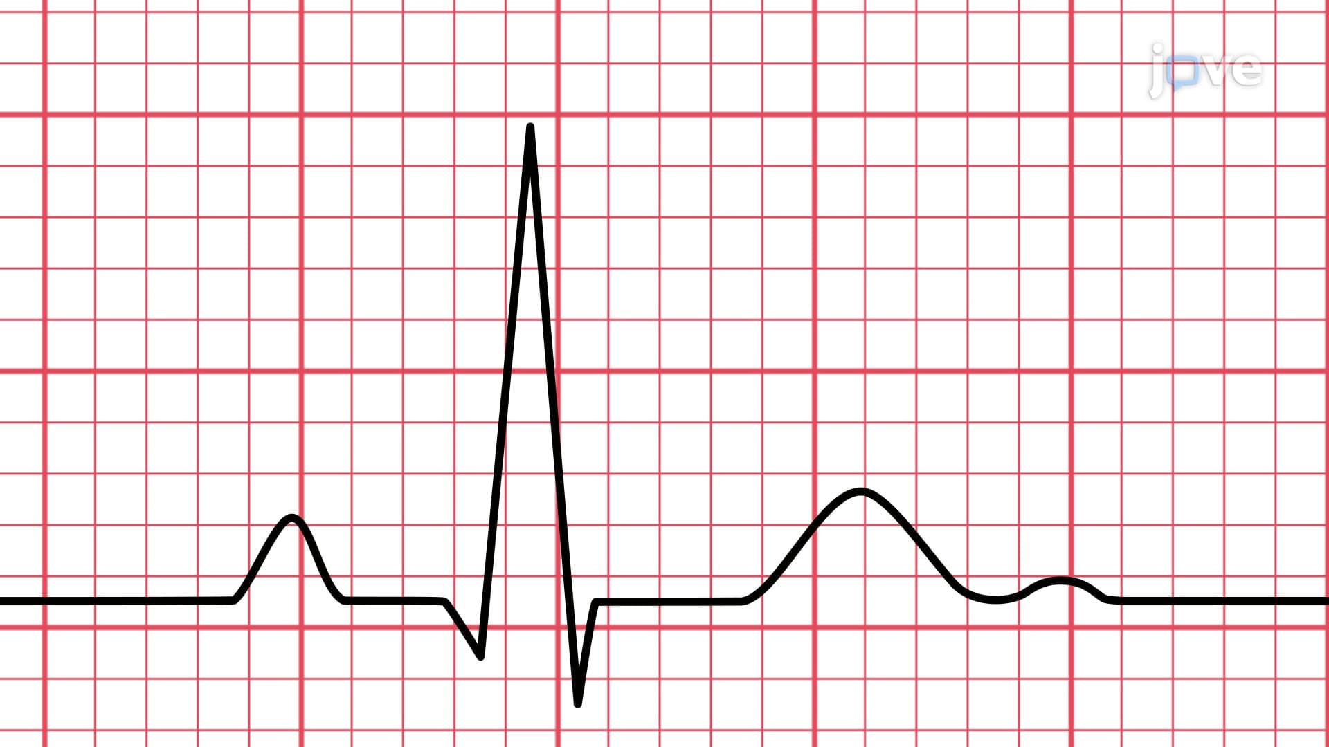
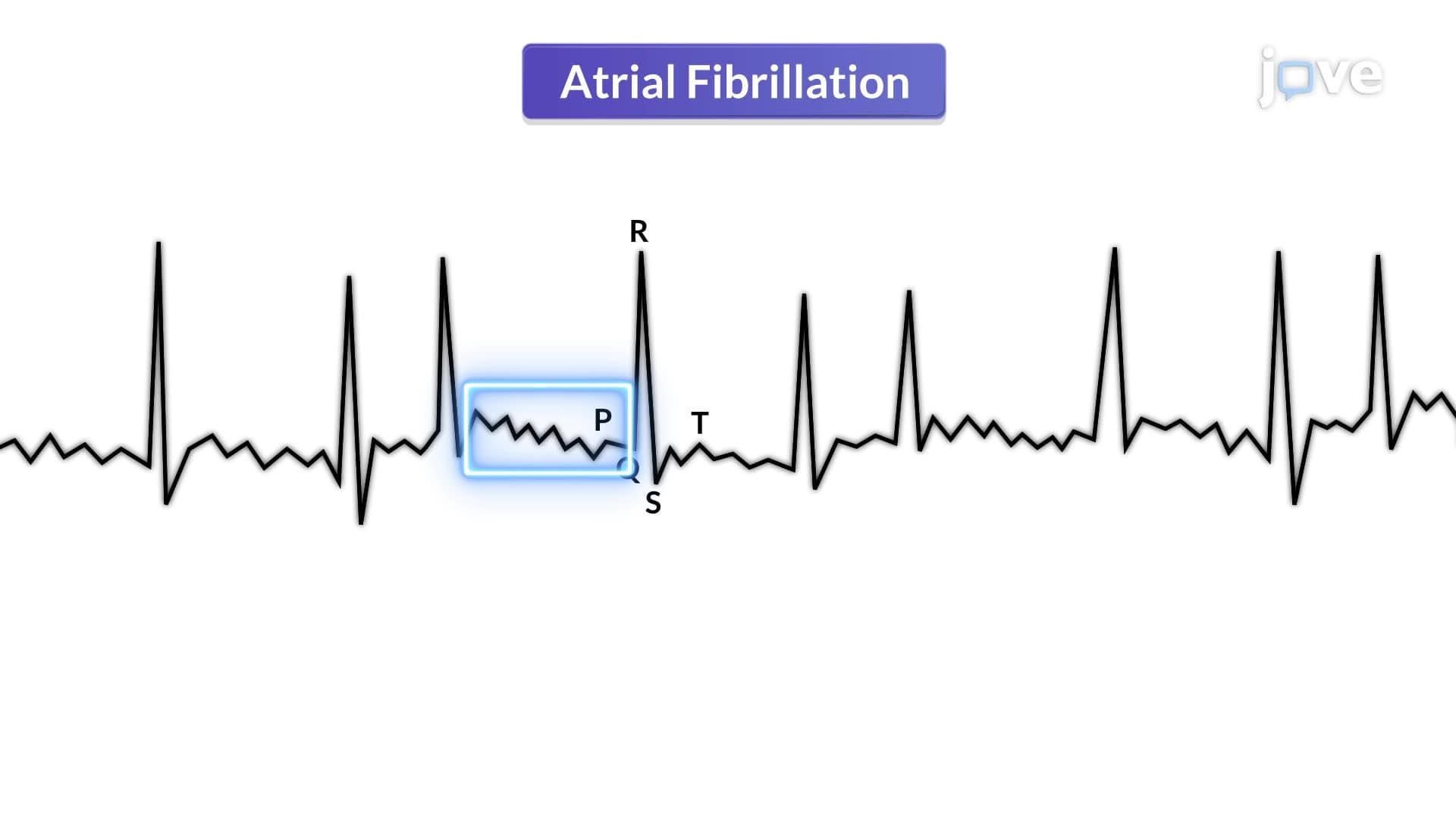
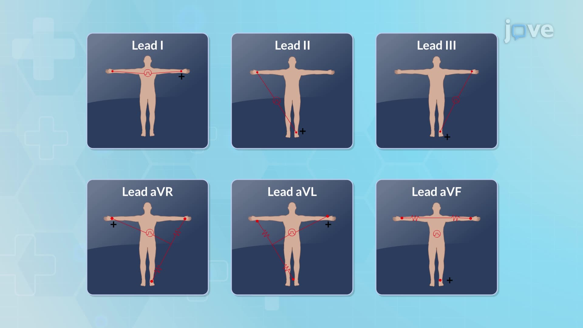
2. Electrocardiogram Fundamentals and Rhythm Interpretation The 12-lead ECG is the cornerstone of cardiac rhythm assessment in US healthcare facilities, utilizing standardized electrode placement including limb leads (I, II, III, aVR, aVL, aVF) and precordial leads (V1-V6). Understanding normal waveform characteristics is essential: P waves represent atrial depolarization, QRS complexes show ventricular depolarization (normally <0.12 seconds), and T waves indicate ventricular repolarization. Heart rate calculation using the 1500 small box method is standard practice. Recognition of common arrhythmias is vital, including sinus bradycardia (<60 bpm), sinus tachycardia (>100 bpm), atrial fibrillation with absent P waves, and dangerous ventricular arrhythmias requiring immediate intervention in American cardiac care units.
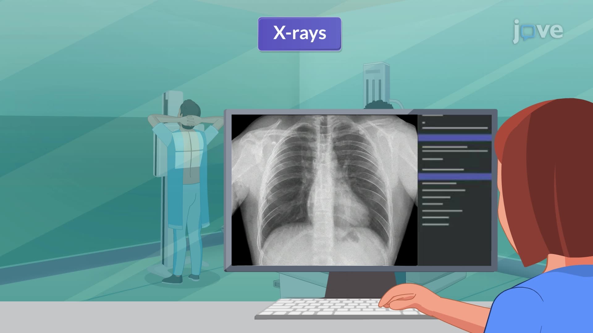
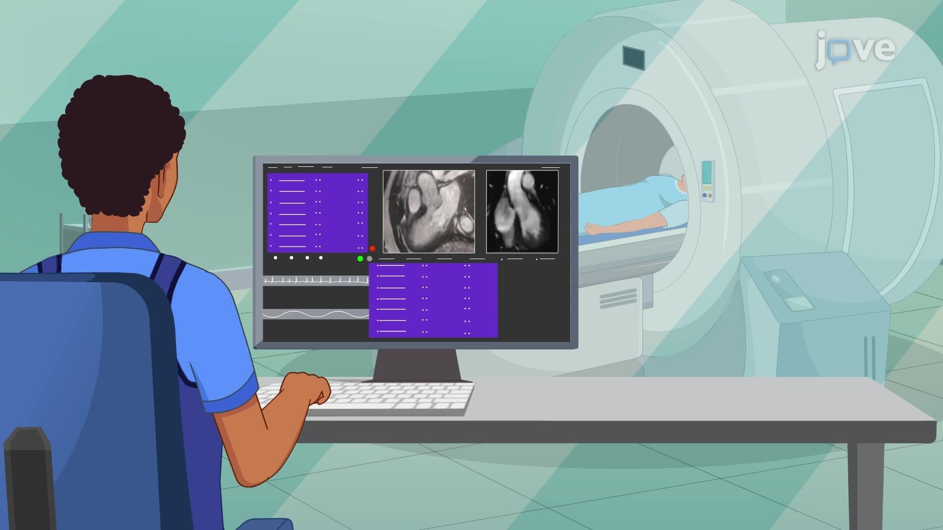
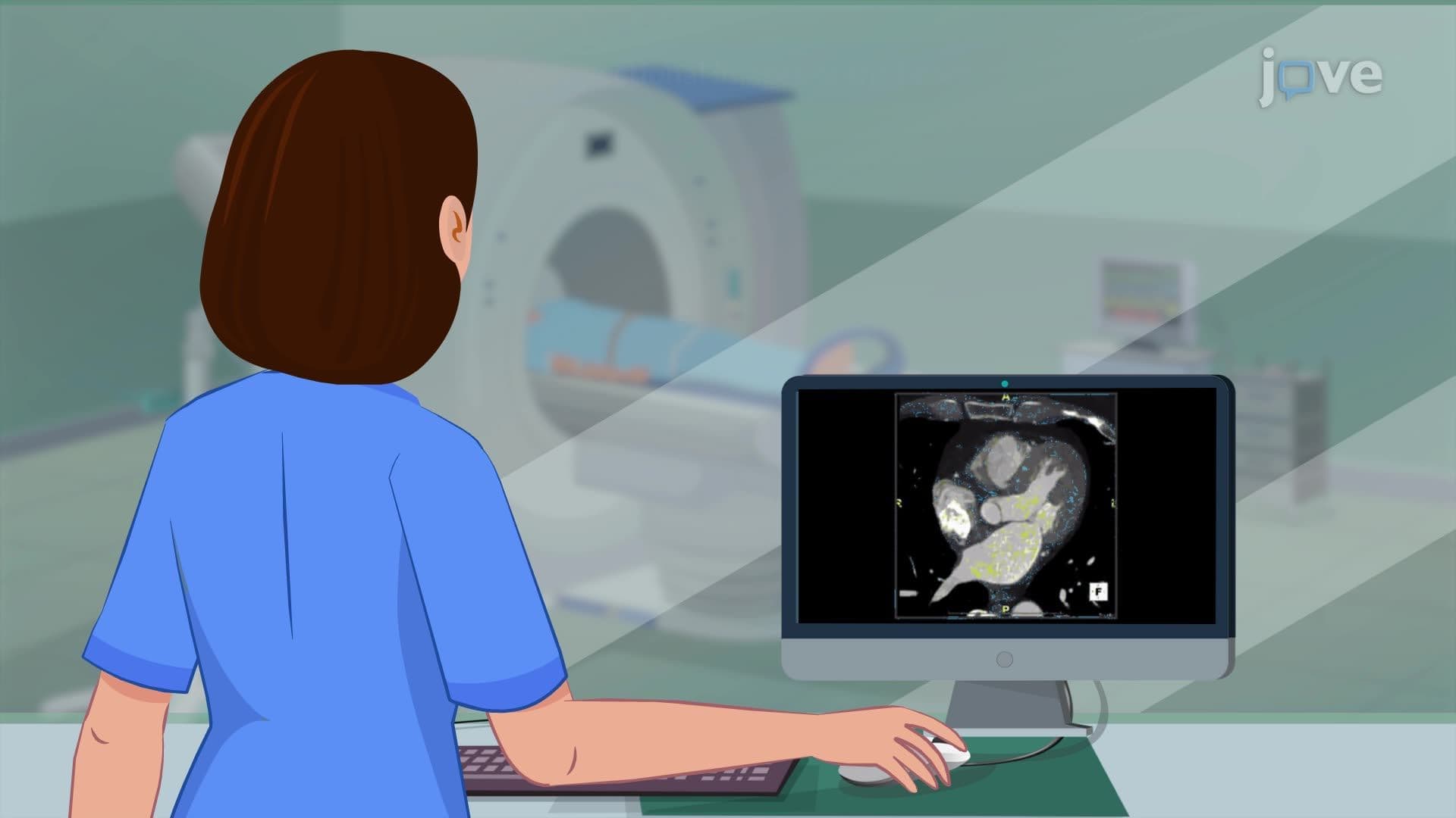
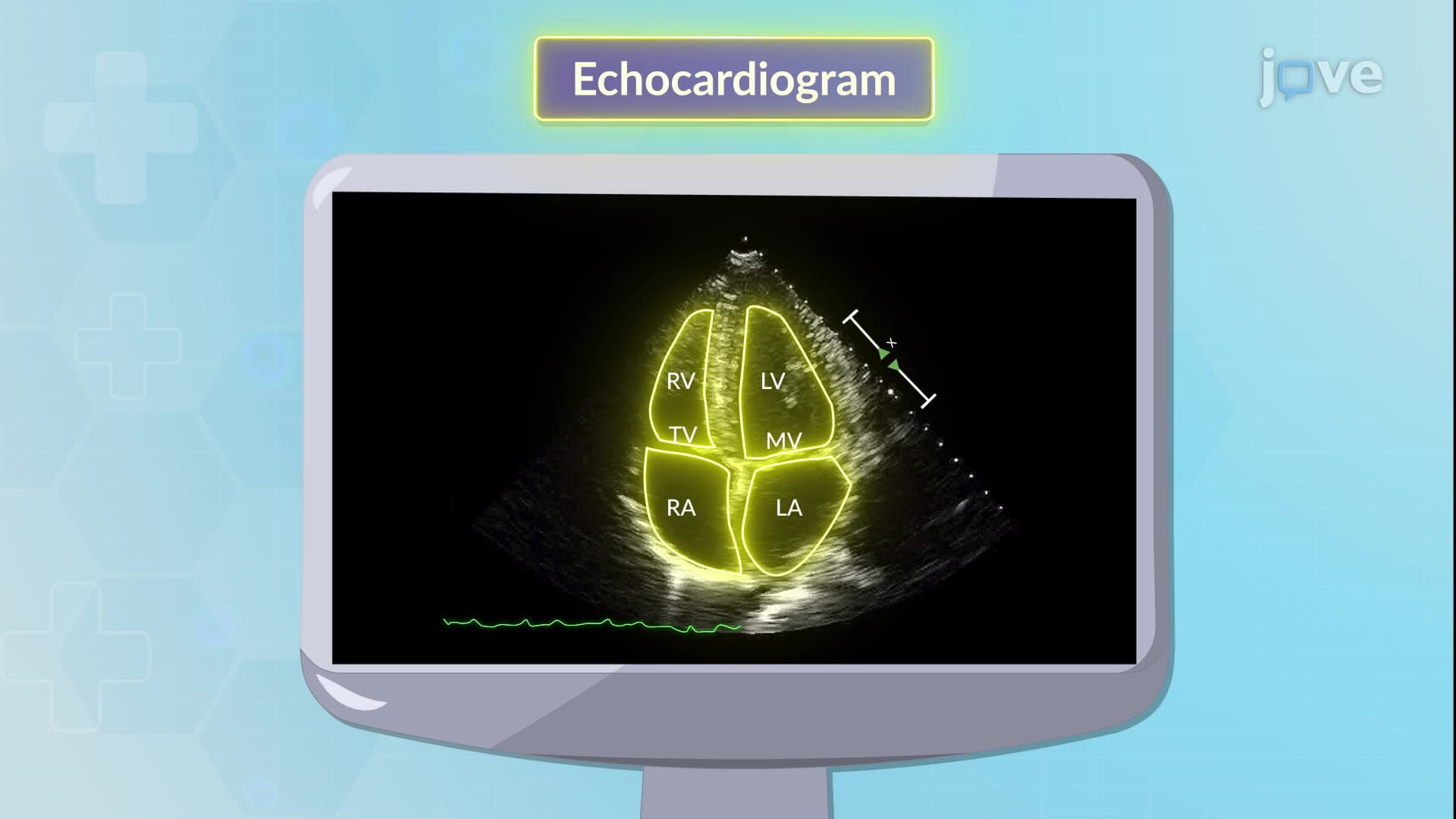
3. Advanced Cardiac Imaging Modalities and Applications Modern cardiovascular imaging in US hospitals encompasses multiple sophisticated techniques. Echocardiography remains the most widely used cardiac imaging modality, with transthoracic echo serving as the first-line approach for assessing wall motion, valve function, and ejection fraction. Transesophageal echocardiography provides superior visualization for valve assessment and blood clot detection. Stress echocardiography, using either exercise or pharmacologic stress with dobutamine, helps diagnose coronary artery disease. Cardiac CT angiography has revolutionized non-invasive coronary assessment, while calcium scoring CT quantifies coronary plaque burden. Cardiac MRI offers the highest soft tissue contrast for evaluating cardiomyopathies and congenital heart disease, though contraindications include non-MRI-safe pacemakers and defibrillators.
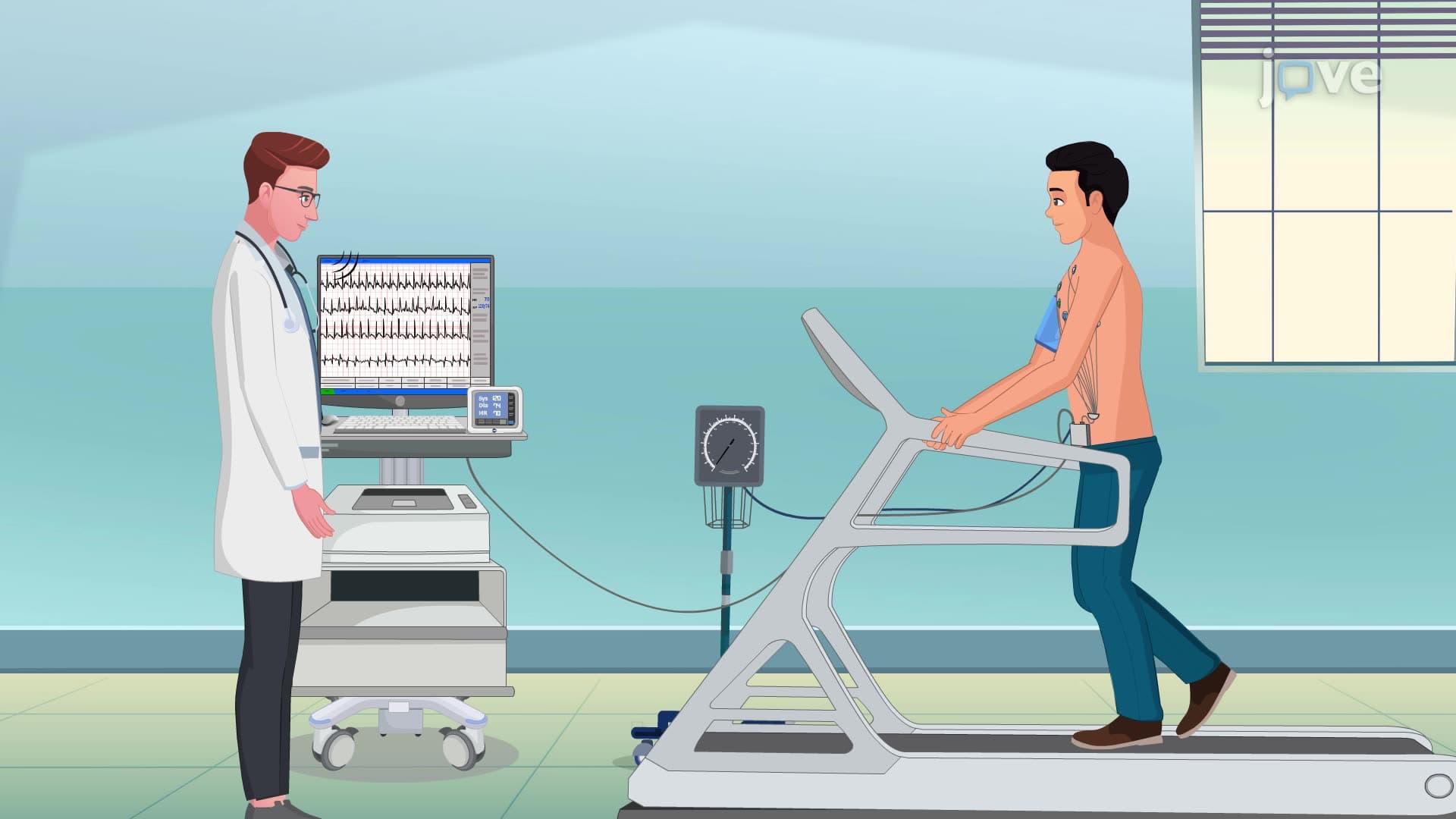
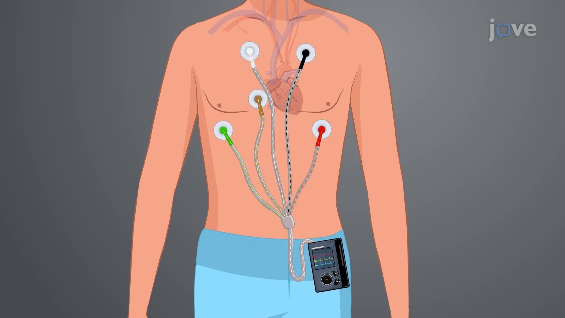
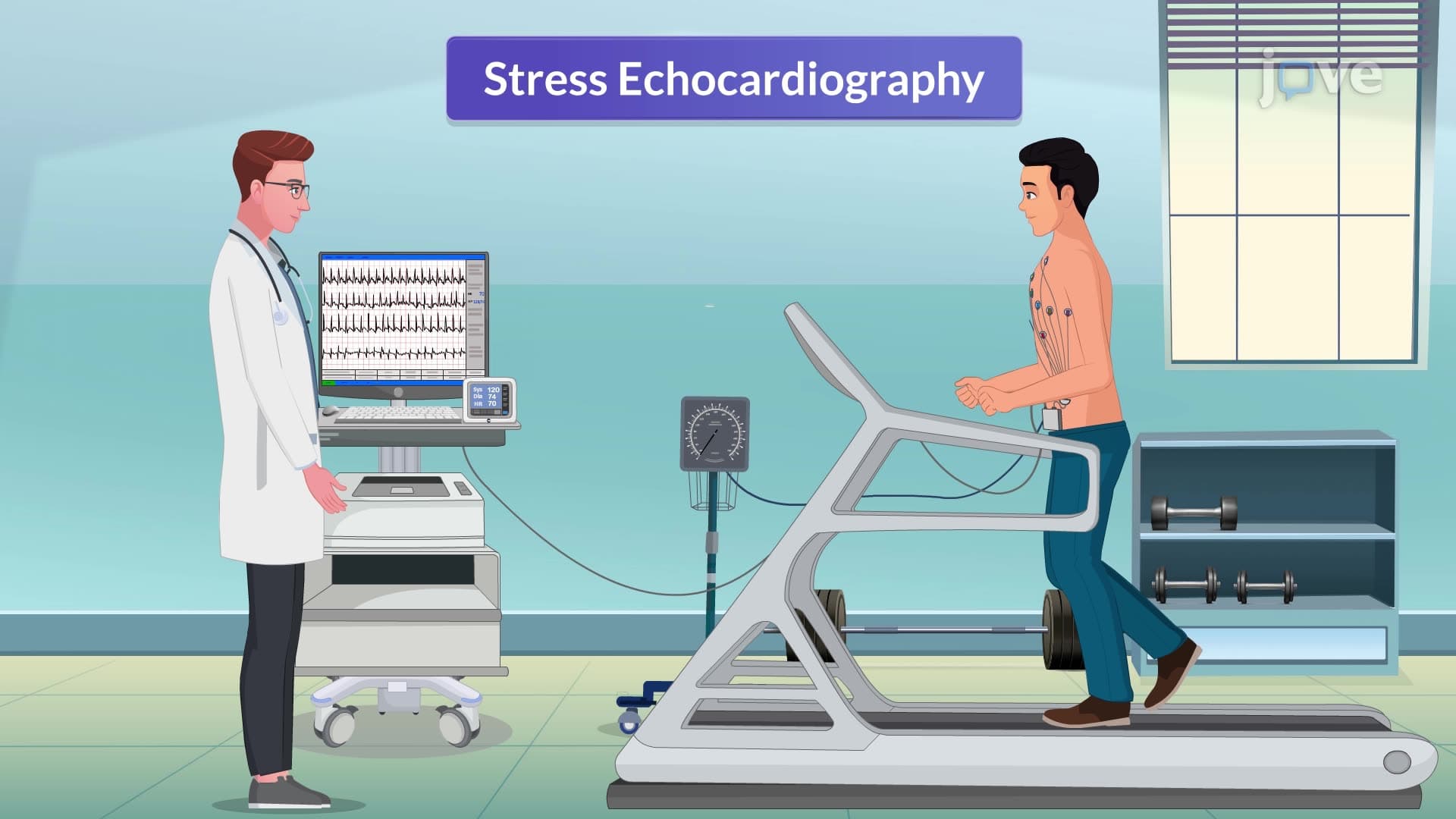
4. Exercise Stress Testing and Continuous Cardiac Monitoring Exercise stress testing is a fundamental diagnostic tool in American cardiology practices for evaluating coronary artery disease and exercise tolerance. Standard protocols involve progressive treadmill or bicycle exercise with continuous ECG monitoring, targeting 85% of age-predicted maximum heart rate. Pre-test preparation includes 3-hour fasting and 24-hour avoidance of stimulants. Positive tests show ST-segment depression ≥1mm, indicating myocardial ischemia requiring further evaluation. Holter monitoring provides 24-48 hour continuous ECG recording for detecting intermittent arrhythmias and silent ischemia. Patients maintain normal activities while wearing the portable device, keeping detailed symptom diaries for correlation with rhythm abnormalities. This outpatient monitoring is particularly valuable for evaluating palpitations, chest pain, and medication effectiveness.
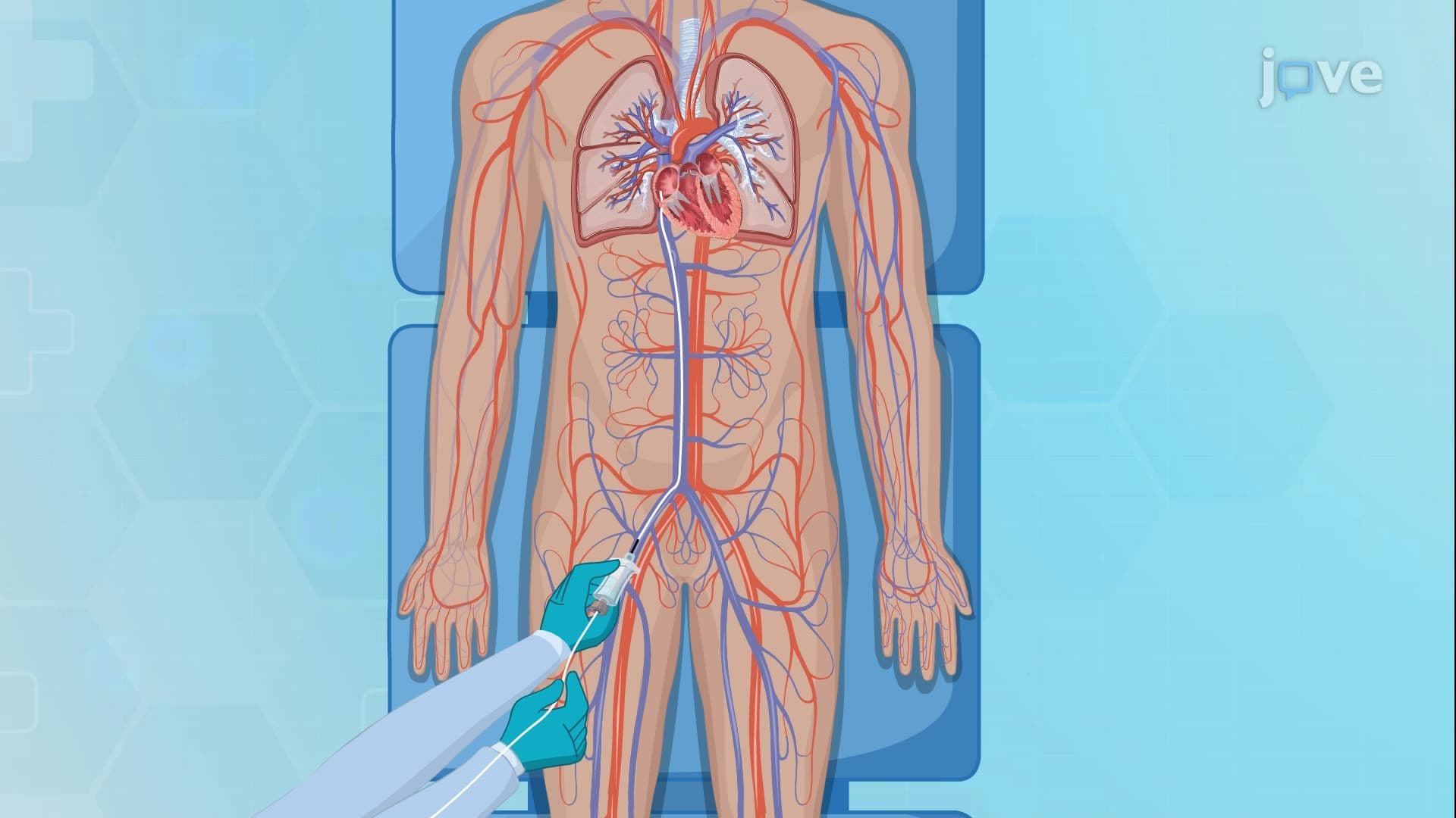
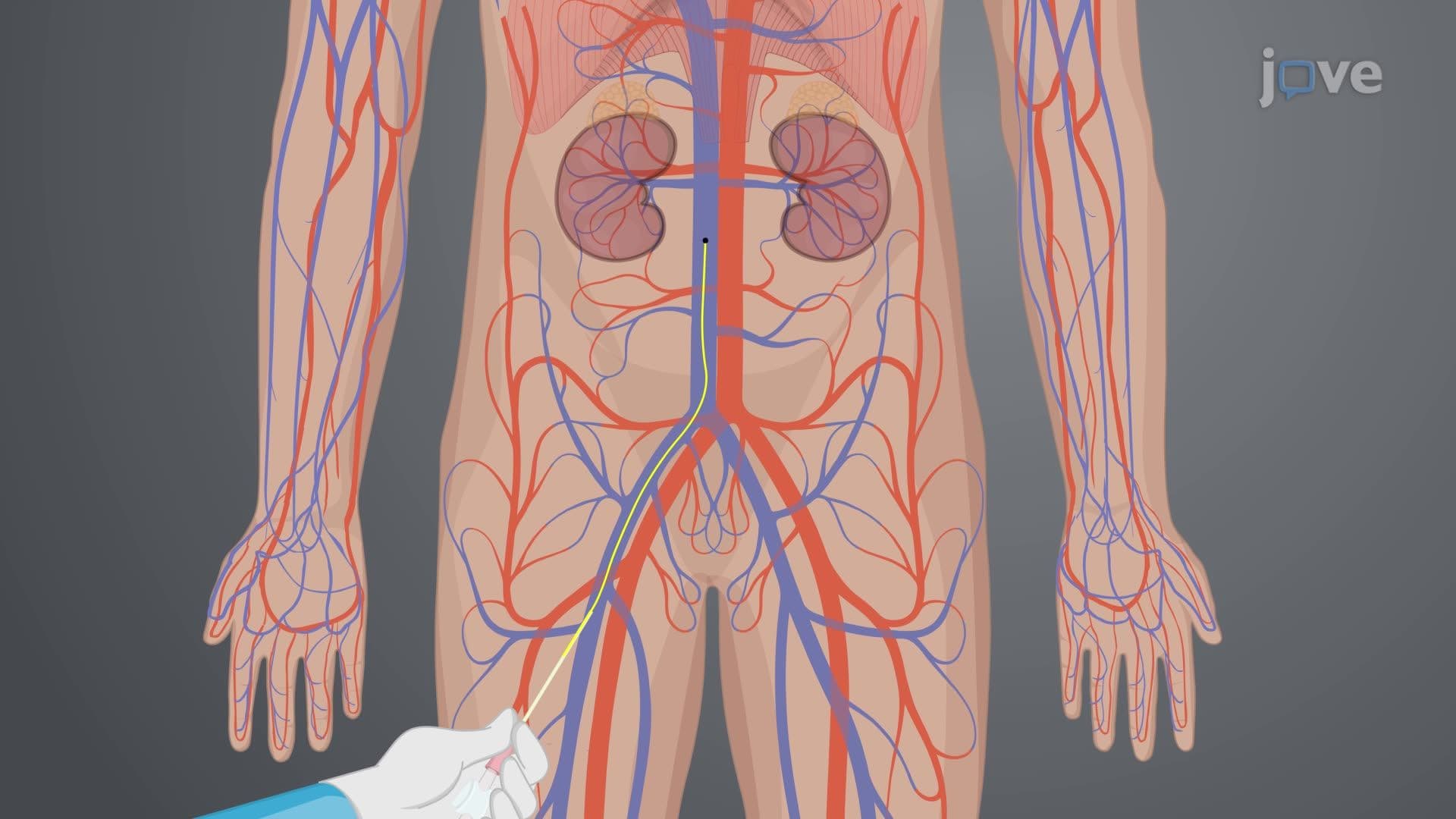
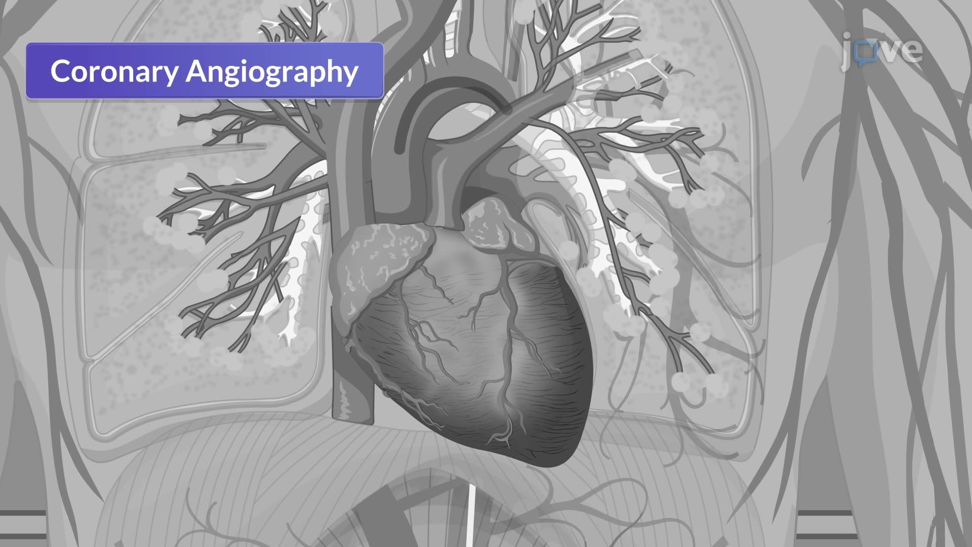
5. Cardiac Catheterization Procedures and Clinical Management Cardiac catheterization represents the gold standard for coronary artery evaluation in US cardiac centers, combining diagnostic and therapeutic capabilities. Right heart catheterization measures pulmonary pressures and cardiac output through venous access via femoral, jugular, or brachial veins. Left heart catheterization evaluates coronary anatomy and left ventricular function through arterial access, commonly via radial or femoral arteries. Coronary angiography involves selective contrast injection into coronary arteries under fluoroscopic guidance. Pre-procedural assessment includes contrast allergy history, renal function evaluation, and coagulation studies. Post-procedural care focuses on access site monitoring, hemostasis maintenance, and complication prevention. Nursing management involves vital sign monitoring, neurovascular assessments, and patient education about activity restrictions and warning signs requiring immediate medical attention.