11 Concepts
5 Concepts
15 Concepts
11 Concepts
7 Concepts
10 Concepts
22 Concepts
11 Concepts
10 Concepts
8 Concepts
14 Concepts
17 Concepts
14 Concepts
19 Concepts
15 Concepts
16 Concepts
15 Concepts
7 Concepts
6 Concepts
7 Concepts
7 Concepts
27 Concepts
8 Concepts
9 Concepts
6 Concepts
10 Concepts
12 Concepts
10 Concepts
Gastric disorders encompass a range of conditions affecting the stomach and duodenum, including gastritis and peptic ulcer disease. This comprehensive course, delivered through JoVE Coach, examines the pathophysiology, clinical manifestations, and evidence-based management strategies essential for healthcare professionals in US clinical settings.
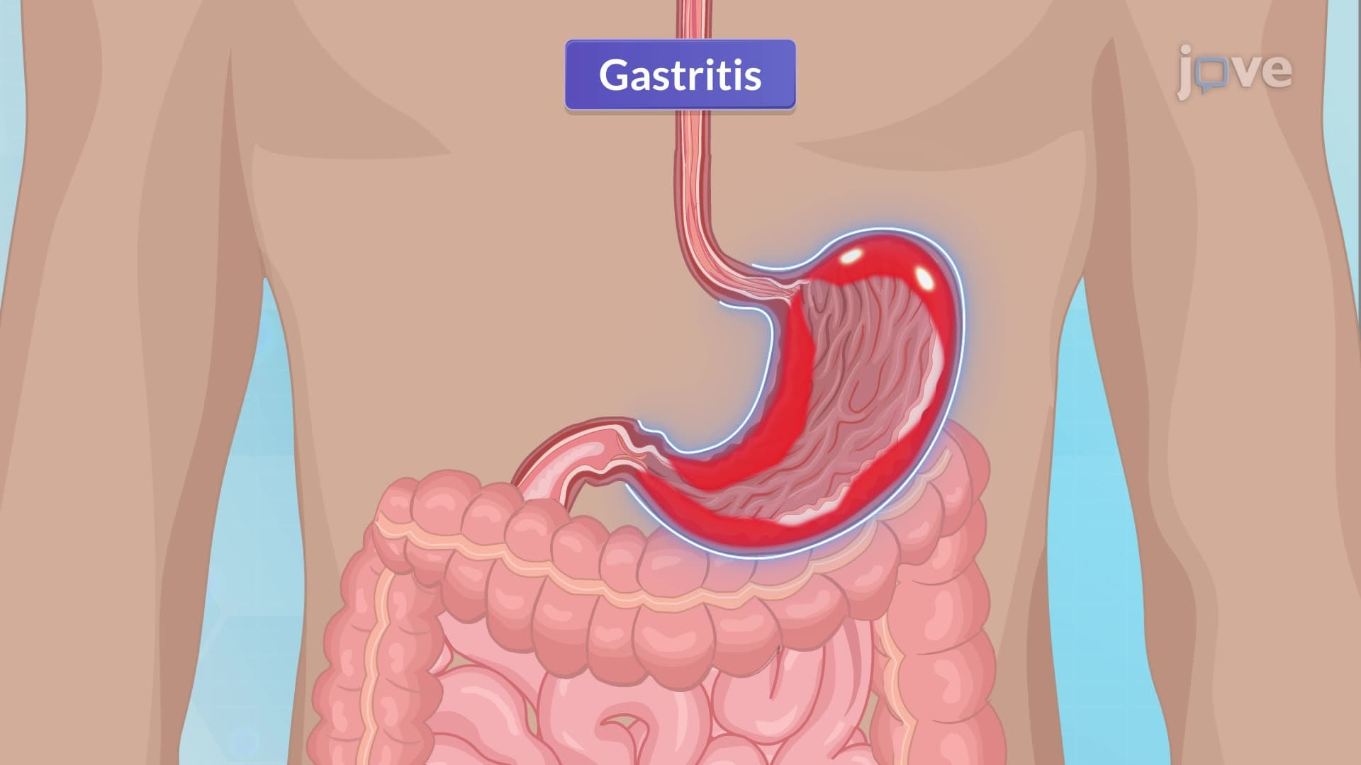
1. Gastritis Classification and Etiology Gastritis represents inflammation of the gastric mucosa, classified into three distinct types. Acute gastritis involves transient mucosal inflammation triggered by corrosive agents, medications like digoxin, alcohol consumption, or severe burns. Chronic gastritis develops gradually over months to years, primarily caused by Helicobacter pylori infections or autoimmune conditions such as Hashimoto's thyroiditis. Reactive gastritis results from prolonged NSAID use, alcohol exposure, or duodenal reflux, often remaining asymptomatic until endoscopic evaluation. Understanding these classifications helps healthcare providers in US hospitals identify appropriate treatment pathways for patients presenting with gastric symptoms.
2. H. pylori Pathophysiology and Adaptation Helicobacter pylori demonstrates remarkable adaptation to the stomach's acidic environment through urease enzyme production. This spiral-shaped bacterium converts urea to ammonia, creating alkaline microenvironments that neutralize gastric acid locally. Once established, H. pylori triggers inflammatory responses while releasing cytotoxins that damage epithelial cells. The organism's flagella enable motility through gastric mucus, allowing colonization of the antrum and corpus regions. This pathophysiology explains why H. pylori infections require combination antibiotic therapy, as seen in treatment protocols used across US medical centers for peptic ulcer disease management.
3. NSAID-Induced Gastric Injury Mechanisms Non-steroidal anti-inflammatory drugs compromise gastric mucosal integrity through cyclooxygenase-1 inhibition, reducing protective prostaglandin synthesis. Prostaglandins normally maintain mucosal blood flow, stimulate mucus and bicarbonate secretion, and promote epithelial cell regeneration. NSAID-induced prostaglandin depletion creates vulnerability to gastric acid damage, leading to erosions and ulceration. This mechanism underlies clinical guidelines recommending proton pump inhibitor co-therapy for patients requiring long-term NSAID treatment. US healthcare providers routinely assess NSAID use history when evaluating patients with gastric symptoms, particularly elderly populations at higher risk.
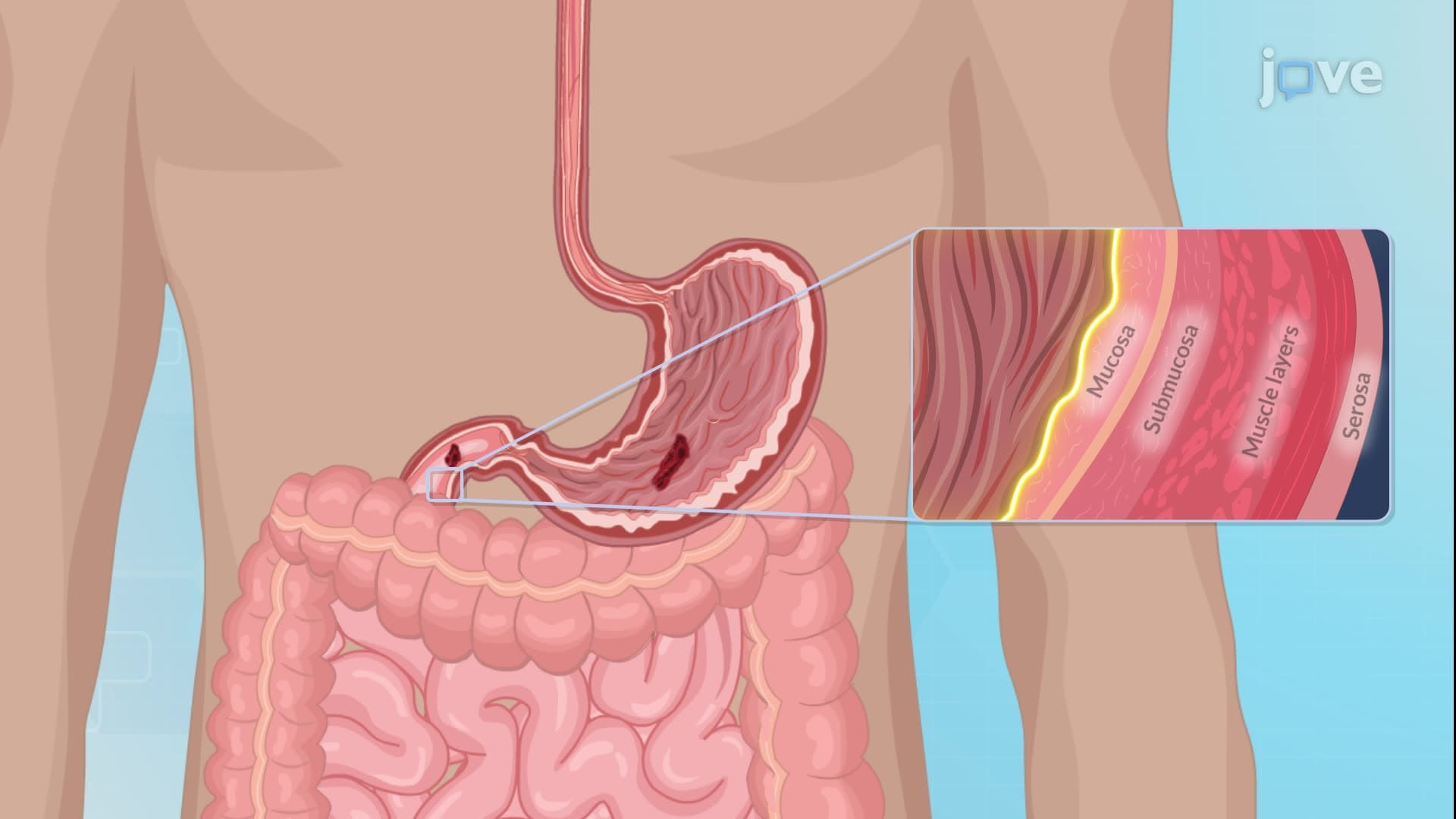
4. Peptic Ulcer Disease Pathogenesis Peptic ulcer formation results from imbalanced aggressive factors and mucosal defense mechanisms. The gastric mucosa maintains a protective gel barrier preventing hydrochloric acid and pepsin penetration under normal conditions. Aggressive factors including H. pylori toxins, NSAIDs, alcohol, and excessive acid secretion can overwhelm defensive mechanisms such as mucus production, bicarbonate buffering, and mucosal blood flow. This imbalance allows hydrogen ion back-diffusion, causing epithelial cell injury and ulcer development. Understanding this pathophysiology guides therapeutic approaches emphasizing both acid suppression and mucosal protection in US clinical practice.
5. Clinical Manifestations and Diagnostic Differentiation Gastric and duodenal ulcers present distinct symptom patterns crucial for accurate diagnosis. Gastric ulcers typically cause burning epigastric pain shortly after meals, while duodenal ulcers produce cramping mid-epigastric pain 2-3 hours post-meal, often relieved by food intake. Complications include hematemesis, melena, perforation with sudden severe abdominal pain, and gastric outlet obstruction causing vomiting. Diagnostic approaches in US hospitals utilize endoscopy for direct visualization, rapid urease testing for H. pylori detection, and imaging studies to identify complications. Early recognition of these manifestations enables prompt intervention, preventing serious complications requiring emergency surgical management.
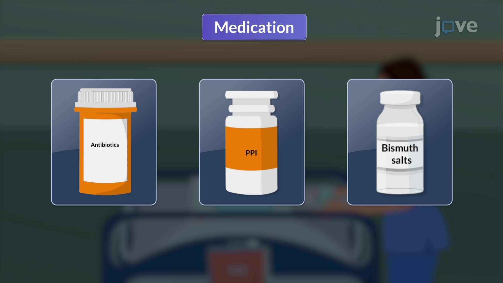
6. Evidence-Based Medical Management Contemporary peptic ulcer disease management follows standardized protocols emphasizing H. pylori eradication when present. Triple therapy combines two antibiotics (typically clarithromycin and metronidazole) with proton pump inhibitors for 10-14 days. Quadruple therapy adds bismuth salts and tetracycline for resistant cases. Zollinger-Ellison syndrome requires specialized management with high-dose PPIs or octreotide for gastrin hypersecretion control. Cytoprotective agents like sucralfate provide additional mucosal protection. US treatment guidelines emphasize antibiotic resistance testing and patient compliance monitoring to optimize eradication rates and prevent recurrence.
7. Surgical Interventions and Techniques Surgical management becomes necessary for complications including perforation, obstruction, or refractory bleeding. Vagotomy procedures reduce gastric acid secretion by transecting vagal nerve branches, often combined with pyloroplasty to prevent gastric stasis. Partial gastrectomy options include antrectomy (distal third removal) or subtotal gastrectomy (middle and distal thirds). Reconstruction involves gastroduodenostomy (Billroth I) or gastrojejunostomy (Billroth II) procedures. These techniques, commonly performed in US surgical centers, require careful patient selection and comprehensive preoperative evaluation to minimize complications and optimize outcomes.
8. Comprehensive Nursing Care and Monitoring Nursing care for gastric and duodenal conditions encompasses comprehensive assessment, intervention, and patient education strategies. Preoperative preparation includes NPO status maintenance, anxiety reduction, and baseline vital sign documentation. Postoperative monitoring focuses on vital signs, nasogastric tube management for gastric decompression, and intake/output recording. Nurses assess for complications including anastomotic leaks (abdominal pain, fever), hemorrhage (hematemesis, hypotension), and infection (surgical site redness, drainage). Patient education addresses dietary modifications, medication compliance, and symptom recognition. This holistic approach, standard in US healthcare facilities, promotes optimal recovery and prevents readmissions.