11 Concepts
5 Concepts
15 Concepts
11 Concepts
7 Concepts
10 Concepts
22 Concepts
11 Concepts
10 Concepts
8 Concepts
14 Concepts
17 Concepts
14 Concepts
19 Concepts
15 Concepts
16 Concepts
15 Concepts
7 Concepts
6 Concepts
7 Concepts
7 Concepts
27 Concepts
8 Concepts
9 Concepts
6 Concepts
10 Concepts
12 Concepts
10 Concepts
Genitourinary system assessment forms a critical foundation for nursing practice in the United States healthcare system. This comprehensive course examines GU anatomy from kidneys to urethra, physiological processes including glomerular filtration and tubular reabsorption, and systematic clinical assessment techniques. Students master inspection, palpation, percussion, and auscultation methods essential for detecting urological disorders in hospital and outpatient settings with JoVE Coach.
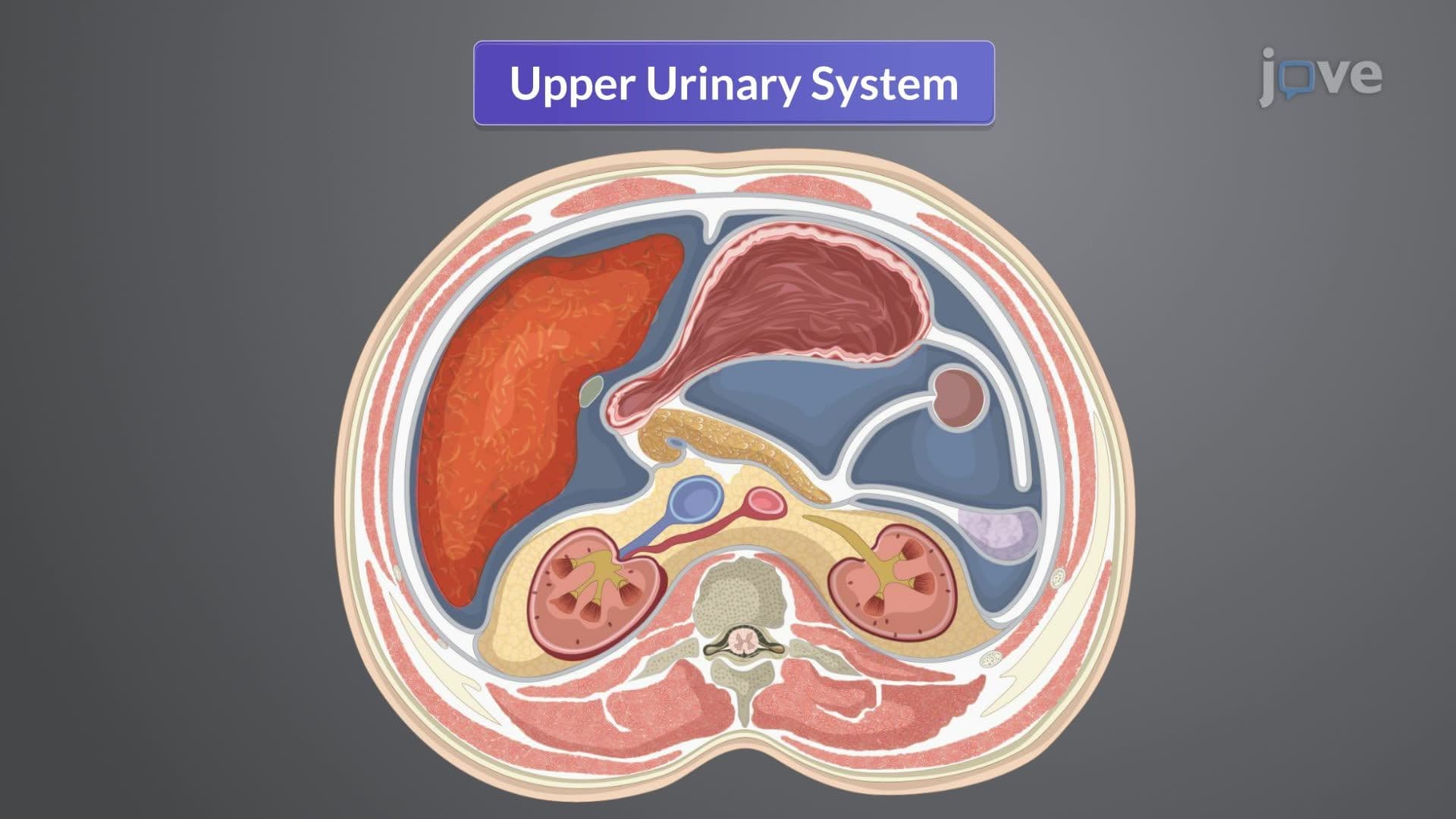
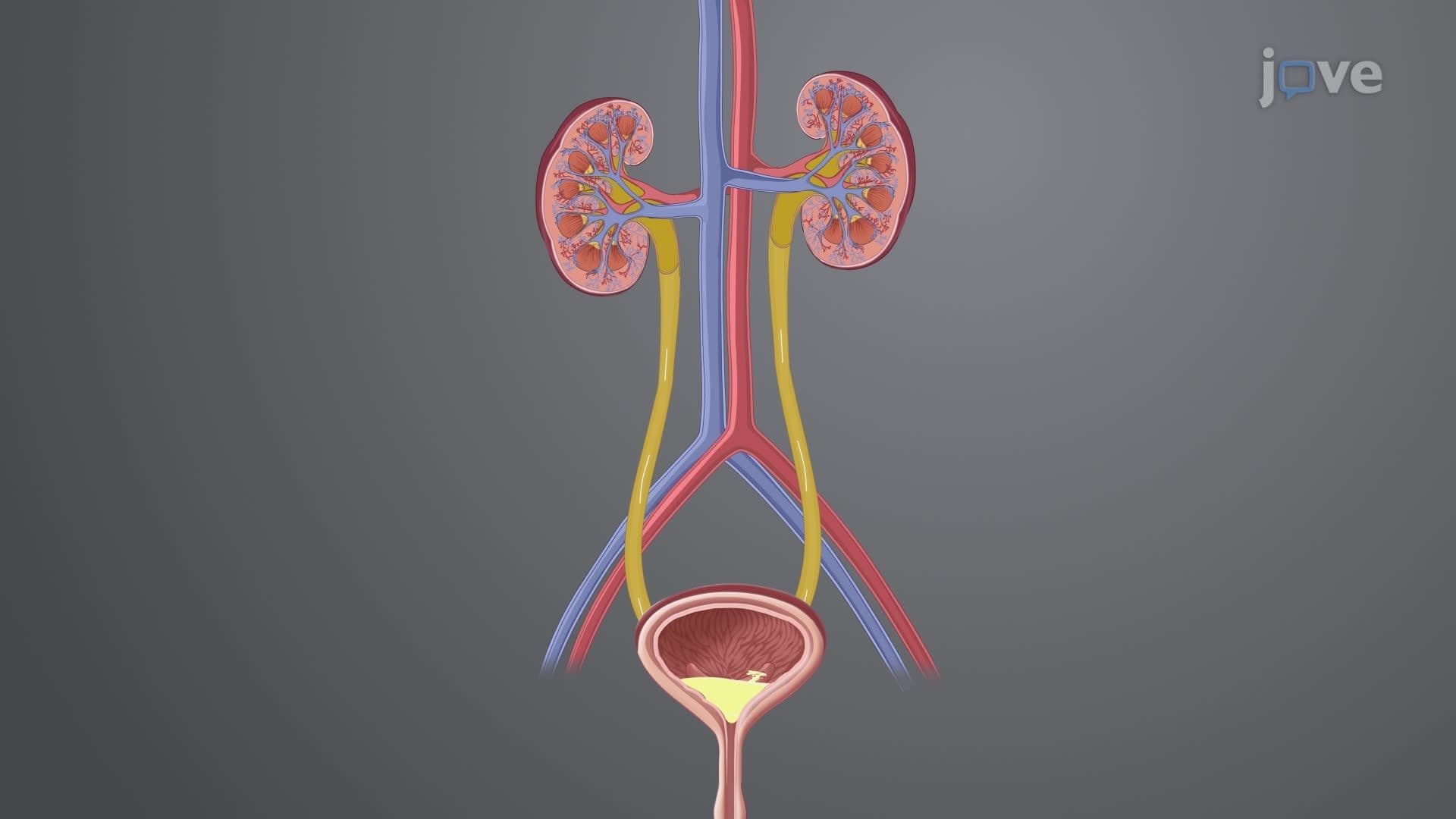
1. Upper Urinary System Anatomy: The kidneys and ureters form the upper urinary tract, with each kidney containing approximately one million nephrons housed within the cortex and medulla. The renal capsule provides protection while the retroperitoneal location shields these vital organs from injury. Each ureter extends 25-30 centimeters from the renal pelvis to the bladder, featuring three distinct layers that facilitate urine transport through peristaltic contractions. Understanding this anatomy helps nurses recognize conditions like kidney stones, which commonly lodge at ureter constriction points during passage to the bladder.
2. Lower Urinary System Structure: The bladder serves as a muscular reservoir capable of storing 400-500 milliliters of urine, with the detrusor muscle contracting during micturition. The trigone region at the bladder base represents a critical anatomical landmark where ureters enter and the urethra exits. Urethral length differs significantly between males (20-25 cm) and females (2.5-5 cm), influencing infection susceptibility and catheterization procedures. The internal and external urethral sphincters provide voluntary and involuntary control over urination, essential knowledge for managing incontinence issues in clinical practice.
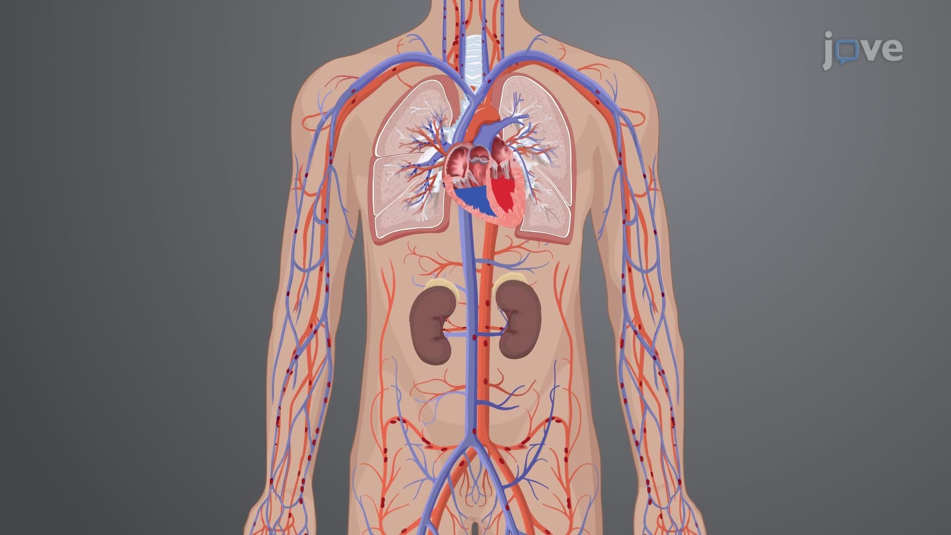
3. Renal Blood Flow and Filtration: The kidneys receive 20-25% of cardiac output, approximately 1200 mL per minute, demonstrating their metabolic importance in maintaining homeostasis. Glomerular filtration rate (GFR) averaging 125 mL per minute serves as a key indicator of kidney function, with values below 60 mL/min/1.73m² indicating chronic kidney disease. The filtration process occurs at the glomerular-capillary membrane, where blood pressure forces water and small solutes into Bowman's capsule. Nurses must understand GFR calculations when administering medications cleared by the kidneys, such as digoxin and certain antibiotics commonly used in US hospitals.
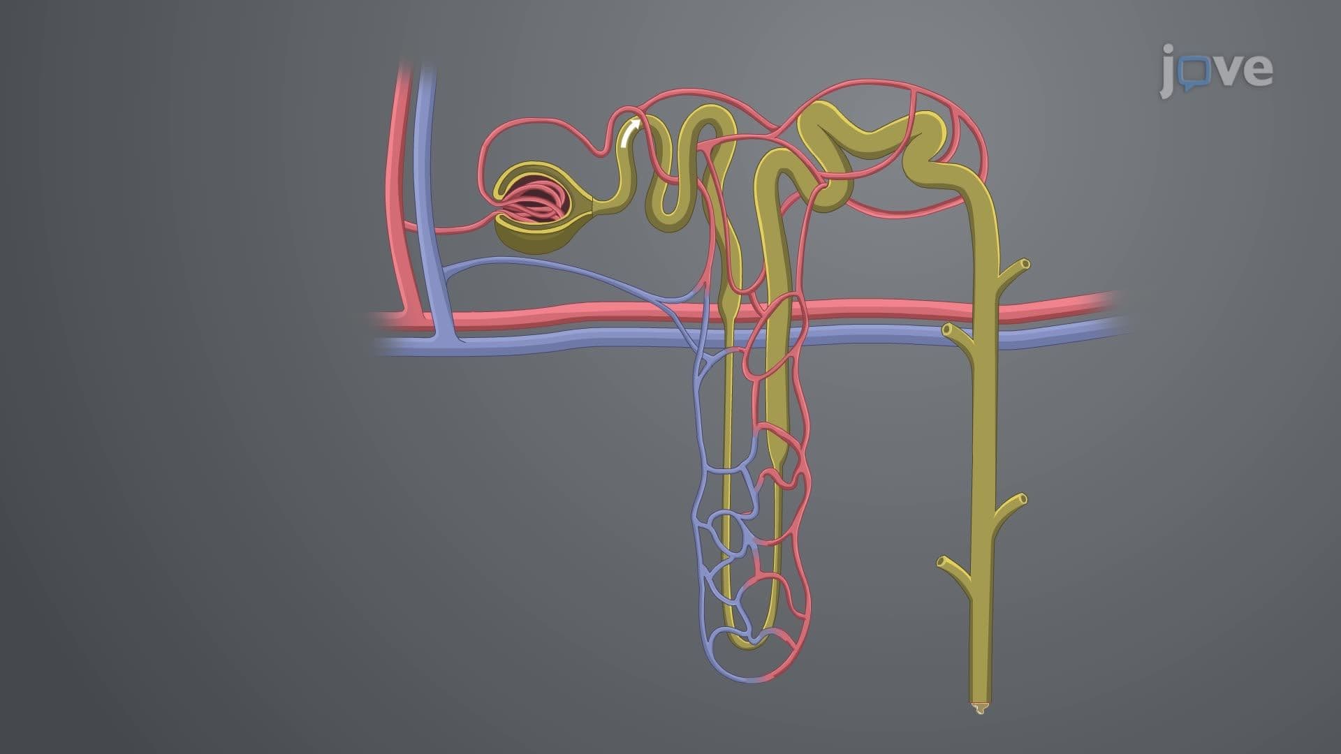
4. Tubular Reabsorption and Secretion Mechanisms: The nephron reabsorbs 99% of the 180 liters of filtrate produced daily, with the proximal convoluted tubule handling glucose, amino acids, and 65% of sodium and water reabsorption. The loop of Henle creates the concentration gradient essential for urine concentration, while the distal convoluted tubule fine-tunes electrolyte balance under hormonal control. Aldosterone regulates sodium retention and potassium excretion, critical for managing patients with heart failure or hypertension. Understanding these processes helps nurses recognize medication side effects and interpret laboratory values like serum creatinine and blood urea nitrogen levels.
5. Urine Concentration and Hormonal Regulation: The countercurrent multiplication system in the loop of Henle establishes the medullary osmotic gradient necessary for concentrating urine during dehydration states. Antidiuretic hormone (ADH) from the hypothalamus increases water reabsorption by inserting aquaporin-2 channels in collecting duct cells, producing concentrated urine with osmolality up to 1200 mOsm/kg. Diabetes insipidus results from ADH deficiency, while syndrome of inappropriate ADH (SIADH) causes excessive water retention. Nurses monitoring patients receiving diuretics or those with fluid restrictions must understand these mechanisms to prevent complications like hyponatremia or dehydration.
6. Comprehensive Health History Assessment: Systematic genitourinary assessment begins with detailed questioning about urinary symptoms, including frequency, urgency, dysuria, hematuria, and nocturia patterns. Sexual history requires sensitivity while gathering information about sexually transmitted infection risks, contraceptive use, and reproductive concerns. Past medical history should explore diabetes mellitus, hypertension, and autoimmune conditions that affect kidney function. Family history screening identifies genetic risks like polycystic kidney disease or hereditary nephritis. Medication reconciliation must include nephrotoxic drugs, herbal supplements, and over-the-counter medications that can impair renal function in US patients.
7. Physical Examination Techniques: Inspection begins with observing for uremic signs like pale, yellow-gray skin discoloration and uremic frost in advanced kidney disease cases. Palpation techniques for kidney assessment require proper hand placement between the 12th rib and iliac crest, with normal kidneys rarely palpable except in thin individuals. Bladder palpation above the symphysis pubis can detect distention exceeding 150 mL capacity. Costovertebral angle tenderness assessment helps identify pyelonephritis or kidney stones, common conditions seen in US emergency departments. These examination skills form the foundation for detecting genitourinary abnormalities before they progress to serious complications.