11 Concepts
5 Concepts
15 Concepts
11 Concepts
7 Concepts
10 Concepts
22 Concepts
11 Concepts
10 Concepts
8 Concepts
14 Concepts
17 Concepts
14 Concepts
19 Concepts
15 Concepts
16 Concepts
15 Concepts
7 Concepts
6 Concepts
7 Concepts
7 Concepts
27 Concepts
8 Concepts
9 Concepts
6 Concepts
10 Concepts
12 Concepts
10 Concepts
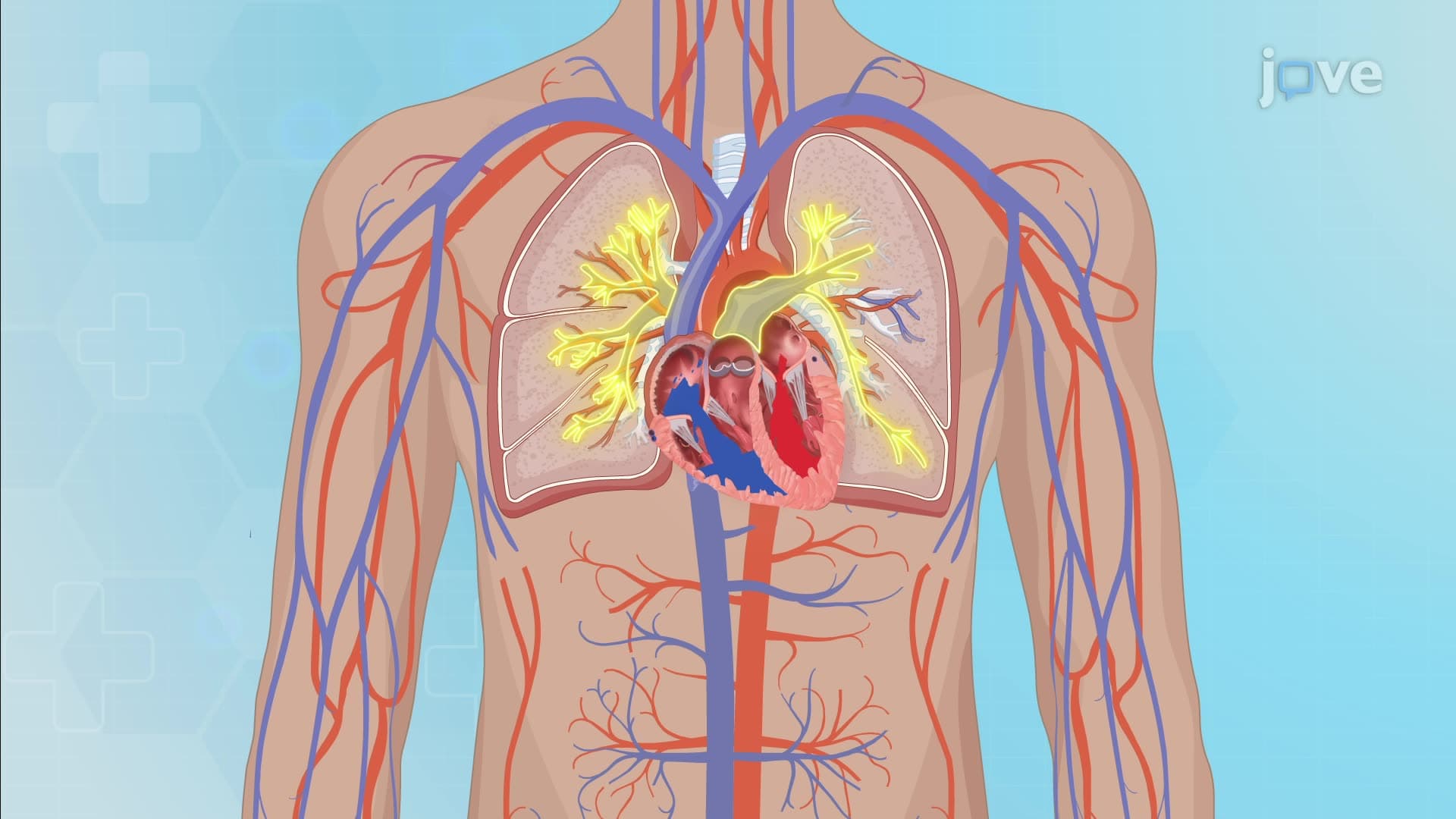
The cardiovascular system forms the foundation of human physiology, encompassing heart anatomy, blood vessel structure, cardiac electrical conduction, and hemodynamic regulation. This comprehensive course combines cardiovascular assessment nursing techniques with clinical overview of the cardiovascular system principles. Students master cardiac assessment skills, ECG interpretation basics, heart sounds identification, and cardiovascular nursing fundamentals essential for healthcare practice. JoVE Coach provides interactive learning experiences that prepare students for real-world clinical applications.
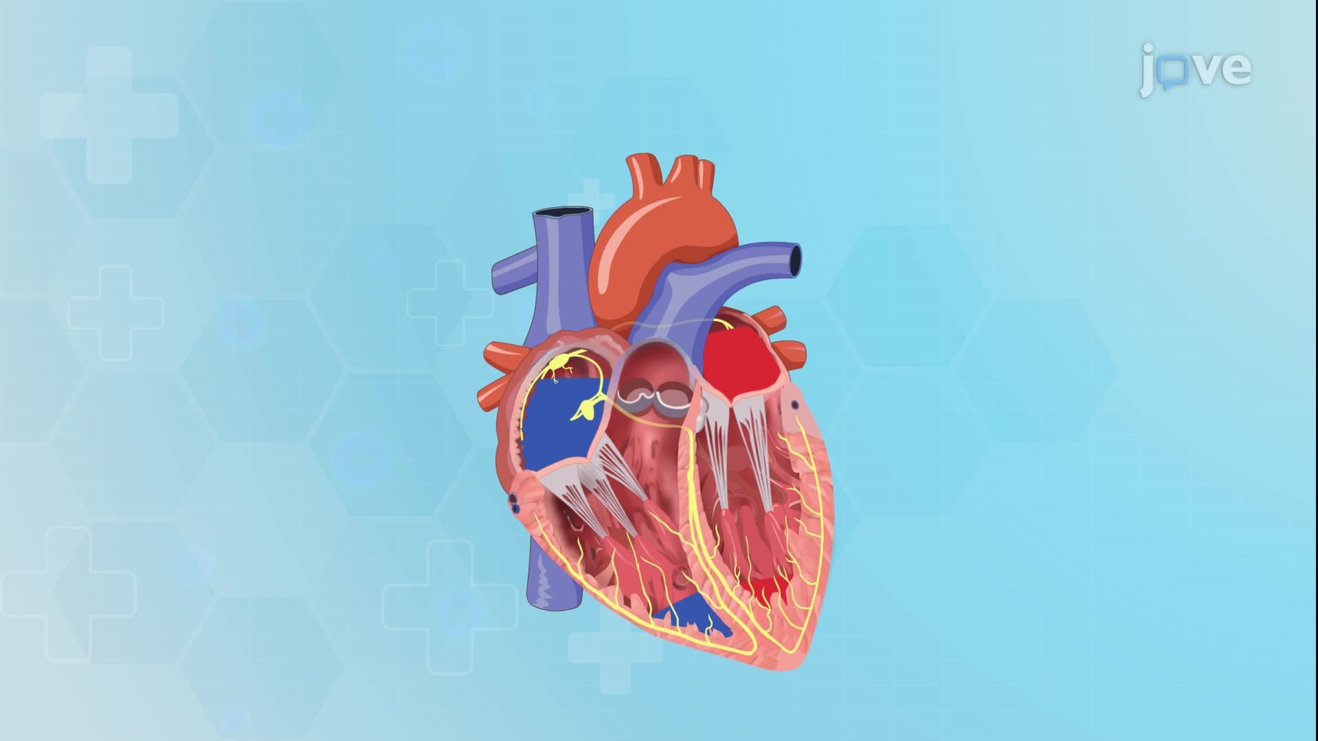
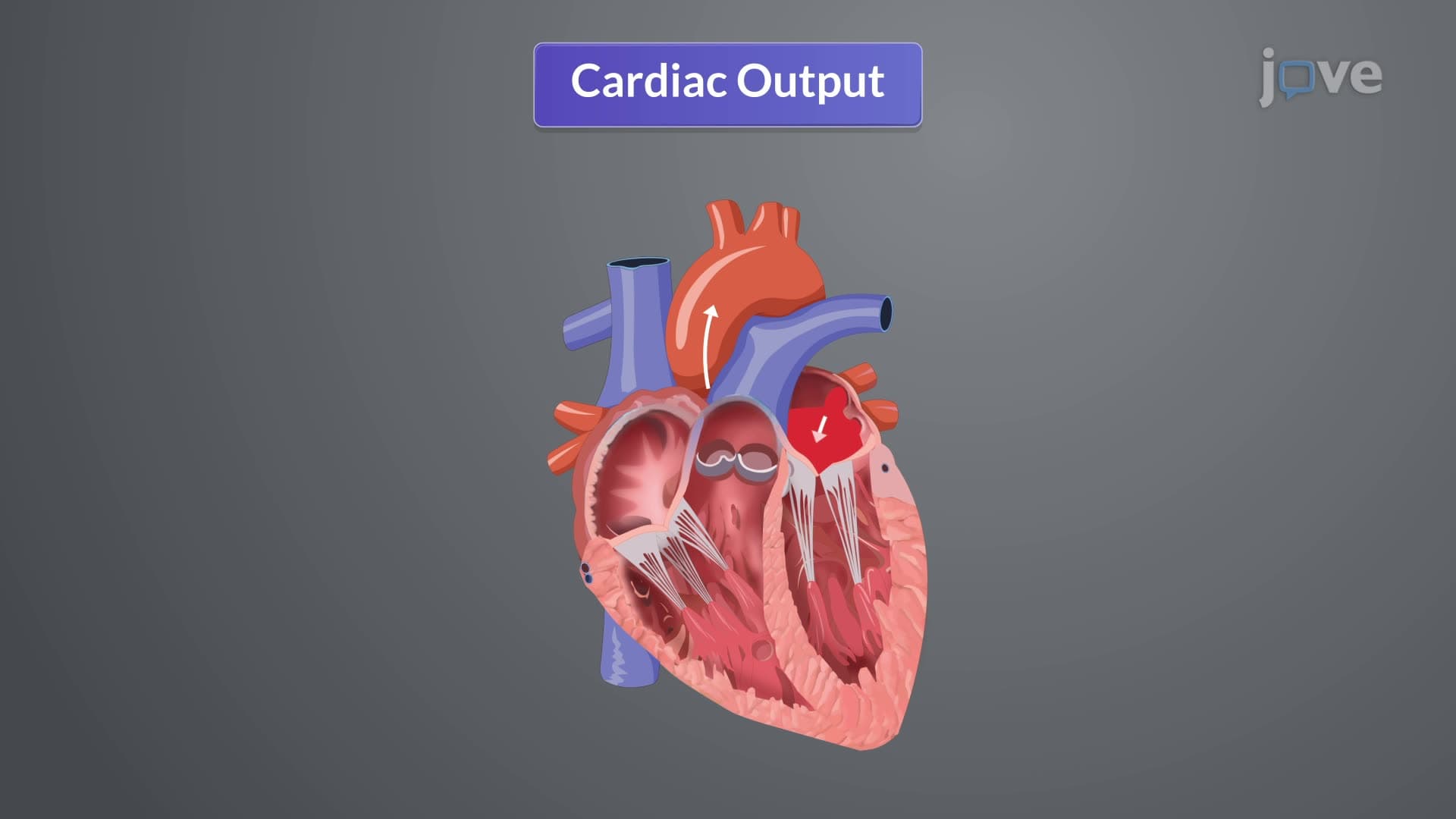
1. Heart Anatomy and Structure: The human heart consists of three distinct layers - epicardium (outer), myocardium (middle muscular), and endocardium (inner lining). Four chambers include right and left atria for blood collection, and right and left ventricles for blood ejection. The tricuspid valve controls flow between right atrium and ventricle, while the mitral valve regulates left-sided flow. Semilunar valves - pulmonary and aortic - prevent backflow during ventricular relaxation. Major vessels include superior and inferior vena cavae returning deoxygenated blood, and pulmonary veins delivering oxygenated blood from lungs.
2. Blood Vessel Organization and Function: The vascular network begins with the aorta, the body's largest artery with thick elastic walls that accommodate high-pressure blood flow. Arteries progressively branch into smaller arterioles that regulate blood pressure and tissue perfusion. Microscopic capillaries facilitate nutrient and gas exchange between blood and tissues through their thin walls. Venules collect deoxygenated blood and merge into larger veins with thinner walls and one-way valves. The superior and inferior vena cavae return blood to the right atrium, completing the circulatory loop essential for maintaining cellular homeostasis.
3. Cardiac Cycle Mechanics: The cardiac cycle represents one complete heartbeat consisting of three phases. Diastole involves ventricular relaxation with AV valves open and semilunar valves closed, allowing passive ventricular filling. Atrial systole follows SA node stimulation, actively pushing additional blood into ventricles. Ventricular systole involves powerful ventricular contraction, closing AV valves and opening semilunar valves to eject blood into pulmonary and systemic circulation. This coordinated sequence ensures unidirectional blood flow and maintains approximately 70 beats per minute in healthy adults at rest.
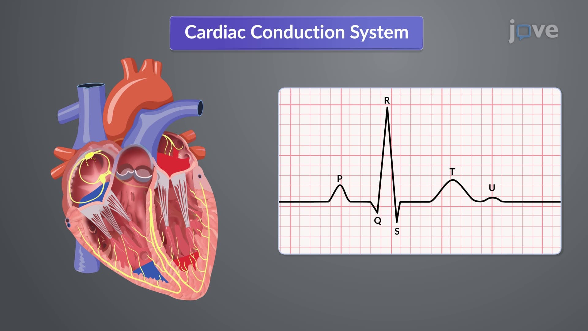
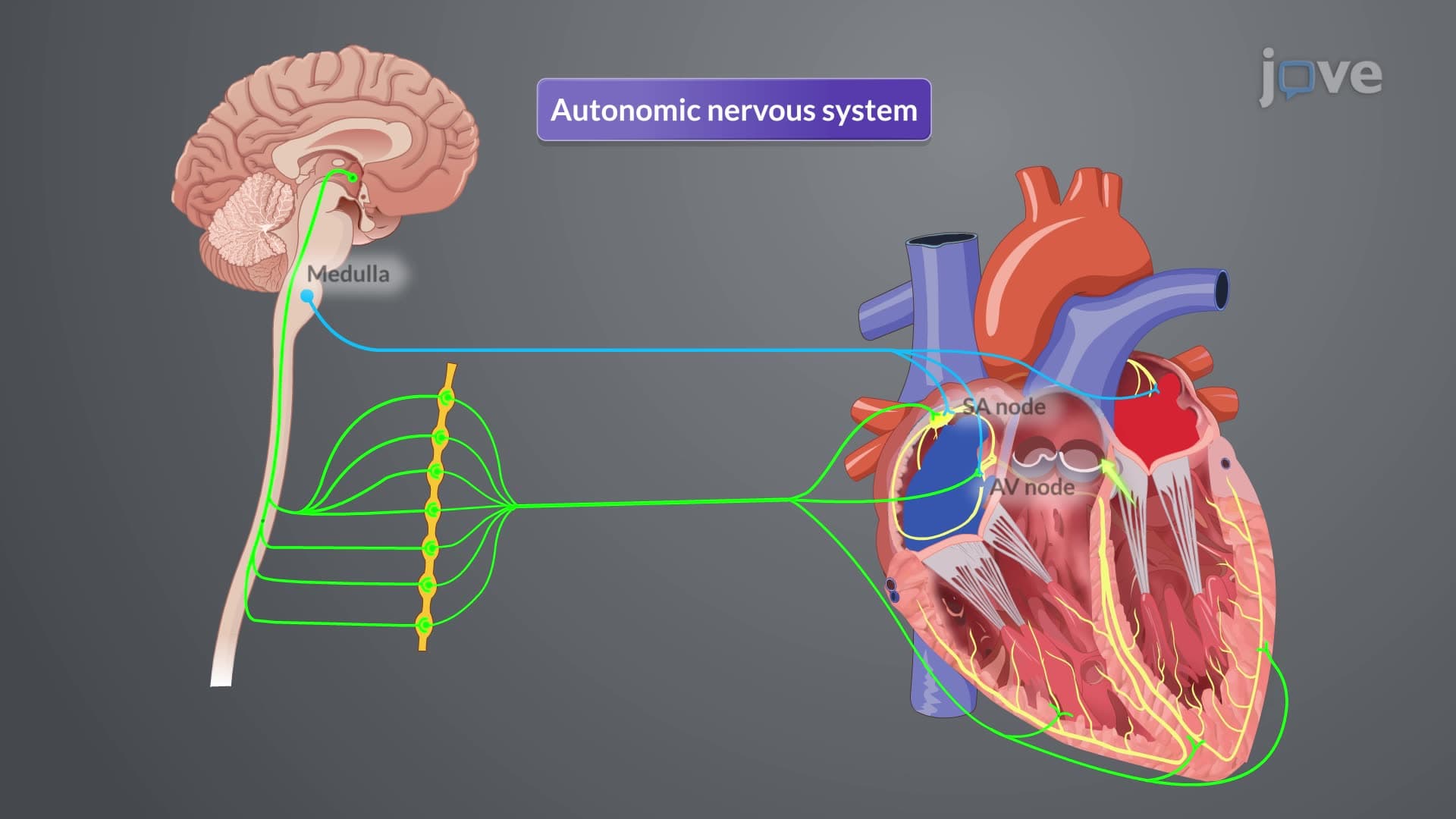
4. Electrical Conduction System: Cardiac electrical activity originates at the sinoatrial (SA) node, located where the superior vena cava meets the right atrium. Electrical impulses spread through internodal pathways and Bachmann's bundle to coordinate atrial contraction. The atrioventricular (AV) node introduces a crucial delay, allowing complete atrial emptying before ventricular activation. Impulses then travel through the Bundle of His, dividing into right and left bundle branches. Purkinje fibers distribute electrical signals throughout ventricular muscle, ensuring synchronized contraction. This system relies on automaticity, excitability, and conductivity properties for proper heart rhythm maintenance.
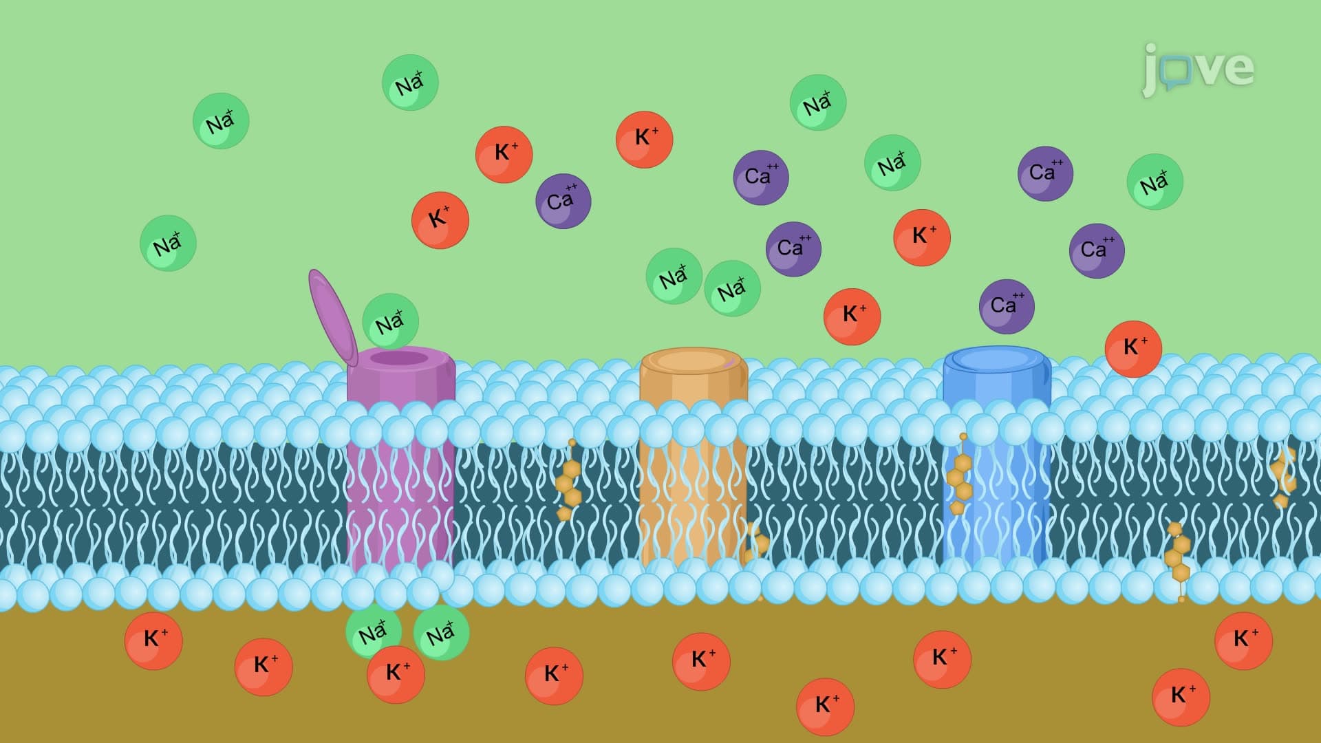
5. Cardiac Action Potential and Ion Movement: Cardiac action potentials involve five distinct phases controlled by ion channel activity. Phase 0 features rapid sodium influx causing depolarization in ventricular myocytes, while nodal cells depolarize more slowly through calcium channels. Phase 1 shows initial repolarization through potassium efflux. Phase 2 creates a plateau through balanced calcium influx and potassium efflux, prolonging contraction. Phase 3 involves final repolarization dominated by potassium outflow. Phase 4 maintains resting potential until the next cycle. Understanding these phases helps explain how medications like calcium channel blockers and beta-blockers affect cardiac function.
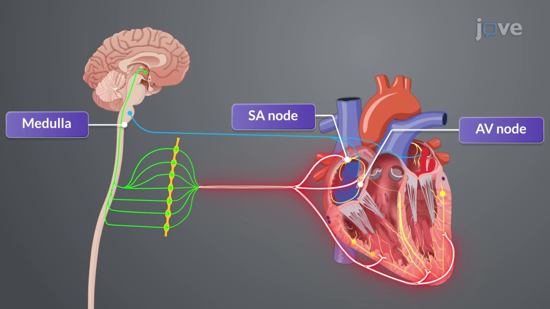
6. Cardiac Output Regulation: Cardiac output equals heart rate multiplied by stroke volume, typically 4-6 liters per minute in adults. Heart rate regulation involves autonomic nervous system balance - parasympathetic stimulation via vagus nerve decreases rate, while sympathetic activation increases it through beta-1 receptors. Baroreceptors in aortic arch and carotid arteries sense pressure changes, triggering compensatory responses. Stroke volume depends on preload (ventricular filling), afterload (ejection pressure), and contractility (muscle strength). Factors like dehydration decrease preload, hypertension increases afterload, and sympathetic stimulation enhances contractility, demonstrating the system's remarkable adaptability.
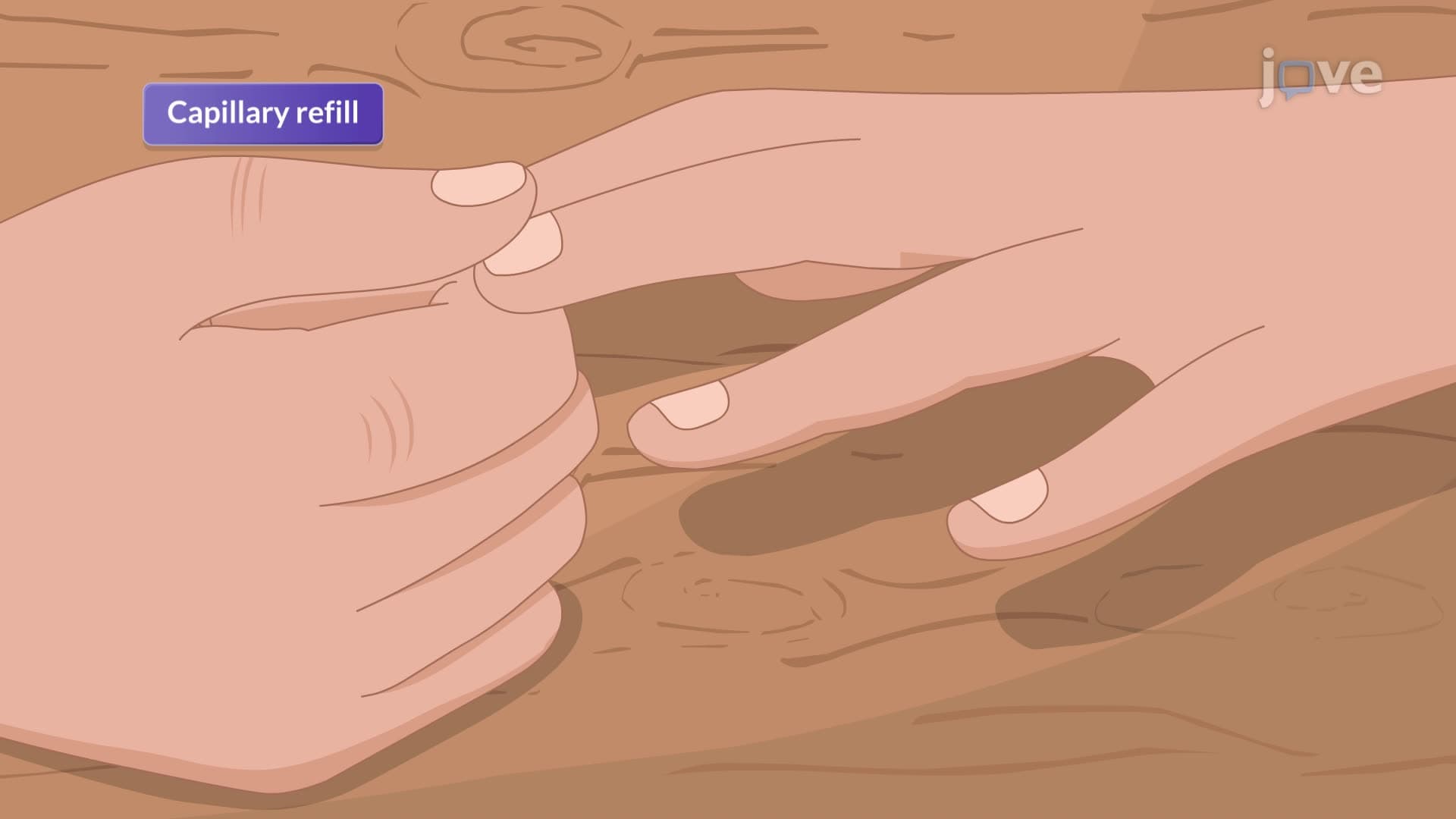
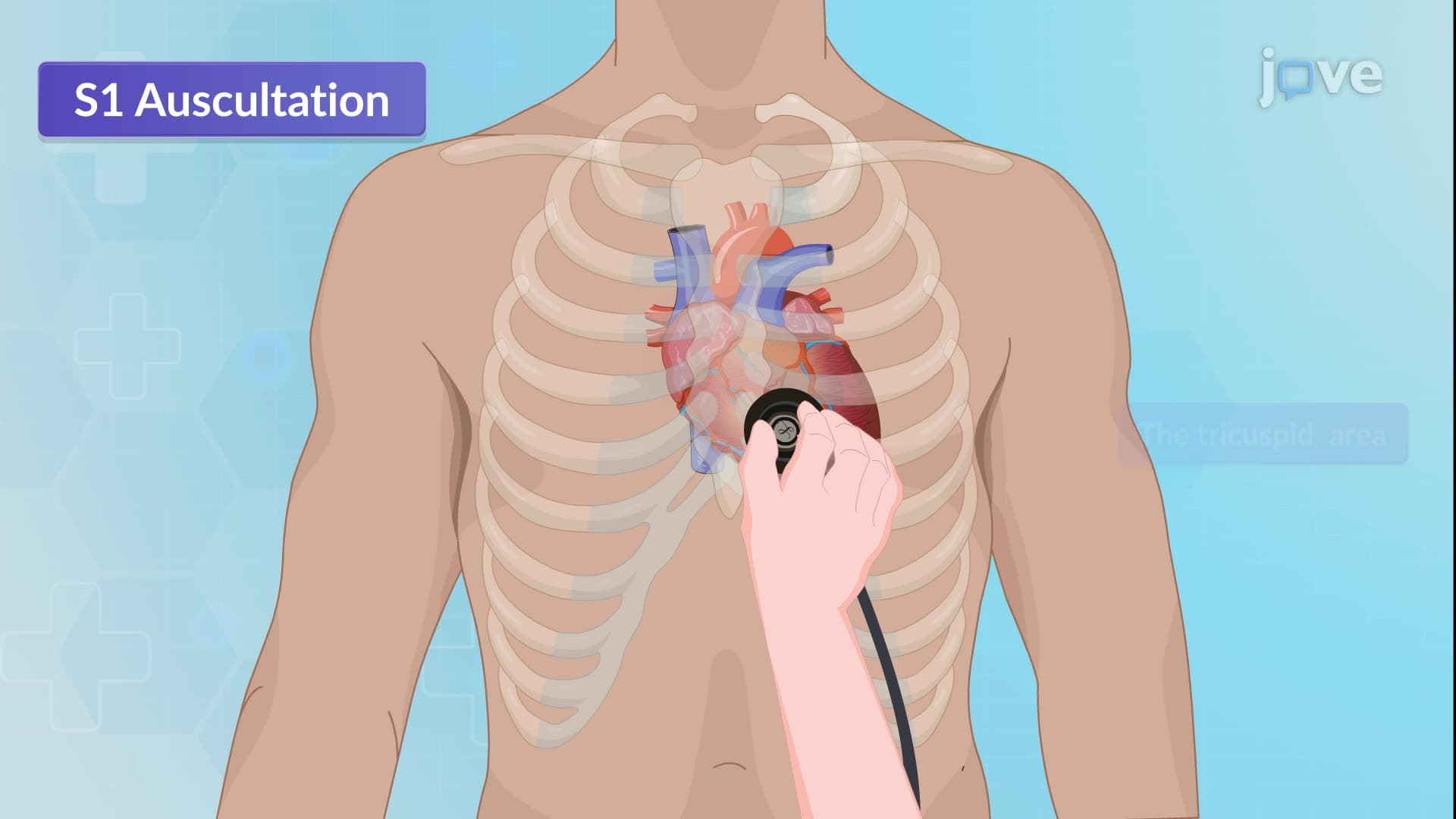
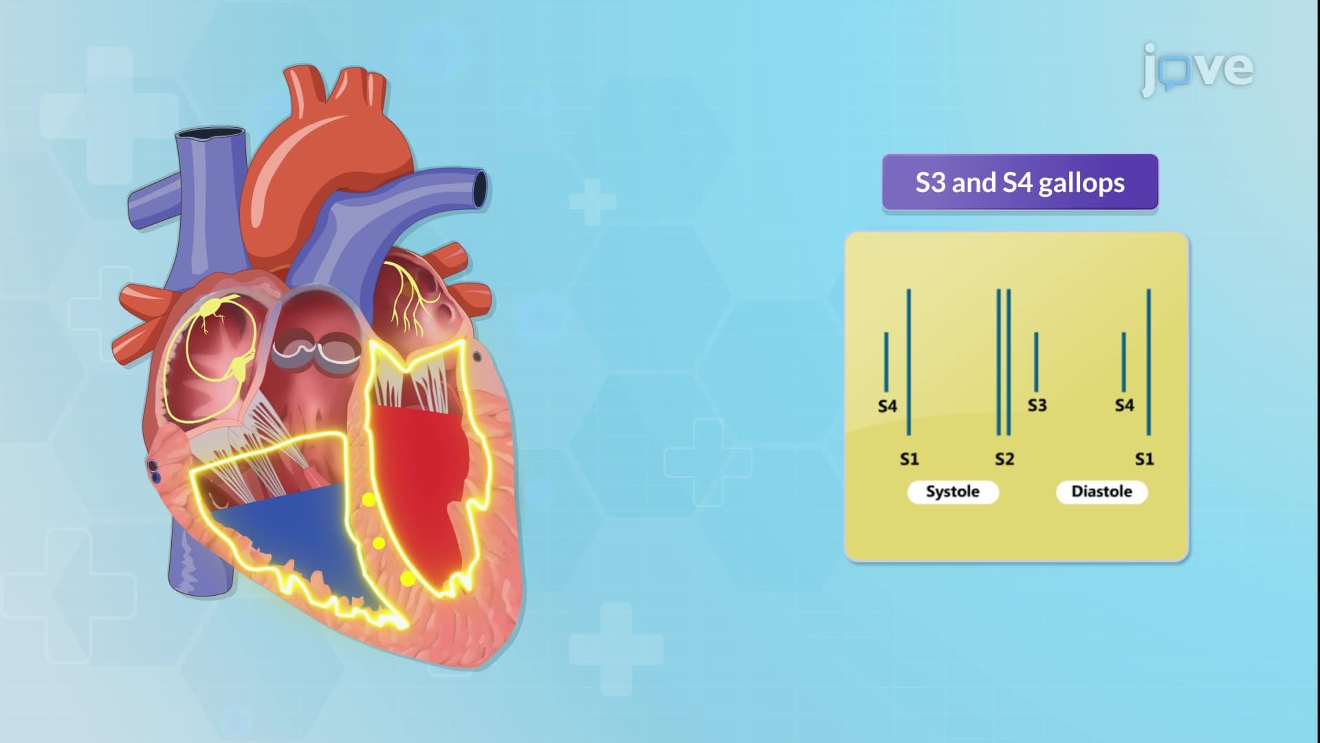
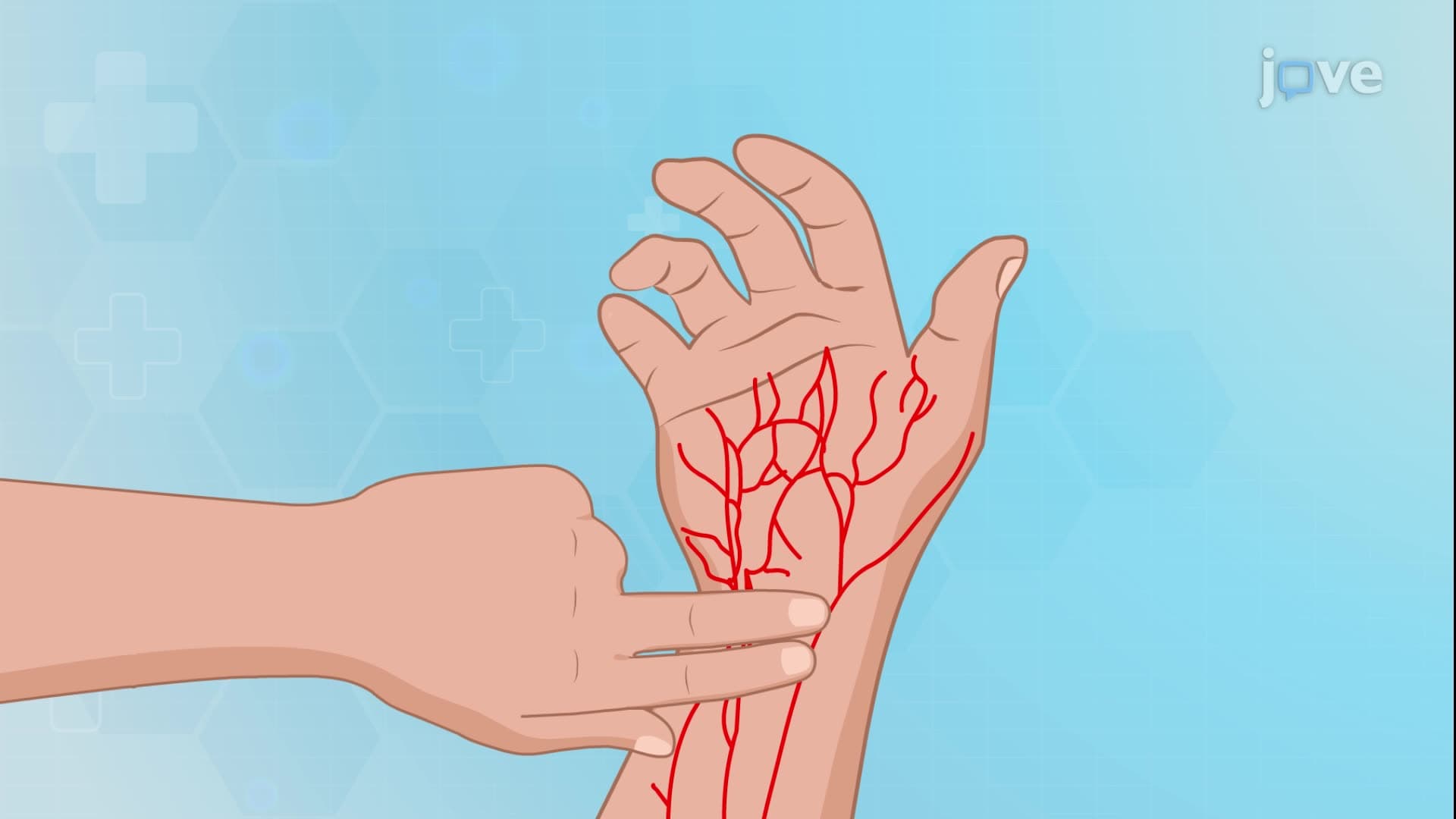
7. Cardiovascular Assessment Techniques: Systematic cardiovascular assessment begins with comprehensive subjective data collection including medical history, current symptoms, medications, and lifestyle factors. Inspection involves observing for distress signs, skin color changes, jugular venous distension, and extremity edema in supine and semi-Fowler positions. Palpation techniques assess jugular venous pressure, point of maximal impulse (PMI) location, pulse characteristics, capillary refill, and edema presence. Auscultation uses stethoscope diaphragm for S1 and S2 heart sounds at specific anatomical landmarks, while the bell detects low-pitched S3 and S4 gallops indicating pathological conditions.
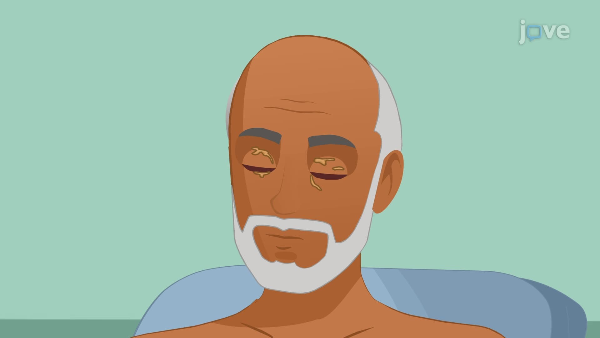
8. Abnormal Cardiovascular Findings: Critical abnormal findings include respiratory distress, cyanosis, and nail clubbing suggesting chronic hypoxia. Xanthelasma indicates hyperlipidemia risk, while splinter hemorrhages suggest infective endocarditis. Displaced PMI beyond the fifth intercostal space indicates cardiac enlargement. Abnormal heart sounds include S3 gallop (heart failure), S4 gallop (hypertension), opening snaps (mitral stenosis), and various murmurs. Systolic murmurs may indicate aortic stenosis or mitral regurgitation, while diastolic murmurs suggest valve insufficiency. Pulse abnormalities include weak, thready, or absent pulses indicating poor perfusion, requiring immediate clinical attention and further diagnostic evaluation.