Loading subject page…


11 Concepts
5 Concepts
15 Concepts
11 Concepts
7 Concepts
10 Concepts
22 Concepts
11 Concepts
10 Concepts
8 Concepts
14 Concepts
17 Concepts
14 Concepts
19 Concepts
15 Concepts
16 Concepts
15 Concepts
7 Concepts
6 Concepts
7 Concepts
7 Concepts
27 Concepts
8 Concepts
9 Concepts
6 Concepts
10 Concepts
12 Concepts
10 Concepts
 17 Concepts
 9.8K Views
 Popular topics







Diagnostic and radiological studies of the GI system encompass comprehensive laboratory tests, imaging procedures, and endoscopic techniques essential for evaluating gastrointestinal disorders. This JoVE Coach course covers serum studies, breath and stool tests, barium studies, ultrasound procedures, advanced imaging like CT and MRI, endoscopic procedures including colonoscopy and EGD, and specialized techniques for complete GI system assessment in clinical practice.
1. Laboratory-Based GI Diagnostics: Serum studies form the foundation of GI system evaluation, including liver function tests measuring ALT, AST, and bilirubin levels to assess hepatic disorders. Elevated amylase and lipase levels indicate pancreatic inflammation, while comprehensive metabolic panels evaluate kidney function, electrolyte balance, and glucose metabolism. Tumor markers like carcinoembryonic antigen (CEA) detect colorectal carcinoma, and CA 19-9 identifies pancreatic malignancies. Breath tests utilize hydrogen measurement for carbohydrate malabsorption and urea testing for H. pylori detection. Stool examinations analyze consistency, occult blood, parasites, and bacterial cultures for comprehensive digestive system assessment.
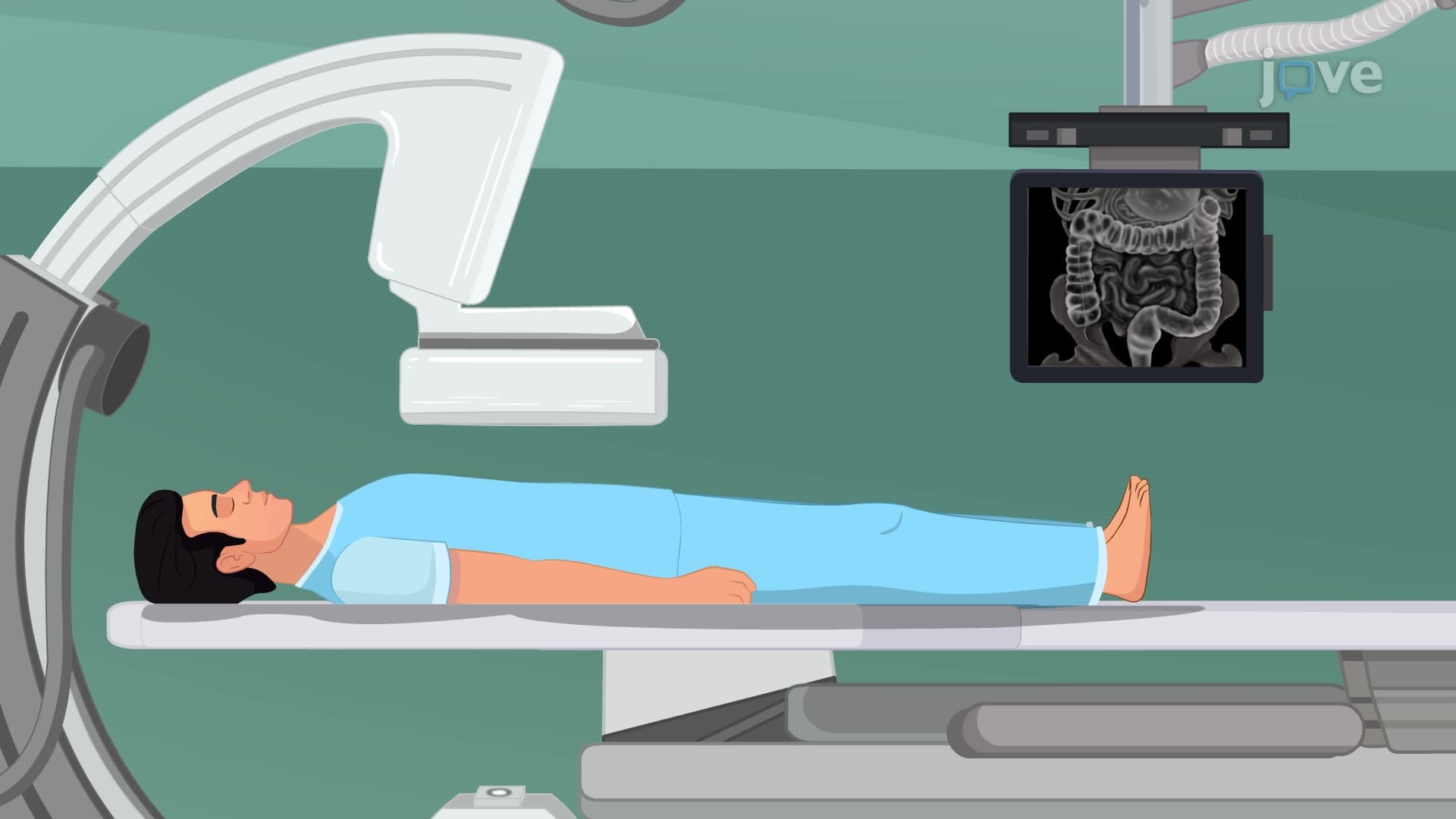
2. Contrast-Enhanced Radiographic Studies: Barium studies utilize radiopaque contrast material to visualize GI tract structures and function. Upper GI series (barium swallow) examines the esophagus, stomach, and duodenum for motility disorders, strictures, and hiatal hernias, requiring 8-hour NPO preparation and fluoroscopic monitoring. Lower GI series (barium enema) evaluates the colon and rectum for colorectal cancer, polyps, and diverticulosis, involving low-residue diet preparation and careful post-procedure barium elimination monitoring. Both procedures require specific patient positioning, clear explanations of discomfort expectations, and thorough post-procedure care including increased fluid intake and laxative administration.
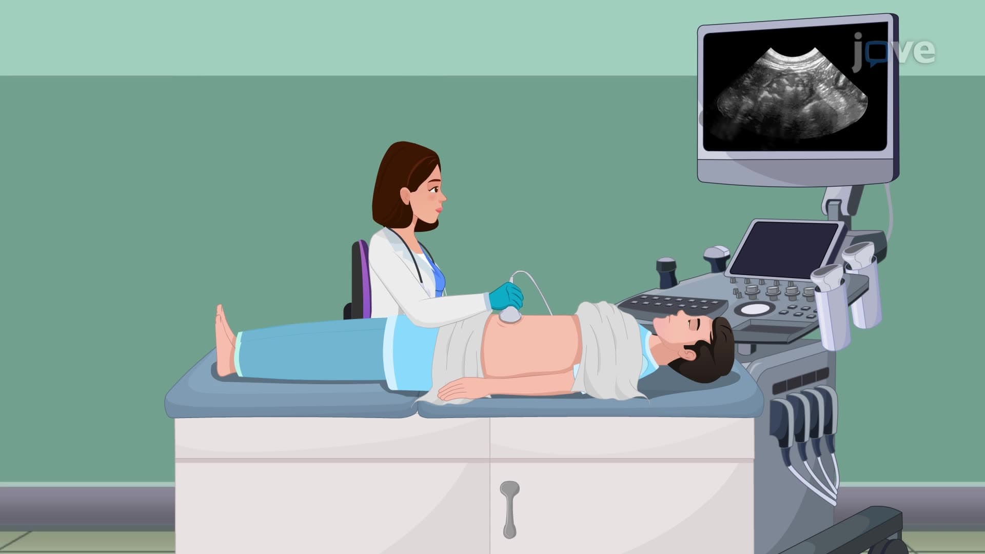
3. Ultrasound-Based Imaging Techniques: Abdominal ultrasonography provides non-invasive visualization of abdominal organs using high-frequency sound waves, requiring 8-12 hours of fasting and supine positioning during the procedure. Endoscopic ultrasound combines endoscopy with ultrasound technology using an echoendoscope to examine digestive tract structures and surrounding tissues, providing detailed images of pancreatic cancer and gastrointestinal tumors. FibroScan technology specifically assesses liver stiffness and fatty changes using low-frequency sound waves, helping diagnose liver fibrosis, cirrhosis, and hepatitis conditions through right mid-axillary line placement.
4. Advanced Cross-Sectional Imaging: CT scans utilize rotating X-ray beams and computer processing to generate detailed cross-sectional images of GI structures, while MRI employs magnetic fields and radio waves for superior soft tissue visualization. PET scans reveal metabolic and biochemical functions using radiotracer injection (typically fluorodeoxyglucose), requiring 30-60 minute waiting periods for tracer distribution before scanning. Scintigraphy involves radioactive isotope labeling of blood cells for monitoring distribution patterns at 24 and 48-hour intervals, highlighting areas of inflammation or abnormal activity throughout the gastrointestinal system.
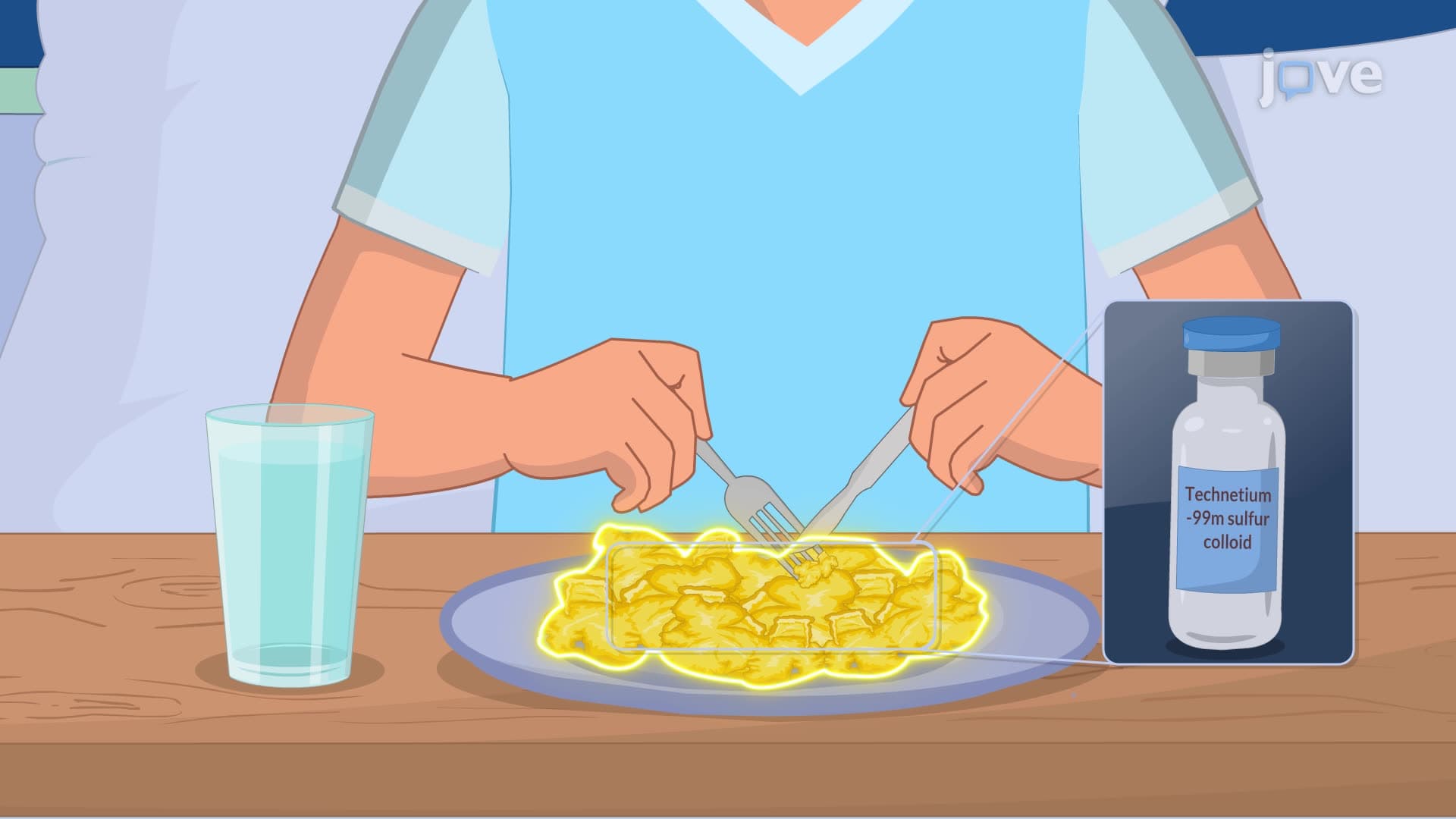
5. Motility and Functional Assessment Studies: Gastric emptying studies evaluate stomach function using technetium-99m sulfur colloid mixed with scrambled eggs, tracked by scintiscanner to diagnose gastroparesis and delayed gastric emptying. Colonic transit studies assess large intestine motility through radionuclide marker capsules, with serial abdominal X-rays over 4-5 days to detect obstructive defecation syndromes. Virtual colonoscopy (CT colonography) provides non-invasive colon examination using air or carbon dioxide insufflation with CT imaging, offering an alternative to traditional colonoscopy for patients unable to tolerate invasive procedures.
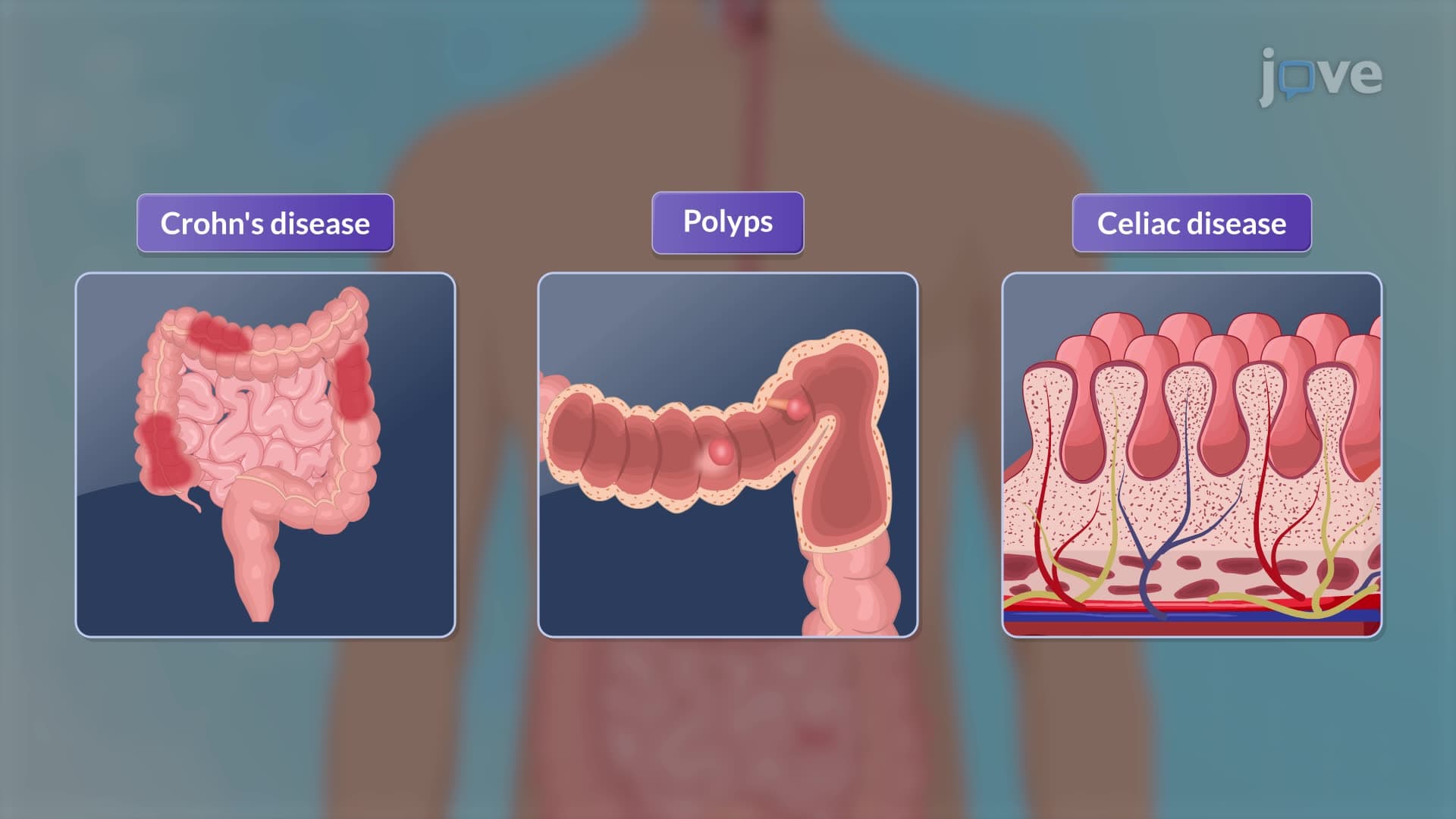
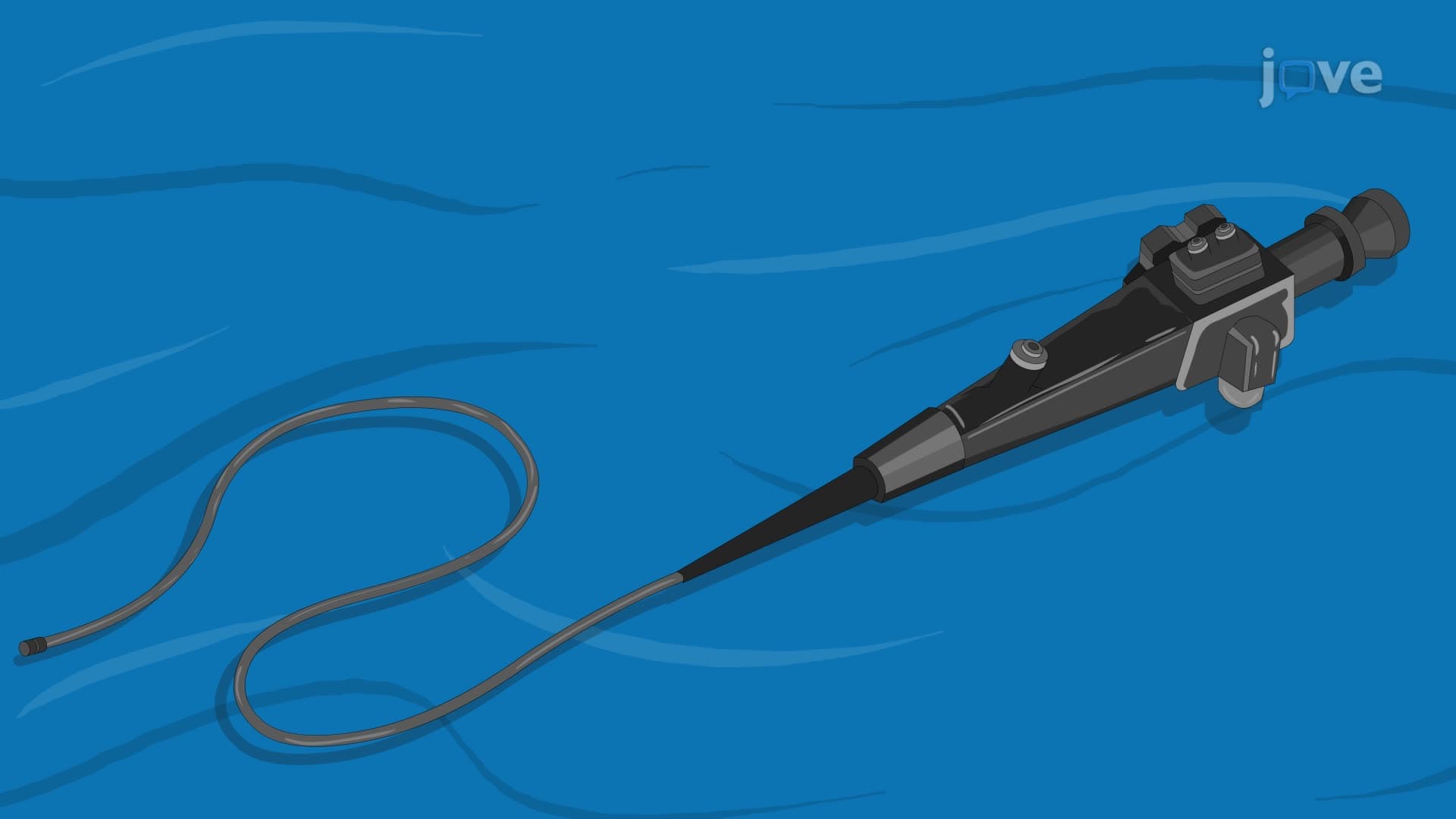
6. Endoscopic Visualization Procedures: Esophagogastroduodenoscopy (EGD) examines the upper GI tract using a flexible camera-equipped endoscope, diagnosing GERD, peptic ulcers, and Barrett's esophagus while allowing therapeutic interventions like biopsy collection and polyp removal. Colonoscopy provides complete colon visualization for colorectal cancer screening and polyp detection, requiring bowel preparation and sedation monitoring. Video capsule endoscopy utilizes a wireless, swallowable camera capsule for small intestine examination, particularly useful for diagnosing Crohn's disease and small bowel pathology in areas inaccessible to traditional endoscopy.
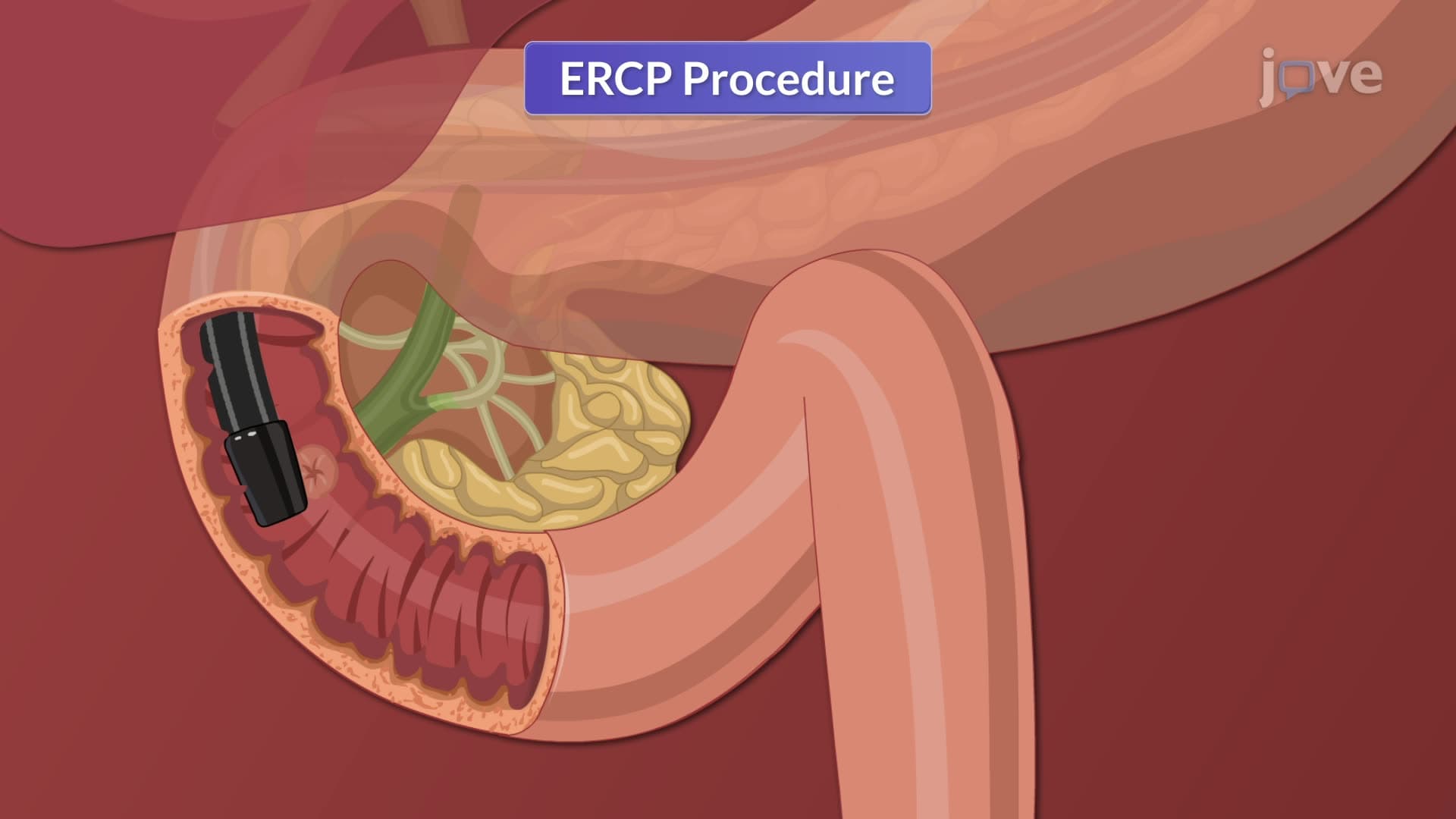
7. Specialized Therapeutic Endoscopic Procedures: ERCP (Endoscopic Retrograde Cholangiopancreatography) combines endoscopy with fluoroscopy to examine biliary and pancreatic ductal systems, using contrast injection and real-time X-ray imaging to diagnose gallstones and biliary strictures. Sigmoidoscopy provides limited colon examination focusing on the rectum and sigmoid colon using flexible sigmoidoscopes. Laparoscopy enables minimally invasive abdominal visualization and surgical procedures through small incisions. These procedures require specific patient preparation, including NPO status, medication management, and careful post-procedure monitoring for complications like perforation.
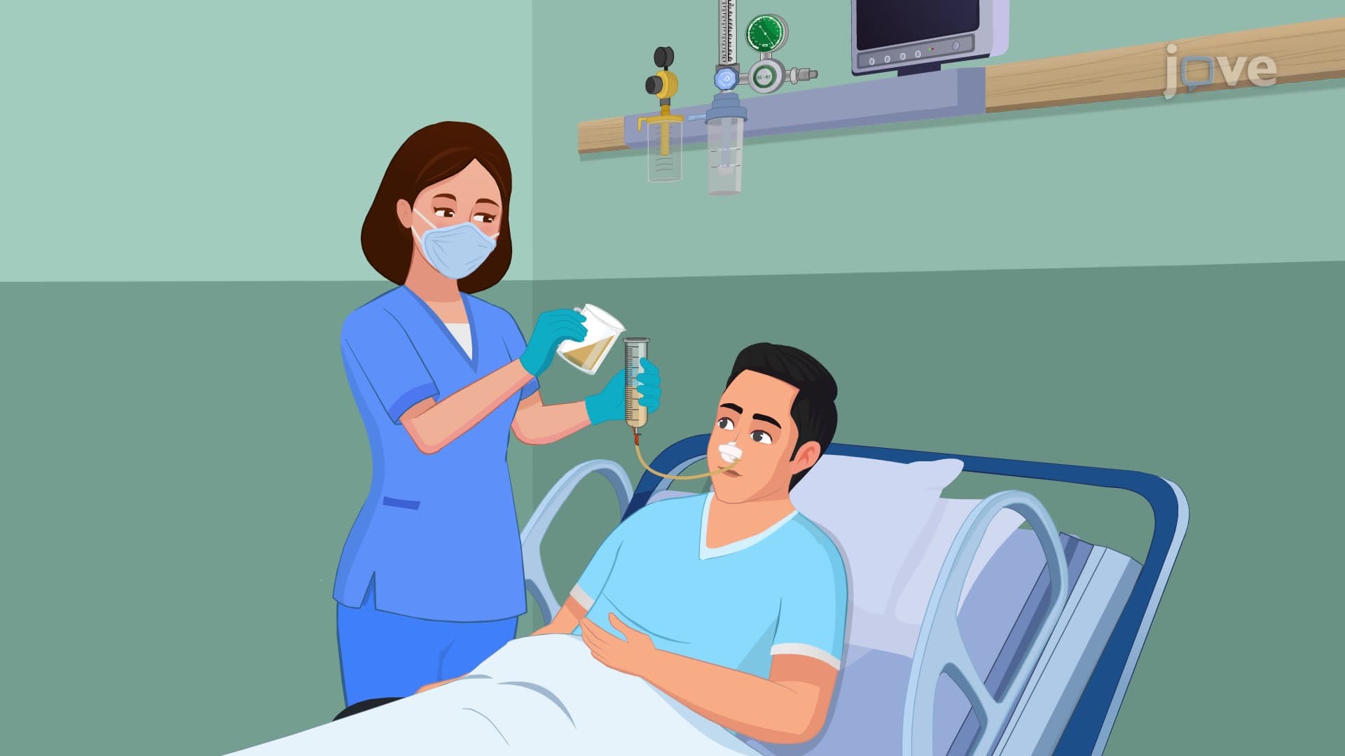
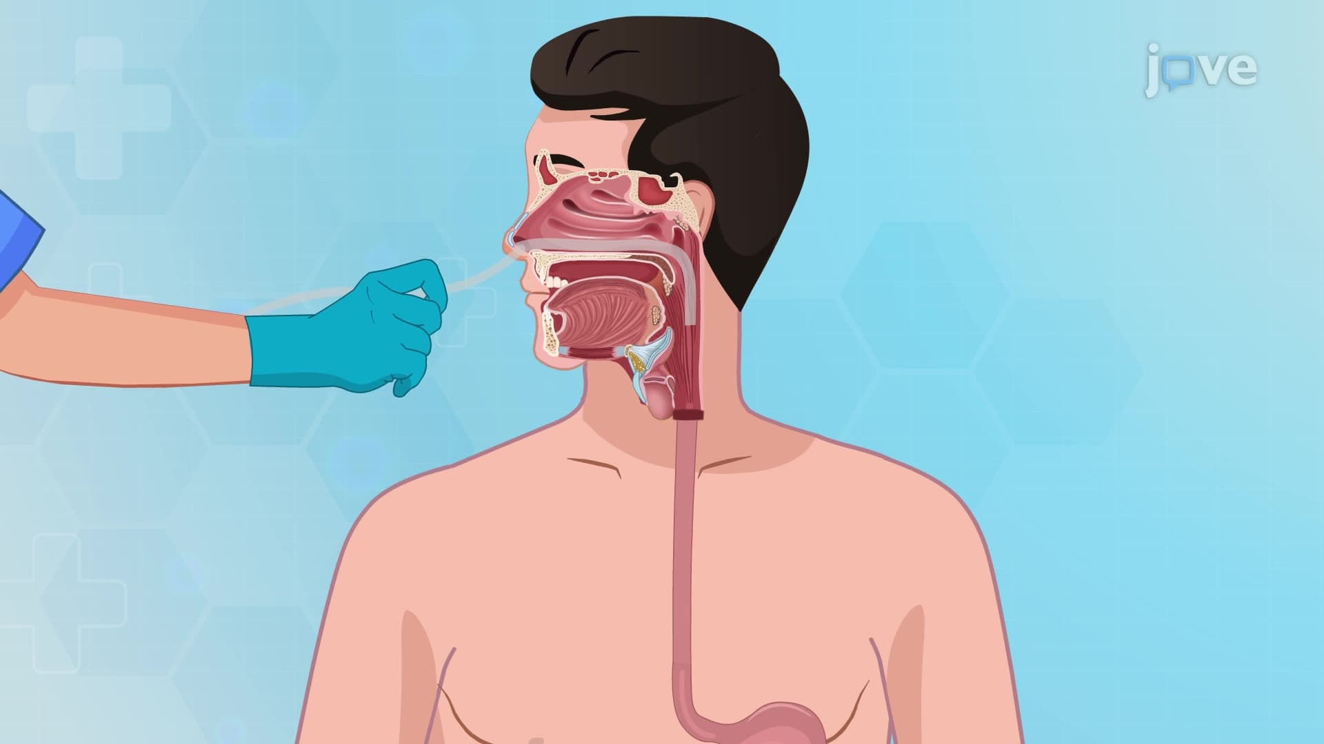
8. Nutritional Support and Ostomy Management: Enteral nutrition delivers nutrients directly through feeding tubes, including nasogastric, orogastric, and nasointestinal routes for short-term feeding, and gastrostomy or jejunostomy for long-term support. Parenteral nutrition bypasses the digestive system entirely, delivering nutrients directly into the bloodstream through central or peripheral venous access. Central parenteral nutrition requires subclavian or internal jugular vein catheterization with X-ray confirmation, while peripheral routes accommodate lower concentration formulas. Ostomy care involves creating artificial openings (stomas) for waste elimination, requiring specialized pouching systems, skin barrier application, and regular monitoring for complications.