Loading video…


 15 Concepts
 10.4K views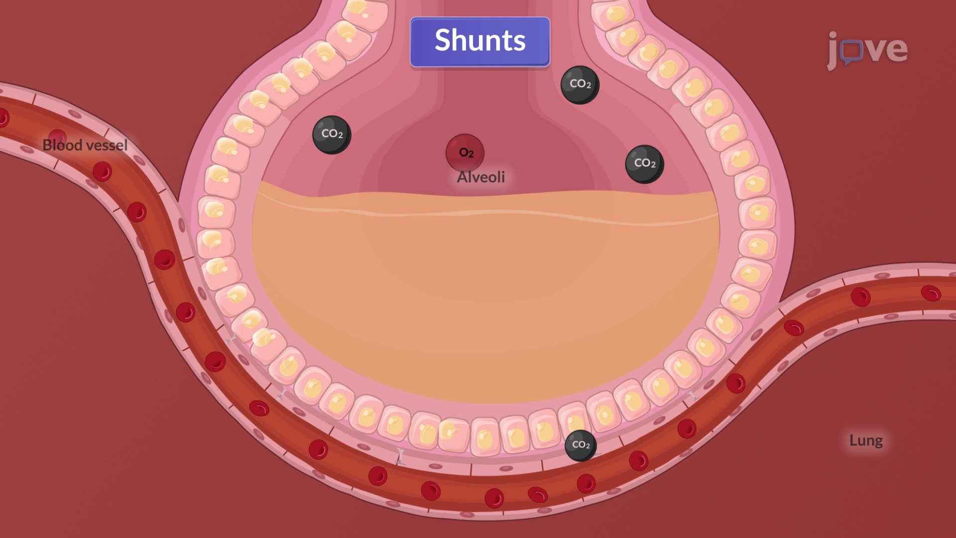

Video Summary: What Is Acute Respiratory Failure Ii
Every 36 seconds, someone dies from respiratory disease in the United States, with acute respiratory failure ii representing one of the most critical emergency conditions in intensive care units. This life-threatening condition occurs when the lungs cannot adequately oxygenate blood, characterized by dangerously low oxygen levels below 60 mmHg. Understanding what is acute respiratory failure ii requires examining four key mechanisms that disrupt normal gas exchange in conditions ranging from pneumonia to pulmonary embolism. Watch the full video on JoVE Coach to master this concept with expert-led visuals and step-by-step explanations.
Acute respiratory failure ii, also known as hypoxemic respiratory failure or Type I respiratory failure, represents a critical medical emergency where the respiratory system fails to maintain adequate blood oxygenation. Unlike Type II respiratory failure which involves CO2 retention, this condition specifically affects oxygen delivery while carbon dioxide elimination remains normal or even enhanced. The acute respiratory failure ii definition centers on arterial oxygen partial pressure (PaO2) falling below 60 mmHg on room air, creating a life-threatening scenario requiring immediate intervention.
This acute respiratory failure ii overview reveals its prevalence in American hospitals, where it accounts for approximately 200,000 ICU admissions annually. The condition carries significant mortality risk, with survival rates depending heavily on rapid recognition and appropriate treatment. Students preparing for the MCAT or advanced placement biology exams must understand that this represents a failure of gas exchange rather than ventilation mechanics.
The acute respiratory failure basics involve four distinct mechanisms that impair oxygenation. Ventilation-perfusion (V/Q) mismatch represents the most common cause, occurring when alveolar ventilation doesn't match pulmonary blood flow. In conditions like asthma attacks common in US emergency departments, bronchospasm creates low V/Q ratios, while pulmonary embolism-affecting 900,000 Americans annually-creates high V/Q ratios through reduced perfusion.
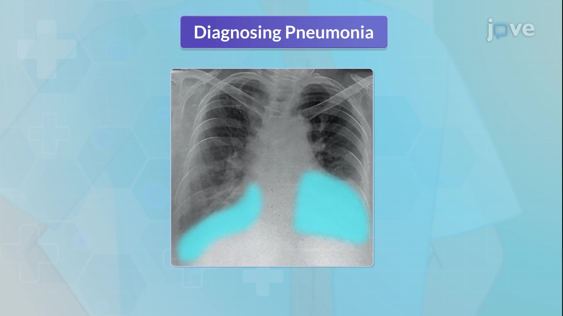
Intrapulmonary shunting occurs when blood bypasses functional gas exchange units, commonly seen in pneumonia where fluid-filled alveoli prevent oxygen uptake. This mechanism explains why pneumonia patients at institutions like Johns Hopkins Hospital often require supplemental oxygen despite normal breathing rates.
Diffusion limitation involves impaired gas transfer across the alveolar-capillary membrane. Conditions like pulmonary edema or acute respiratory distress syndrome (ARDS) thicken this barrier, creating an understanding acute respiratory failure ii scenario where oxygen cannot adequately cross into the bloodstream despite adequate ventilation and perfusion.
This acute respiratory failure ii concept appears frequently in USMLE Step 1 questions and NCLEX-RN examinations, particularly in critical care scenarios. Students encounter this material in college-level anatomy and physiology courses, where understanding the distinction between oxygenation failure and ventilation failure proves crucial.
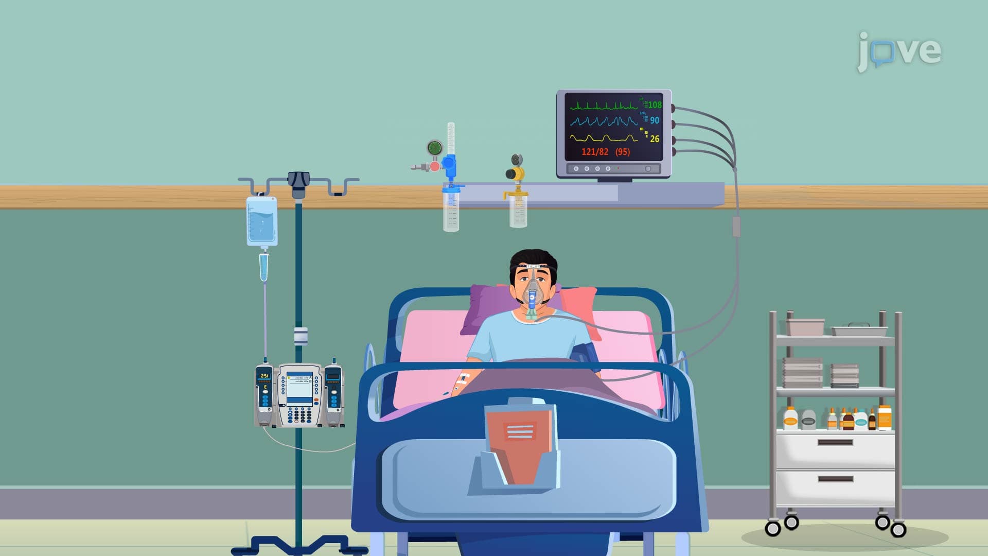
The acute respiratory failure ii study guide approach should emphasize recognizing clinical presentations. For instance, a patient presenting to Massachusetts General Hospital's emergency department with pneumonia might demonstrate normal CO2 levels but critically low oxygen saturation, indicating Type I rather than Type II respiratory failure. This distinction influences treatment protocols, with Type I requiring aggressive oxygenation support rather than ventilation assistance.
Healthcare students must also understand that acute respiratory failure ii can progress rapidly, sometimes within minutes in cases of massive pulmonary embolism or severe pneumothorax. This time-sensitive nature makes early recognition skills essential for future healthcare providers working in American medical systems.
Related Micro-courses