Loading video…


 15 Concepts
 10.4K views

Video Summary: What Is Acute Respiratory Failure Iv
When a patient struggles to breathe and their oxygen levels drop dangerously low, acute respiratory failure IV represents the most critical stage of respiratory compromise. This life-threatening condition affects thousands of Americans daily in emergency departments across hospitals like Johns Hopkins and Mayo Clinic. Understanding what is acute respiratory failure IV helps students recognize when the body's breathing mechanisms fail completely, requiring immediate medical intervention. Watch the full video on JoVE Coach to master this concept with expert-led visuals and step-by-step explanations.
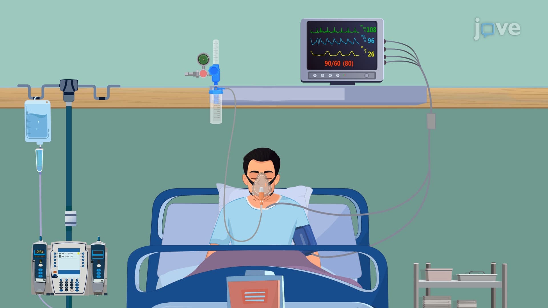
Acute respiratory failure IV represents the most severe classification of respiratory compromise, where the lungs cannot adequately oxygenate blood or remove carbon dioxide from the body. This critical condition affects over 500,000 Americans annually and carries significant mortality risk without immediate intervention. Unlike milder forms of respiratory distress, this stage indicates complete failure of compensatory mechanisms, requiring advanced life support measures in intensive care units.
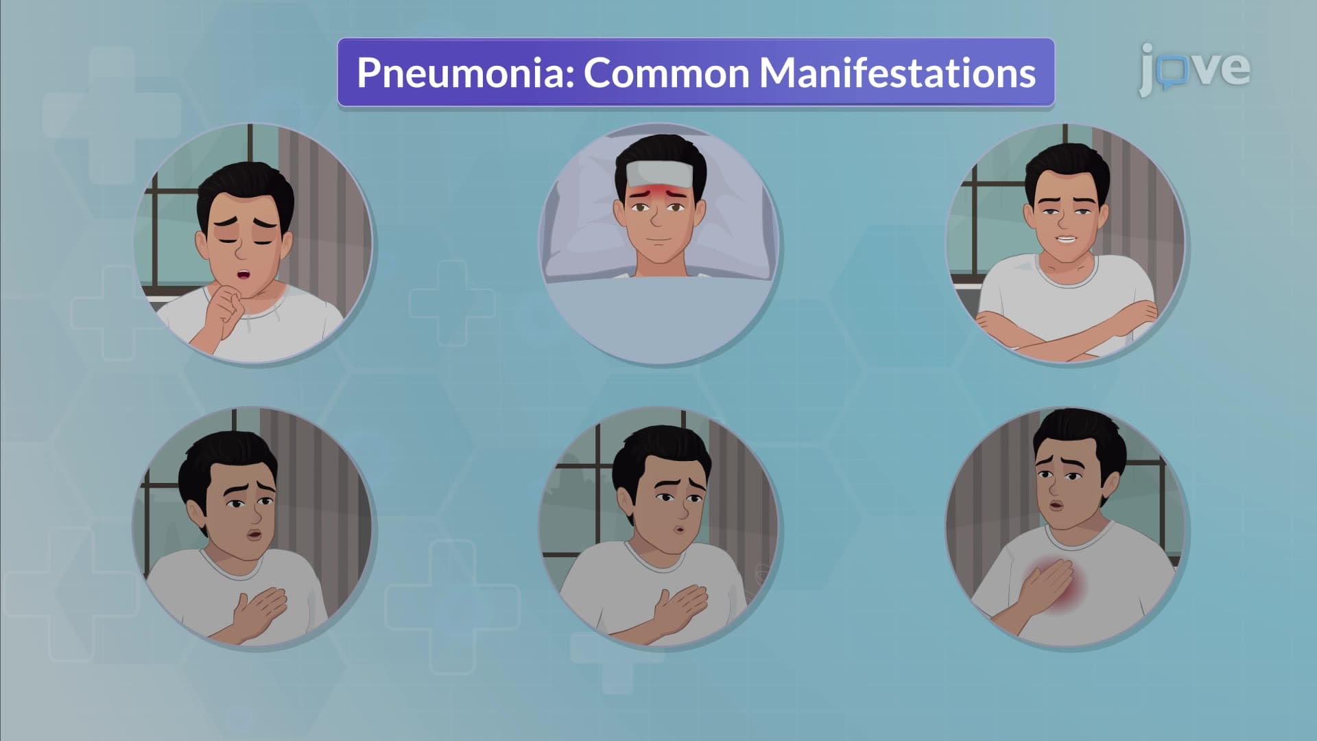
The progression to acute respiratory failure IV begins with subtle neurological symptoms that many students overlook on standardized exams like the NCLEX or MCAT. Initial manifestations include restlessness and confusion, which occur because the brain receives inadequate oxygen delivery. Morning headaches and bradypnea (abnormally slow breathing) signal problems with carbon dioxide removal, creating a dangerous cycle where waste gases accumulate in the bloodstream.
Healthcare professionals in major medical centers like Cleveland Clinic and Massachusetts General Hospital train extensively to recognize these early indicators. Students preparing for AP Biology or college physiology courses should understand that these neurological changes precede the more obvious respiratory symptoms, making early recognition crucial for patient survival.
As respiratory failure progresses, the body activates multiple compensatory mechanisms that create observable clinical signs. Tachycardia (rapid heart rate) and tachypnea (rapid breathing) represent the cardiovascular and respiratory systems working harder to maintain oxygen delivery. Pallor indicates poor circulation, while dramatically increased work of breathing (WOB) reflects the enormous effort required for each breath.
Patient positioning becomes a critical assessment tool that nursing students encounter frequently on the HESI A2 and TEAS exams. Moderate respiratory distress patients often prefer sitting upright to improve lung expansion, while those with severe compromise adopt the classic tripod position-leaning forward with arms supported on an overbed table or knees. This positioning maximizes the mechanical advantage of respiratory muscles and appears in countless clinical scenarios on standardized medical exams.
Pursed-lip breathing represents another compensatory mechanism where patients instinctively slow their respirations with prolonged expiration phases. These patients typically speak only 2-3 words before pausing to breathe, a pattern that emergency physicians at hospitals like Johns Hopkins use for rapid severity assessment. When primary respiratory muscles (diaphragm and intercostals) become insufficient, the body recruits accessory muscles including neck, shoulder, and abdominal muscles to assist with breathing.
The most ominous sign-paradoxical breathing-occurs in end-stage respiratory failure where the chest moves inward during inspiration and outward during expiration, opposite to normal patterns. This indicates complete exhaustion of respiratory muscles and typically requires immediate mechanical ventilation. Pre-med students studying for the MCAT should recognize this pattern as a medical emergency requiring intensive care unit admission.
Related Micro-courses