Loading video…


 15 Concepts
 10.4K views

Video Summary: What Is Acute Respiratory Failure V
Every year, over 190,000 Americans require mechanical ventilation in ICUs due to acute respiratory failure v, making it one of the most critical medical emergencies healthcare professionals encounter. Acute respiratory failure v occurs when the lungs cannot adequately oxygenate blood or remove carbon dioxide, requiring immediate intervention to prevent organ damage. For instance, COVID-19 patients at Johns Hopkins Hospital often experienced this condition, necessitating complex treatment protocols. Understanding acute respiratory failure v management involves mastering collaborative care strategies that combine respiratory therapy, pharmacological interventions, and supportive care measures. Watch the full video on JoVE Coach to master this concept with expert-led visuals and step-by-step explanations.
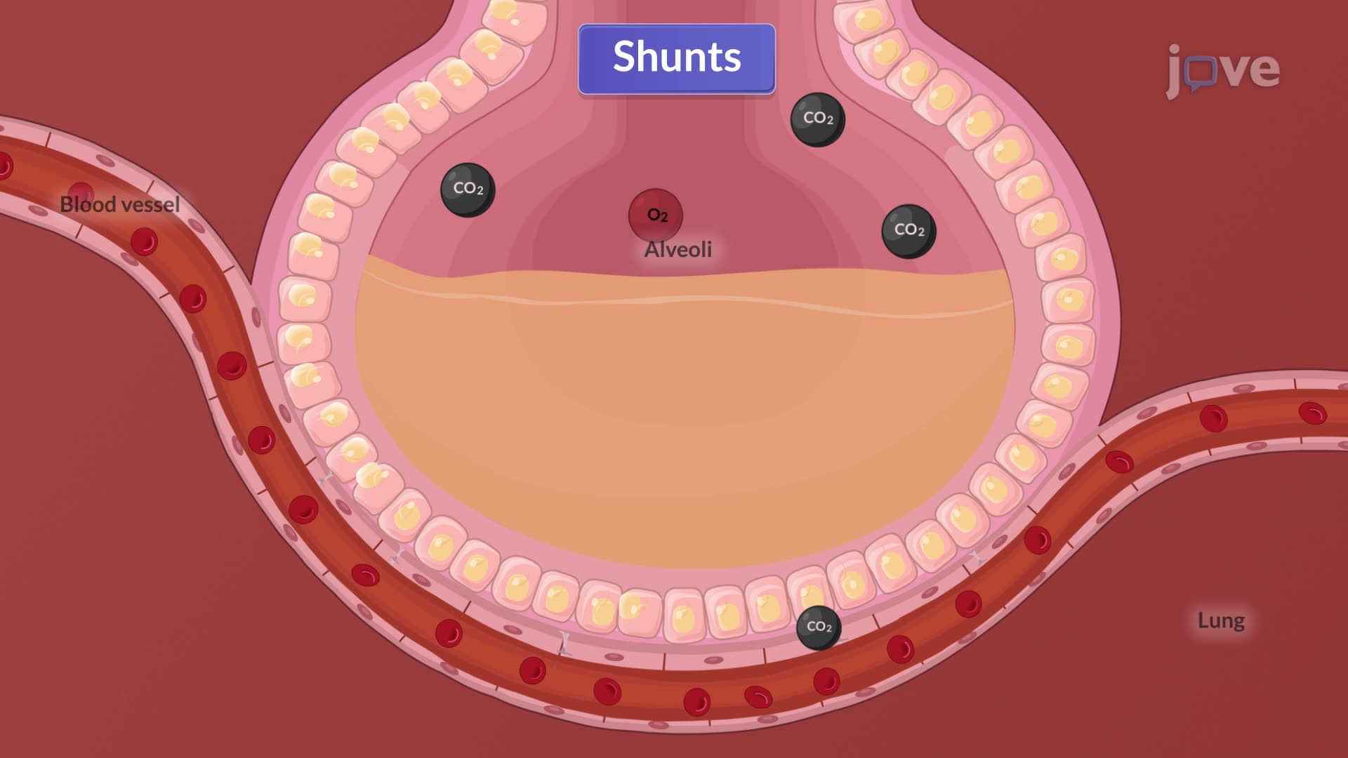
Acute respiratory failure v represents a life-threatening condition where the respiratory system fails to maintain adequate gas exchange, leading to hypoxemia, hypercapnia, or both. This condition affects approximately 137 per 100,000 adults annually in the United States, with mortality rates ranging from 26% to 58% depending on underlying causes. The pathophysiology involves ventilation-perfusion mismatches, diffusion impairments, or complete respiratory muscle failure, requiring immediate medical intervention to prevent multi-organ dysfunction.
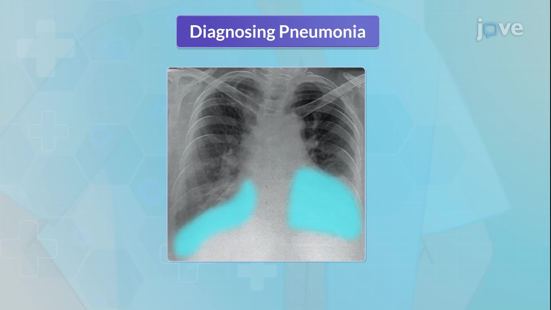
Effective management of acute respiratory failure v demands seamless coordination between respiratory therapists, physicians, nurses, and nutritionists. At major US medical centers like Mayo Clinic and Cleveland Clinic, standardized protocols ensure optimal patient outcomes through evidence-based interventions. The approach begins with rapid assessment using arterial blood gas analysis, chest imaging, and pulmonary function evaluation to determine the severity and underlying etiology.
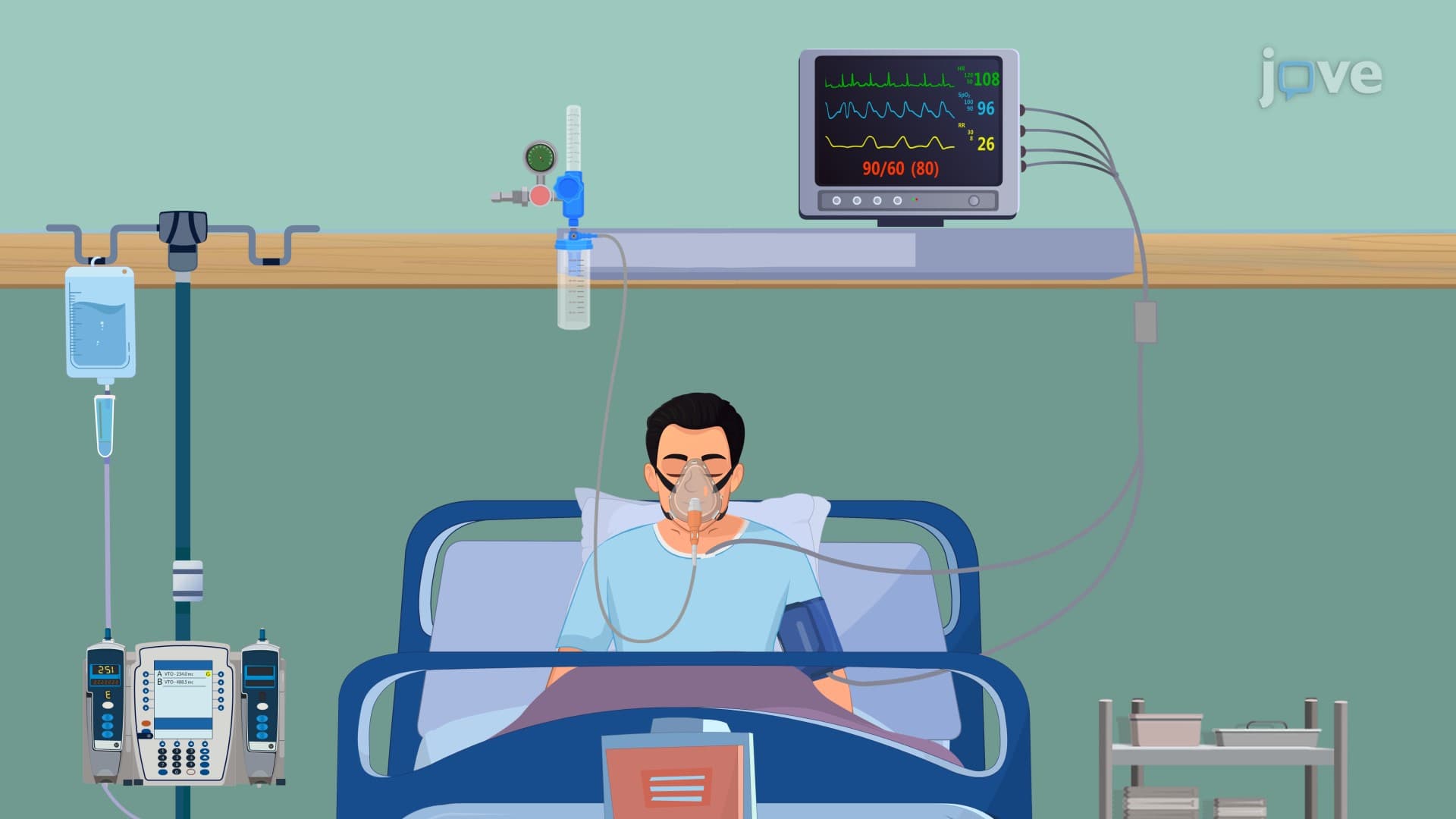
Treatment escalation follows a systematic approach, starting with supplemental oxygen through nasal cannulas or Venturi masks for mild cases. Severe presentations may require non-invasive positive pressure ventilation (BiPAP) or intubation with mechanical ventilation. Pharmacological management includes beta-2 agonists like albuterol for bronchodilation, systemic corticosteroids such as methylprednisolone to reduce inflammation, and loop diuretics like furosemide for pulmonary edema. Students preparing for the MCAT or NCLEX-RN examinations frequently encounter questions about these therapeutic hierarchies and drug mechanisms.
Successful recovery often depends on effective secretion management through chest physiotherapy techniques, including percussion, vibration, and postural drainage. Modern approaches incorporate high-frequency chest wall oscillation devices and mechanical insufflation-exsufflation systems. Adequate hydration (2-3 liters daily unless contraindicated) helps maintain optimal mucus viscosity, while nebulized normal saline or mucolytic agents like acetylcysteine facilitate expectoration. Nutritional support becomes crucial for recovery, with protein requirements increasing to 1.2-2.0 g/kg body weight to support respiratory muscle function and tissue repair. These concepts frequently appear in AP Biology coursework and college-level pathophysiology examinations.
Related Micro-courses