Loading video…


 15 Concepts
 10.4K views

Video Summary: What Is Pneumonia Ii Pathophysiology
Did you know that pneumonia kills more Americans annually than car accidents? Understanding pneumonia II pathophysiology reveals how pathogens like *Streptococcus pneumoniae* transform healthy lung tissue into fluid-filled sacs, disrupting the oxygen exchange that keeps us alive. When pneumonia strikes patients at Houston Methodist Hospital, this precise pathophysiological cascade determines treatment success. Watch the full video on JoVE Coach to master this concept with expert-led visuals and step-by-step explanations.
Pneumonia II pathophysiology represents a complex interplay between pathogen virulence and host immune response that determines disease severity and outcomes. This pathophysiological framework is essential for MCAT preparation and advanced placement biology courses, as it demonstrates fundamental concepts of immunology, respiratory physiology, and microbiology in clinical context.
The pathophysiology begins with pathogen invasion through three distinct mechanisms. Inhalation represents the most common route, particularly relevant in healthcare settings like those studied at Johns Hopkins School of Medicine. Aspiration of oropharyngeal flora occurs frequently in elderly patients or those with swallowing disorders, while hematogenous spread typically involves *Staphylococcus aureus* in patients with underlying bloodstream infections. Understanding these entry mechanisms helps predict which pathogens are most likely in specific clinical scenarios-knowledge crucial for USMLE Step 1 success.
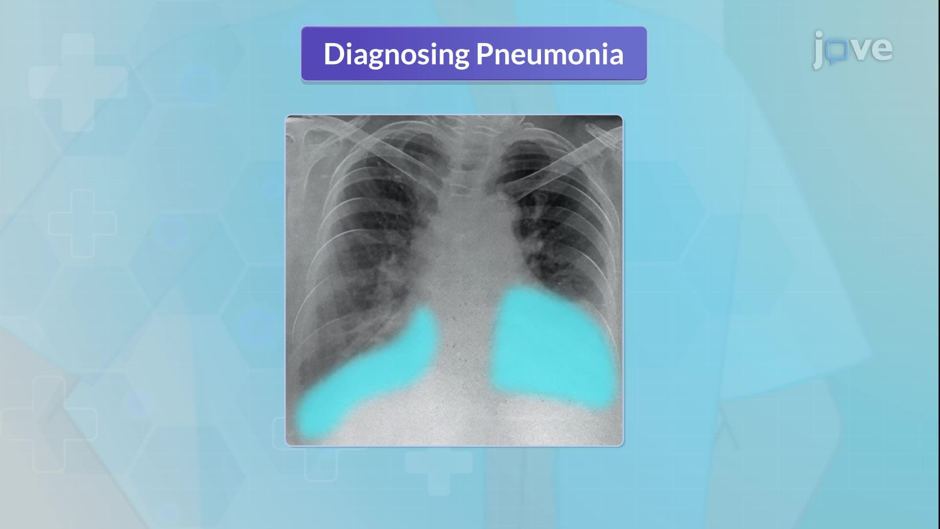
Once pathogens reach the alveolar level, they encounter alveolar macrophages-the lung's first line of defense. However, successful pathogens like *Streptococcus pneumoniae* possess capsular polysaccharides that resist phagocytosis. This leads to bacterial multiplication and release of inflammatory mediators, triggering neutrophil recruitment. The resulting inflammatory exudate contains white blood cells, fibrin, and cellular debris that fills alveolar spaces, creating the characteristic "hepatization" seen in pathology specimens at medical schools like Harvard Medical School.
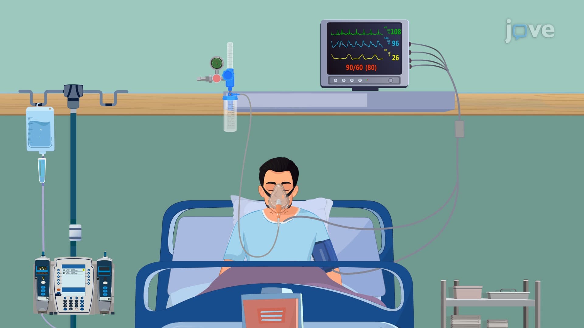
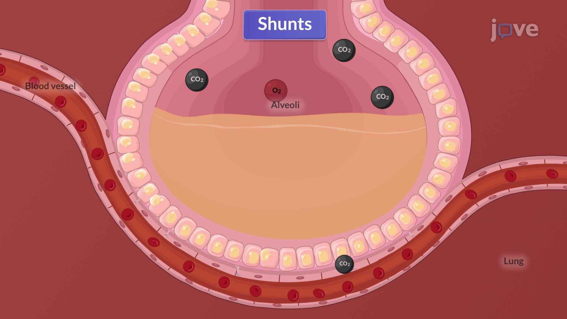
The accumulation of inflammatory exudate fundamentally alters pulmonary mechanics and gas exchange. Normal alveolar-capillary oxygen diffusion becomes impaired as fluid replaces air-filled spaces. This pathophysiological change manifests clinically as hypoxemia, dyspnea, and compensatory tachypnea. The increased capillary permeability-a hallmark finding tested on NCLEX examinations-allows protein-rich fluid to leak into alveolar spaces, further compromising respiratory function. Students preparing for AP Biology exams should understand how this represents a failure of normal homeostatic mechanisms that maintain optimal oxygen-carbon dioxide exchange ratios essential for cellular metabolism.
Related Micro-courses