Loading video…


 15 Concepts
 10.4K views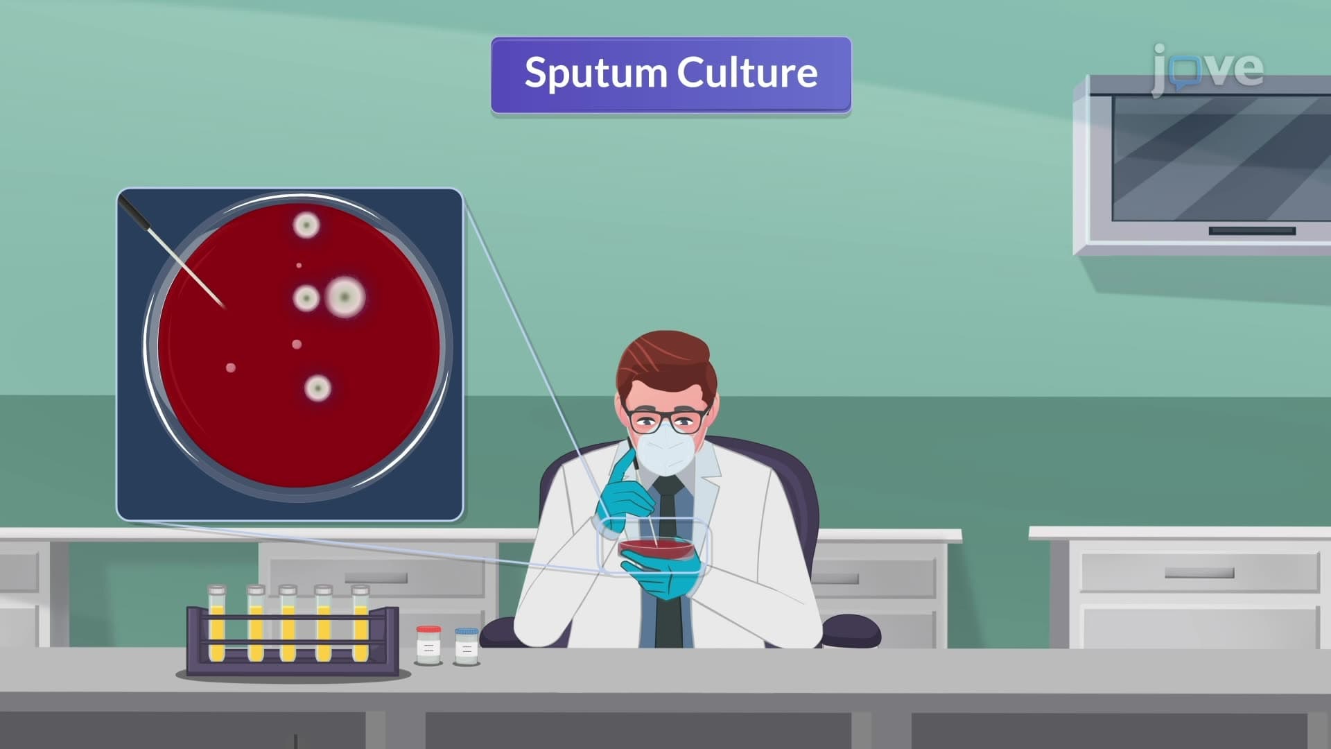

Video Summary: What Is Pulmonary Tuberculosis Iv
Every 18 seconds, someone dies from tuberculosis worldwide, yet many students struggle to understand pulmonary tuberculosis IV diagnostic methods that could save lives. Pulmonary tuberculosis IV encompasses advanced diagnostic techniques used by healthcare professionals at institutions like Johns Hopkins Hospital to accurately identify this deadly respiratory infection. From the Mantoux skin test showing positive results in as little as 48 hours to sophisticated interferon-gamma release assays, understanding What is Pulmonary Tuberculosis IV becomes crucial for future healthcare workers. Watch the full video on JoVE Coach to master this concept with expert-led visuals and step-by-step explanations.
Pulmonary tuberculosis IV represents the fourth installment in understanding tuberculosis diagnosis, focusing specifically on the multi-step diagnostic process that healthcare professionals use to confirm infection. This systematic approach combines traditional screening methods with modern molecular techniques, creating a comprehensive diagnostic strategy essential for controlling one of the world's most persistent infectious diseases.
The Mantoux test serves as the cornerstone of tuberculosis screening programs across US healthcare systems, from community health centers in rural Mississippi to major academic medical centers like UCLA. This intradermal injection of tuberculin-purified protein derivative creates a measurable immune response within 48-72 hours. The interpretation requires careful consideration of patient risk factors: immunocompromised individuals, including HIV patients treated at facilities like San Francisco General Hospital, require only 5mm of induration for a positive result, while healthy individuals need 15mm. This risk-stratified approach reflects the reality that compromised immune systems may produce weaker responses despite active infection.
Interferon-gamma release assays (IGRAs) represent a significant advancement in tuberculosis diagnostics, measuring specific immune cell responses to Mycobacterium tuberculosis proteins. These blood tests, commonly used in US hospitals and clinics, offer advantages over skin tests by eliminating false positives from BCG vaccination and reducing the need for return visits. Chest imaging remains crucial, with characteristic findings including upper lobe infiltrates and cavitations that radiologists at institutions like Mayo Clinic routinely identify. The presence of mediastinal lymphadenopathy often suggests active disease requiring immediate treatment.
Sputum culture provides definitive bacterial identification, though results may take weeks due to the slow growth characteristics of Mycobacterium tuberculosis. Modern laboratories across the US, from Quest Diagnostics to hospital-based facilities, use specialized media and incubation techniques to maximize detection rates. For extrapulmonary cases, CT and MRI imaging help identify involvement of organs beyond the lungs, crucial for comprehensive treatment planning. Students preparing for exams like the MCAT or NCLEX must understand these diagnostic sequences, as tuberculosis remains a high-yield topic in medical education and clinical practice.
Related Micro-courses