Loading subject page…


35,512 views
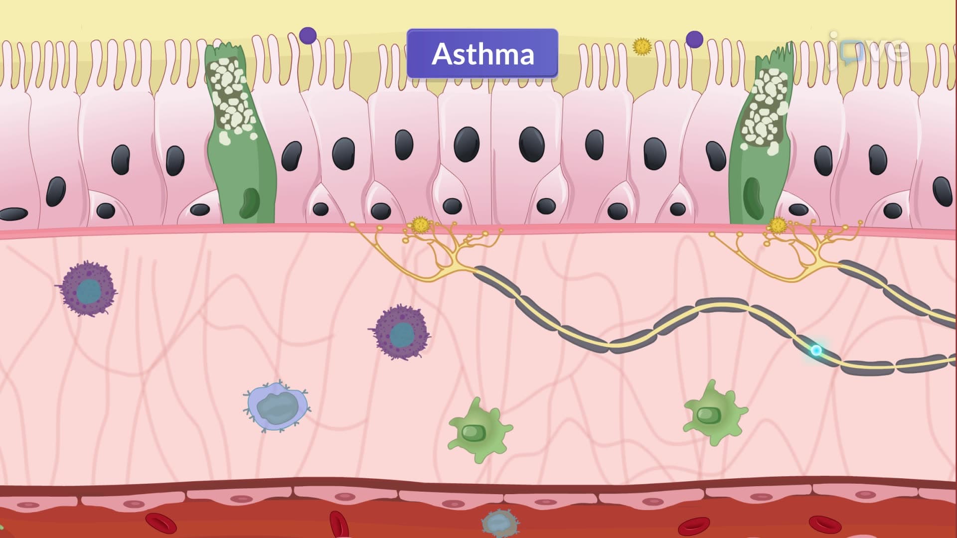

Video Summary: Asthma Part Ii Classification and Pathophysiological Mechanisms
Did you know that asthma affects over 25 million Americans, with symptoms triggered by everything from pollen to cold air? Asthma II pathophysiology and classification reveals the complex immune cascade behind airway inflammation and obstruction. When allergens like ragweed pollen enter the respiratory system, they trigger a multi-step inflammatory response involving IgE antibodies, mast cells, and inflammatory mediators that cause the characteristic wheezing and shortness of breath. Watch the full video on JoVE Coach to master this concept with expert-led visuals and step-by-step explanations.
Asthma represents one of the most complex respiratory conditions, affecting approximately 8% of the US population. The pathophysiology involves a sophisticated interplay between genetic predisposition, environmental triggers, and immune system dysfunction that results in chronic airway inflammation and hyperresponsiveness.
The pathophysiological journey begins when genetically susceptible individuals encounter allergens such as dust mites, pet dander, or seasonal pollens common throughout American households. Upon initial exposure, antigen-presenting cells process these allergens and present them to T-helper cells, specifically Th2 cells. This interaction stimulates B lymphocytes to differentiate into plasma cells that produce allergen-specific IgE antibodies.
These IgE antibodies circulate through the bloodstream and bind to high-affinity receptors on mast cells and basophils, effectively "arming" these cells for future encounters. When the same allergen is encountered again, it cross-links with IgE antibodies on mast cell surfaces, triggering rapid degranulation and release of preformed inflammatory mediators including histamine, tryptase, and chemotactic factors.
The initial mast cell response represents just the beginning of a complex inflammatory cascade. Released mediators recruit additional inflammatory cells including eosinophils, neutrophils, and activated T lymphocytes to the airways. Eosinophils, particularly important in allergic asthma, release major basic protein and eosinophil cationic protein, which damage airway epithelium and perpetuate inflammation.
Simultaneously, activated cells produce newly synthesized mediators including leukotrienes (particularly LTC4, LTD4, and LTE4) and prostaglandins. These lipid mediators are potent bronchoconstrictors and vasodilators, contributing to the classic asthmatic triad of bronchoconstriction, vascular congestion, and increased mucus secretion. Students preparing for the MCAT or AP Biology exams should understand how these mediators work synergistically to create airway obstruction.
Perhaps most concerning is the development of airway remodeling in persistent asthma cases. Chronic inflammation triggers structural changes including subepithelial fibrosis, smooth muscle hypertrophy and hyperplasia, mucus gland enlargement, and increased angiogenesis. These changes, commonly seen in patients treated at major medical centers like Johns Hopkins or Mayo Clinic, can become irreversible and contribute to declining lung function over time.
The neural component adds another layer of complexity, with inflammatory mediators sensitizing airway nerves and increasing acetylcholine release, further promoting bronchoconstriction and mucus secretion. This neurogenic inflammation helps explain why asthmatic patients often experience symptoms in response to non-allergic triggers like cold air or exercise.
Related Micro-courses