Loading subject page…

- Medical Surgical Nursing
- Obstructive Pulmonary Diseases
- Chronic Obstructive Pulmonary Disease-II: Pathophysiology
Micro-course: Obstructive Pulmonary Diseases
35,512 views
Learn key concepts and earn your certificate.






Chronic Obstructive Pulmonary Disease-II: Pathophysiology

00:00
01:20
Video Summary: Chronic Obstructive Pulmonary Disease Ii Pathophysiology Explained
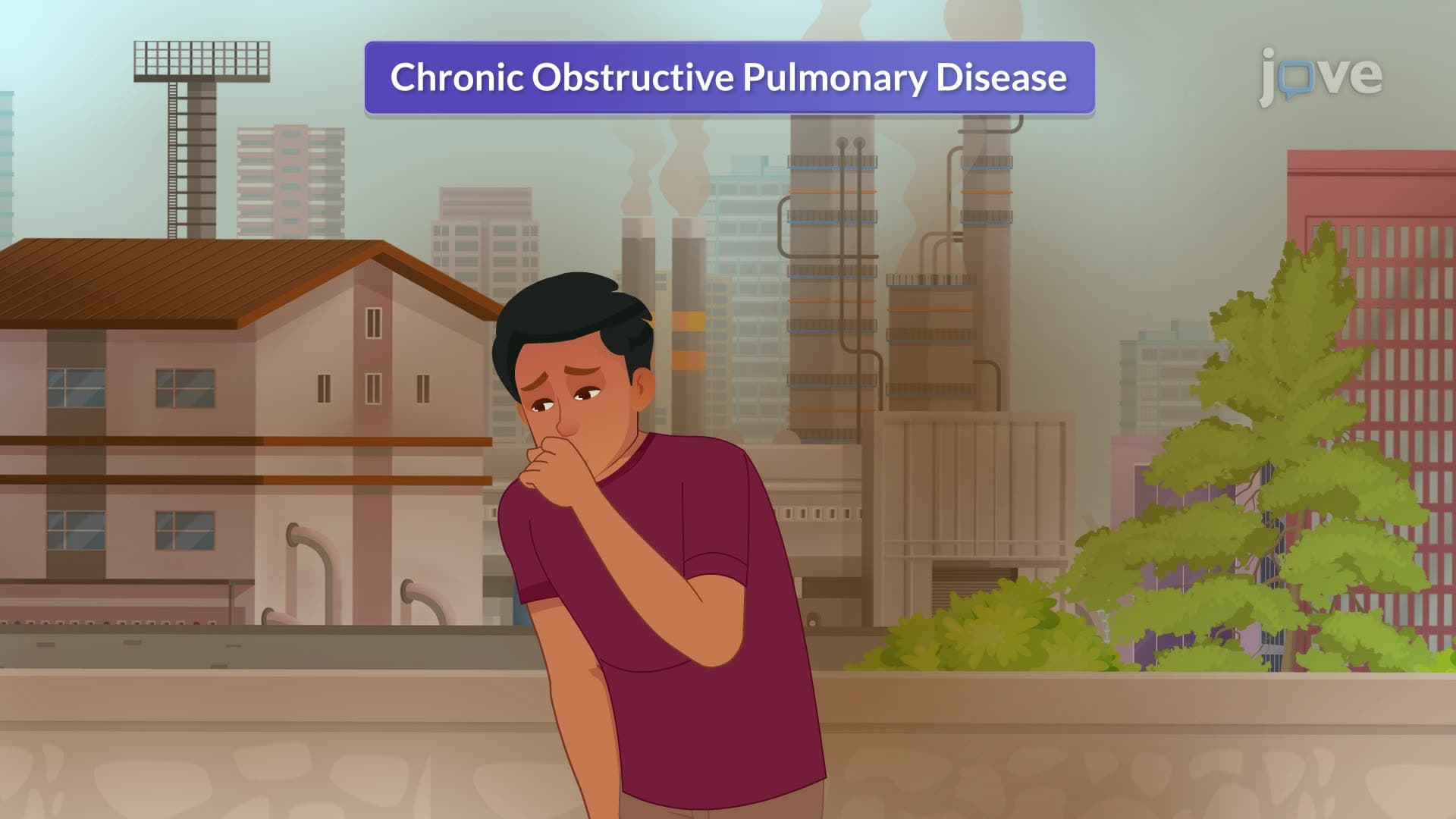
Every year, approximately 16 million Americans live with chronic obstructive pulmonary disease (COPD), making it the third leading cause of death nationwide. Understanding chronic obstructive pulmonary disease II pathophysiology reveals how inflammatory cells like neutrophils and macrophages systematically destroy lung tissue through protease-antiprotease imbalances. For instance, coal miners in West Virginia face heightened COPD risk due to chronic dust exposure triggering this devastating inflammatory cascade. Watch the full video on JoVE Coach to master this concept with expert-led visuals and step-by-step explanations.
- Understand chronic obstructive pulmonary disease II pathophysiology as a progressive inflammatory process affecting airway structure and function
- Identify the primary inflammatory cells involved in COPD progression, including lymphocytes, macrophages, and neutrophils
- Analyze how protease-antiprotease imbalances lead to alveolar destruction and loss of lung elasticity
- Recognize the role of oxidative stress and free radical formation in COPD tissue damage
- Evaluate how chronic inflammation contributes to airway narrowing and excessive mucus production
- Apply knowledge of inflammatory mediators like leukotrienes and cytokines to understand disease progression
- Compare normal lung tissue homeostasis with pathological changes seen in COPD patients
- Explore the connection between environmental irritants and chronic airway inflammation patterns
Understanding Chronic Obstructive Pulmonary Disease II Pathophysiology
Chronic obstructive pulmonary disease II pathophysiology represents a complex interplay of inflammatory processes that fundamentally alter lung structure and function. Unlike acute respiratory conditions, COPD develops through sustained inflammatory responses triggered by long-term exposure to noxious particles and gases. The pathophysiology centers on an aberrant immune response that, rather than protecting the lungs, systematically destroys healthy tissue through uncontrolled inflammation.
Inflammatory Cell Infiltration and Mediator Release
The pathological cascade begins when irritants like cigarette smoke or occupational dust penetrate deep into lung tissue. This triggers massive recruitment of inflammatory cells-primarily neutrophils, macrophages, and CD8+ T lymphocytes-into the airway walls. These cells release a destructive cocktail of inflammatory mediators, including tumor necrosis factor-alpha, interleukin-8, and leukotriene B4. In healthy individuals, this inflammatory response would be temporary and controlled. However, in COPD patients, particularly those with genetic predispositions like alpha-1 antitrypsin deficiency (affecting 1 in 2,500 Americans), this inflammation becomes chronic and self-perpetuating.
Protease-Antiprotease Imbalance: The Key Destructive Mechanism
The hallmark of COPD pathophysiology lies in the disruption of the protease-antiprotease balance. Normally, proteases (tissue-degrading enzymes) like neutrophil elastase and matrix metalloproteinases are carefully regulated by antiproteases such as alpha-1 antitrypsin and secretory leukoprotease inhibitor. In COPD, oxidative stress inactivates these protective antiproteases while simultaneously upregulating destructive proteases. This imbalance results in unchecked degradation of elastin and collagen fibers in alveolar walls, leading to emphysematous changes and loss of lung recoil.
Oxidative Stress and Tissue Damage
Oxidative stress amplifies COPD pathophysiology through multiple mechanisms. Inhaled oxidants and those produced by activated inflammatory cells overwhelm the lung's antioxidant defense systems. This oxidative burden not only damages cellular structures directly but also activates transcription factors like nuclear factor-kappa B, which promotes further inflammatory gene expression. The result is a vicious cycle where inflammation begets more inflammation, progressively destroying lung architecture and impairing gas exchange capacity.
Students preparing for the MCAT or advanced placement biology exams should focus on understanding these interconnected pathways, as questions often test the ability to trace inflammatory cascades and predict downstream effects on lung function.
Frequently Asked Questions
Chronic obstructive pulmonary disease II pathophysiology describes the complex inflammatory processes that cause progressive lung damage in COPD patients. It involves chronic inflammation triggered by irritants, leading to protease-antiprotease imbalances that destroy alveolar walls and impair breathing. This pathophysiology explains how environmental exposures translate into the characteristic airflow obstruction seen in COPD. Understanding these mechanisms is crucial for developing targeted treatments and preventive strategies.
MCAT questions typically test your understanding of inflammatory cascades, enzyme regulation, and cellular damage mechanisms in COPD pathophysiology. You'll need to identify key inflammatory cells, explain protease-antiprotease imbalances, and predict how oxidative stress affects lung tissue. Practice connecting molecular-level changes to clinical symptoms like dyspnea and reduced lung function. Focus on understanding cause-and-effect relationships rather than memorizing isolated facts.
COPD pathophysiology directly relates to AP Biology Unit 2 (Cell Structure and Function) and Unit 3 (Cellular Energetics), particularly enzyme regulation and cellular respiration. It also connects to immune system function and homeostasis concepts. Students should understand how enzyme imbalances affect tissue structure and how chronic inflammation disrupts normal cellular processes. This topic frequently appears in free-response questions about disease mechanisms and homeostatic disruption.
Healthcare providers use COPD pathophysiology knowledge to develop personalized treatment plans for patients at major medical centers like Johns Hopkins and Mayo Clinic. Understanding inflammatory mechanisms guides medication choices, such as corticosteroids to reduce inflammation or bronchodilators to counter airway narrowing. Pulmonologists also use this knowledge to counsel patients about smoking cessation and environmental modifications. The pathophysiology directly influences diagnostic approaches, including spirometry interpretation and biomarker analysis.
While COPD pathophysiology involves complex mechanisms, it builds logically on basic biology concepts you already know. You need familiarity with cell biology, enzyme function, and immune responses, typically covered in introductory biology courses. The key is understanding how normal protective mechanisms become harmful when dysregulated. Start with basic inflammatory processes and gradually build understanding of specific pathways. Most high school students with solid biology backgrounds can master these concepts with focused study.
Create concept maps linking inflammatory triggers to cellular responses to tissue damage to clinical symptoms. Practice drawing the protease-antiprotease balance under normal versus COPD conditions. Use active recall by explaining the pathophysiology pathway out loud without notes. Focus on understanding why each step occurs rather than memorizing sequences. Connect molecular mechanisms to patient symptoms you might encounter in clinical scenarios or case studies.
Build on this foundation by exploring related respiratory pathophysiology topics like asthma mechanisms, pulmonary fibrosis, and acute respiratory distress syndrome. Study pharmacological interventions that target specific pathways you've learned about in COPD. Investigate other chronic inflammatory diseases to see similar patterns in different organ systems. Advanced students should explore current research on COPD biomarkers and novel therapeutic approaches targeting specific inflammatory mediators.
College-level courses delve deeper into molecular mechanisms, including specific signaling pathways, genetic factors, and epigenetic modifications in COPD development. You'll study detailed structure-function relationships in lung tissue and quantitative aspects of gas exchange impairment. Advanced coursework covers current research methodologies, clinical trial design, and emerging therapeutic targets. Students often analyze primary literature and case studies to understand how pathophysiology translates to patient care and treatment outcomes.
Related Micro-courses

7 Concepts

10 Concepts