Loading subject page…

- Medical Surgical Nursing
- Obstructive Pulmonary Diseases
- Chronic Obstructive Pulmonary Disease-IV: Assessement and Diagnostic Studies
Micro-course: Obstructive Pulmonary Diseases
35,512 views
Learn key concepts and earn your certificate.


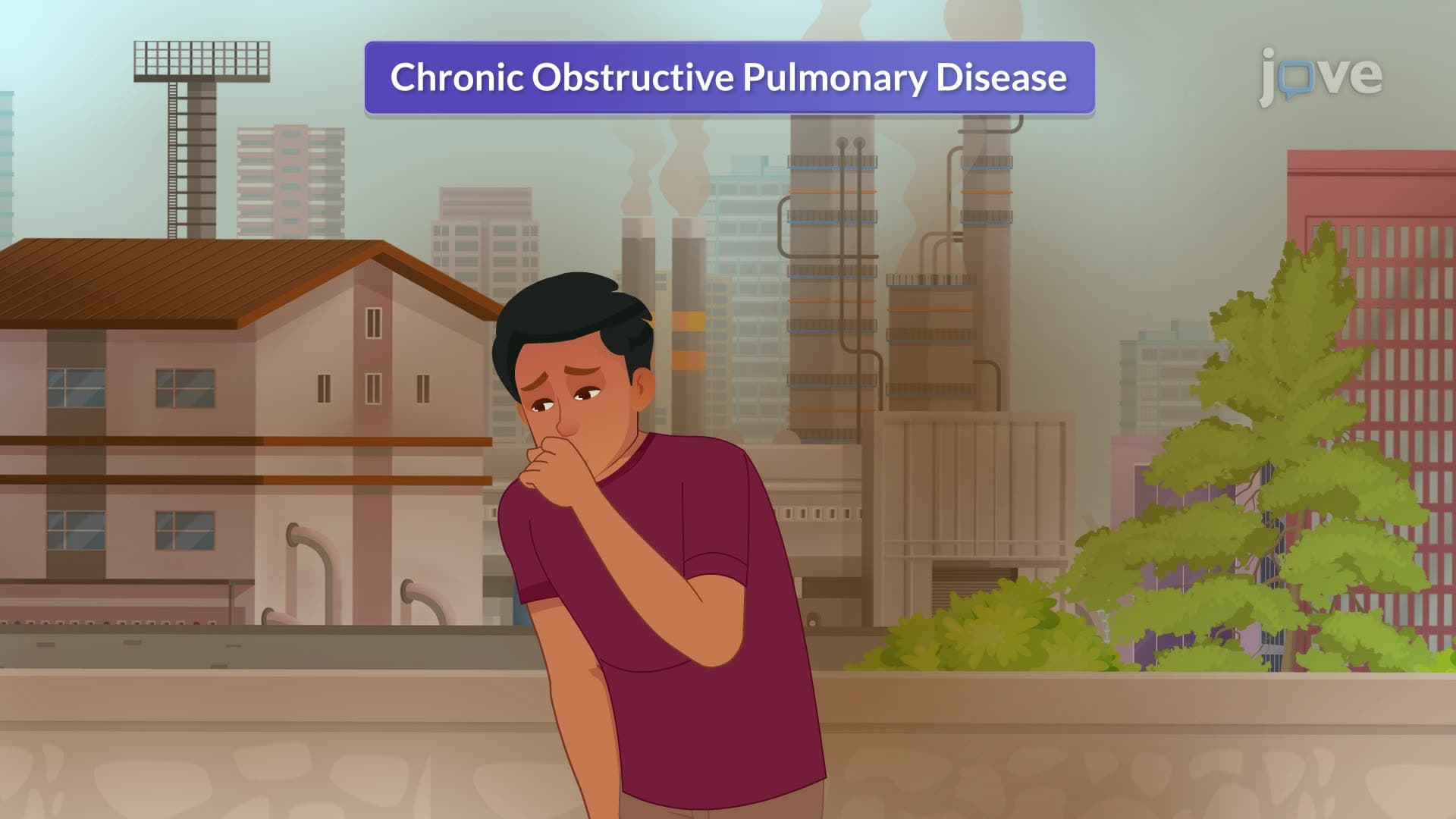
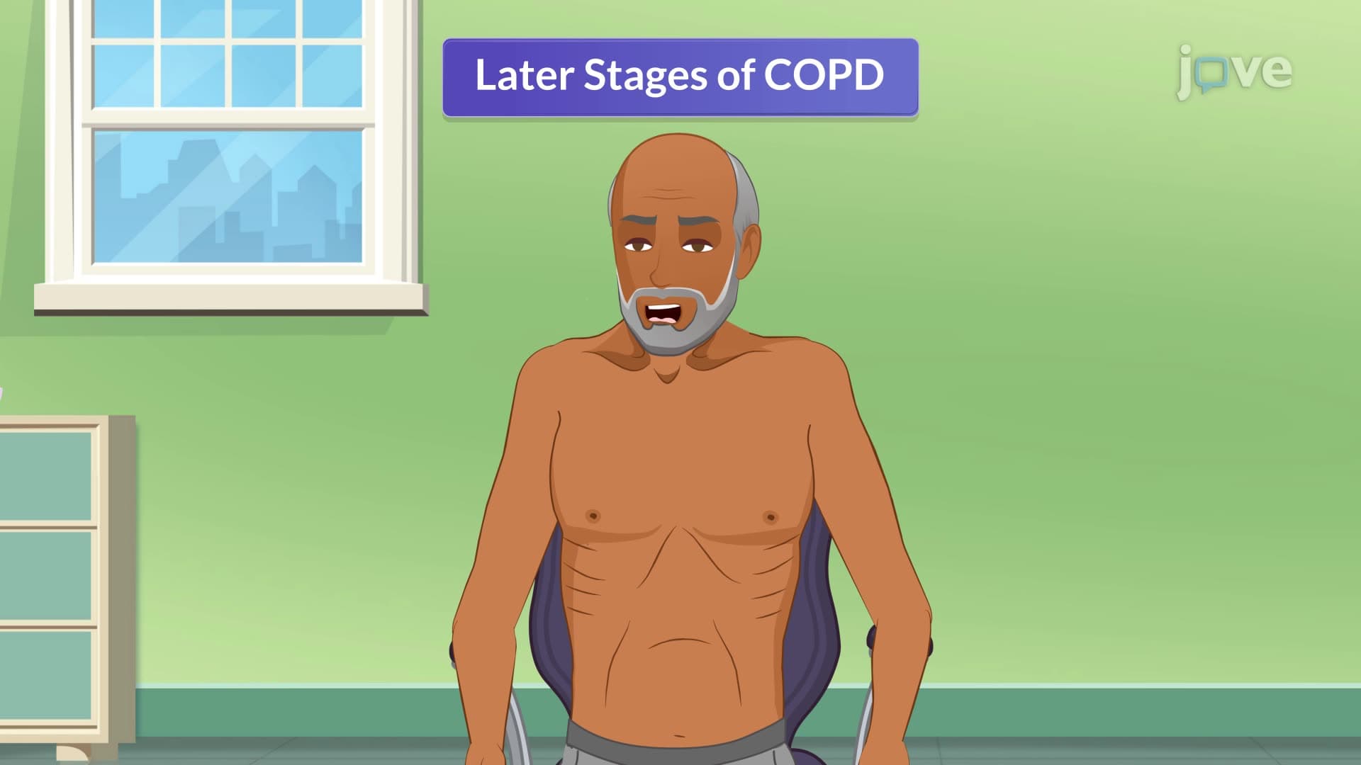
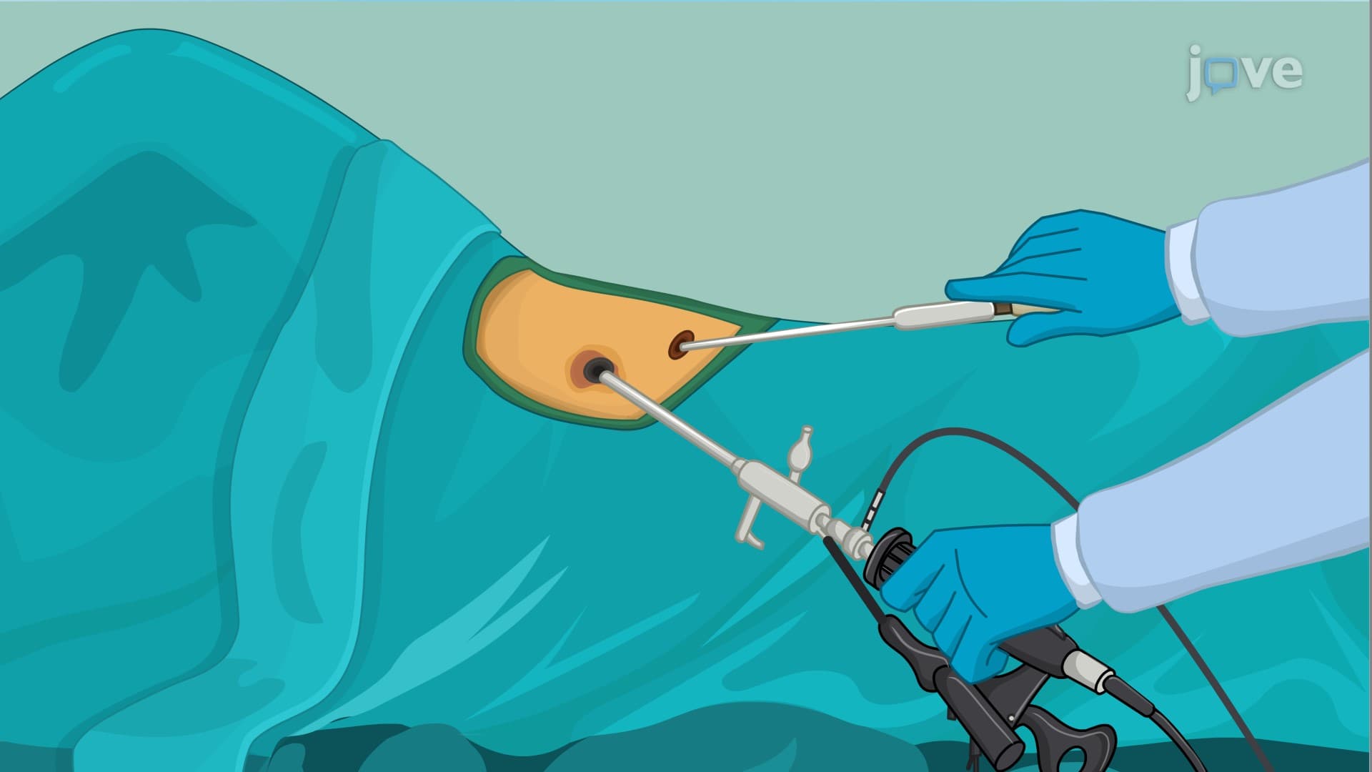




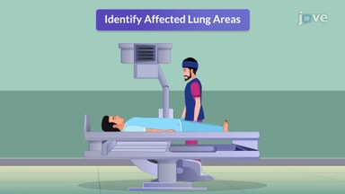
Chronic Obstructive Pulmonary Disease-IV: Assessement and Diagnostic Studies

00:00
01:27
Video Summary: Chronic Obstructive Pulmonary Disease Iv Explained
Did you know that over 16 million Americans live with chronic obstructive pulmonary disease (COPD), yet many cases remain undiagnosed until advanced stages? Chronic Obstructive Pulmonary Disease IV focuses on the critical assessment and diagnostic procedures that healthcare providers use to identify and evaluate this progressive respiratory condition. From spirometry tests measuring lung function to arterial blood gas analysis, these diagnostic tools are essential for patients like those treated at Mayo Clinic's pulmonology department. Watch the full video on JoVE Coach to master this concept with expert-led visuals and step-by-step explanations.
- Understand the comprehensive assessment process for Chronic Obstructive Pulmonary Disease IV, including patient history evaluation and symptom identification
- Identify key physical examination findings such as barrel chest, cyanosis, and abnormal breathing patterns in COPD patients
- Analyze pulmonary function test results, particularly spirometry measurements of FEV1/FVC ratios below 70%
- Evaluate arterial blood gas analysis results to determine oxygen and carbon dioxide levels in COPD patients
- Recognize diagnostic imaging findings on chest X-rays and CT scans that indicate lung hyperinflation and structural changes
- Apply knowledge of sputum testing procedures used during COPD exacerbations to identify bacterial infections
- Compare different diagnostic methods and their specific roles in confirming COPD diagnosis
- Explore the relationship between patient history factors like smoking and environmental exposures in COPD development
Understanding Chronic Obstructive Pulmonary Disease IV Assessment
Chronic Obstructive Pulmonary Disease IV represents the advanced diagnostic phase where healthcare providers systematically evaluate patients suspected of having COPD. This comprehensive assessment goes far beyond simple observation, requiring a multi-faceted approach that combines clinical history, physical examination, and sophisticated diagnostic testing. The process is crucial because early detection can significantly impact patient outcomes and quality of life.
Patient History and Clinical Presentation
The diagnostic journey begins with a thorough patient history focusing on hallmark symptoms. Healthcare providers specifically investigate chronic cough patterns, sputum production characteristics, and progressive dyspnea (shortness of breath). Smoking history remains paramount, as cigarette smoking causes 85-90% of COPD cases in the United States. Environmental and occupational exposures, such as those experienced by construction workers exposed to silica dust or coal miners developing pneumoconiosis, provide additional diagnostic clues. Family history of genetic conditions like alpha-1 antitrypsin deficiency, which affects approximately 100,000 Americans, can indicate hereditary predisposition to emphysema.
Physical Examination Techniques and Findings
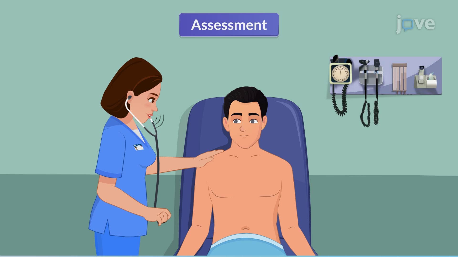
The physical examination reveals characteristic signs that distinguish COPD from other respiratory conditions. Healthcare providers observe breathing patterns, noting the use of accessory muscles during respiration and pursed-lip breathing techniques that patients unconsciously develop. Cyanosis, particularly around the lips and fingernails, indicates inadequate oxygenation. The classic "barrel chest" appearance results from lung hyperinflation and diaphragm flattening. Lung auscultation reveals decreased breath sounds, expiratory wheezes, and crackles that help differentiate between emphysema and chronic bronchitis components.
Advanced Diagnostic Testing and Interpretation
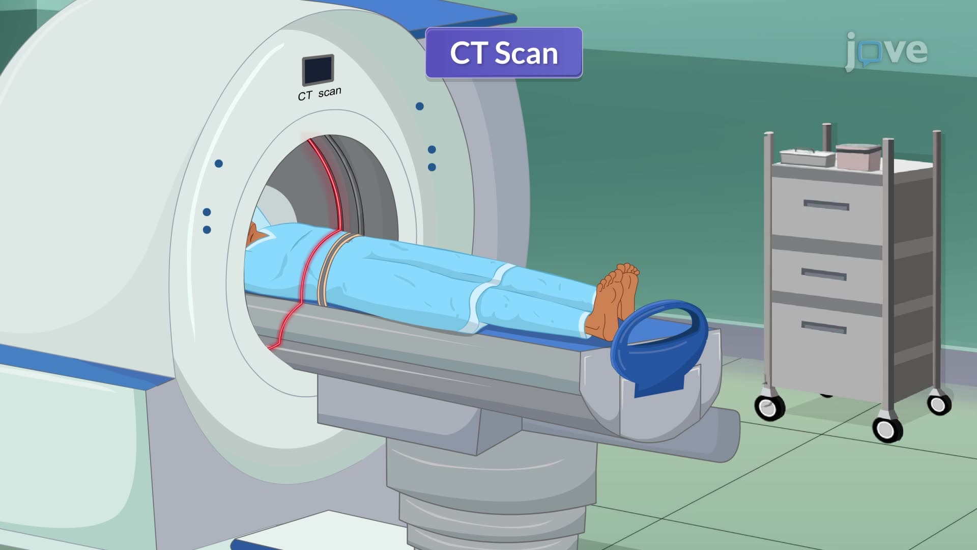
Pulmonary function tests, particularly spirometry, serve as the gold standard for COPD diagnosis. The critical measurement involves the FEV1/FVC ratio, where values below 70% indicate airflow obstruction. This test appears frequently on MCAT practice exams and USMLE Step 1 questions, making it essential knowledge for pre-med students. Arterial blood gas analysis provides crucial information about gas exchange efficiency, often revealing hypoxemia and hypercapnia in advanced cases. Chest imaging, including X-rays showing diaphragm flattening and CT scans detecting emphysematous changes, provides structural evidence of disease progression. During exacerbations, sputum cultures help identify bacterial pathogens like Streptococcus pneumoniae or Haemophilus influenzae, guiding targeted antibiotic therapy.
Frequently Asked Questions
Chronic Obstructive Pulmonary Disease IV refers to the comprehensive diagnostic and assessment phase of COPD evaluation, not a disease stage itself. This phase involves systematic testing including spirometry, imaging, and blood gas analysis to confirm diagnosis and determine disease severity. Unlike the GOLD staging system (stages 1-4), this represents the diagnostic process that determines which stage a patient has reached.
MCAT questions frequently test spirometry interpretation, particularly FEV1/FVC ratios below 70% indicating airflow obstruction. Students must understand the relationship between smoking history and COPD development, plus differentiate between emphysema and chronic bronchitis presentations. USMLE Step 1 often includes arterial blood gas interpretation showing respiratory acidosis with metabolic compensation in advanced COPD patients.
Nursing exams emphasize patient assessment skills, including recognizing barrel chest appearance, pursed-lip breathing, and use of accessory muscles. Students must understand oxygen therapy considerations, medication administration timing, and patient education about spirometry procedures. HESI A2 and TEAS exams may include respiratory system anatomy and physiology related to COPD pathophysiology.
Emergency departments use rapid diagnostic approaches including arterial blood gas analysis to assess oxygenation status, chest X-rays to rule out pneumonia or pneumothorax, and sputum cultures when bacterial infection is suspected. Peak flow measurements provide quick airflow assessment, while patient history of previous COPD diagnoses and current medications guide immediate treatment decisions.
Basic understanding of gas exchange principles and acid-base balance helps, but most COPD diagnostic concepts are accessible to high school students taking AP Biology or college-level anatomy and physiology. The mathematical calculations involved in spirometry interpretation use simple percentages and ratios that don't require calculus or advanced statistics.
Create comparison charts distinguishing emphysema versus chronic bronchitis characteristics, practice interpreting spirometry values with FEV1/FVC ratios, and memorize normal versus abnormal arterial blood gas ranges. Use case study approaches connecting smoking history to specific diagnostic findings, and understand how environmental exposures contribute to occupational lung diseases.
Progress to asthma diagnosis and differentiation from COPD, explore pulmonary rehabilitation techniques, and study medication classifications including bronchodilators and corticosteroids. Advanced topics include mechanical ventilation principles for respiratory failure and lung transplantation criteria for end-stage COPD patients.
Related Micro-courses
7 Concepts

10 Concepts