- Pathophysiology
- Nervous System Disorders
Micro-courses:5

Nervous System Disorders

1. Stroke: Introduction and Types
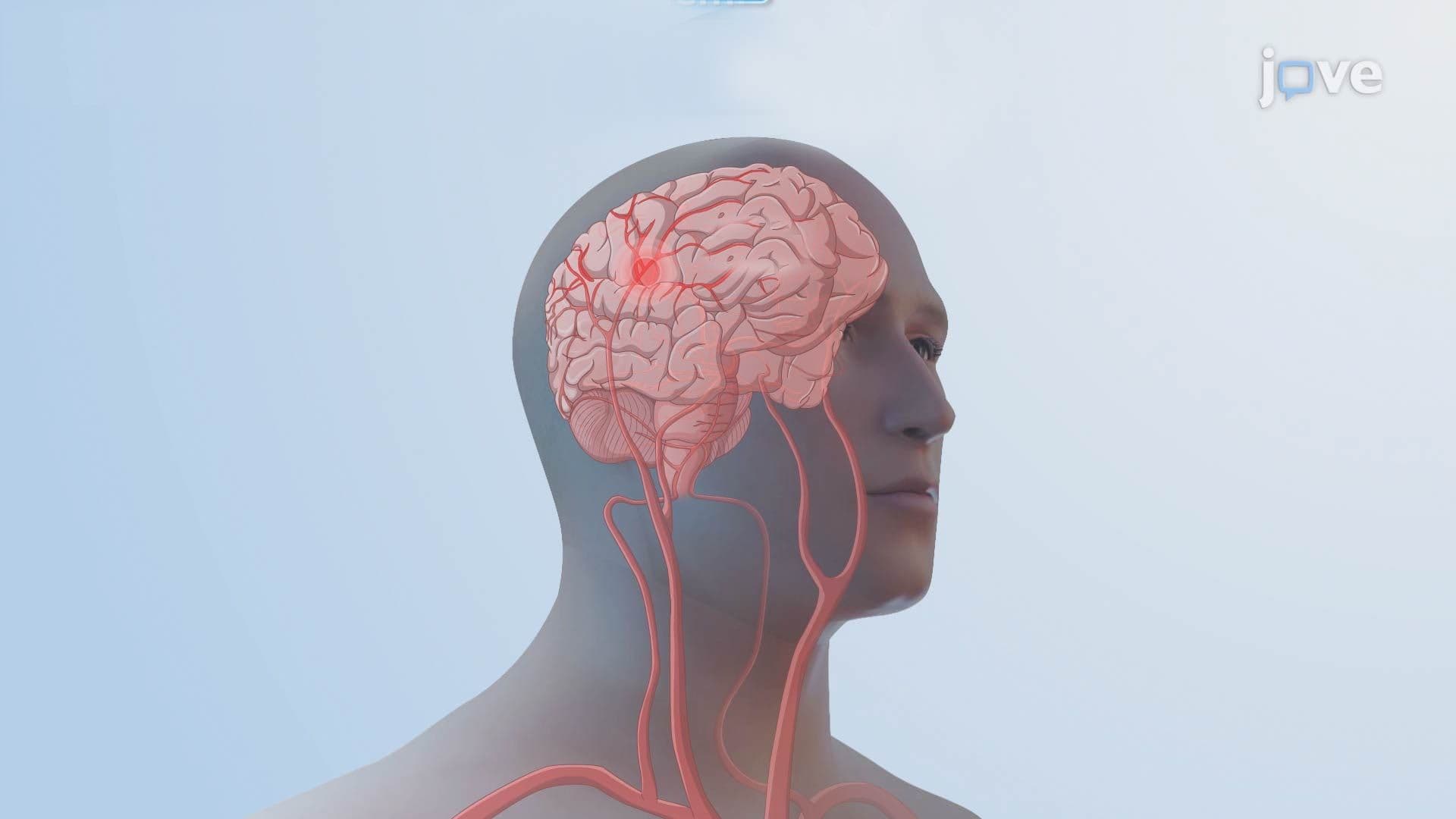
2. Ischemic Stroke l: Introduction
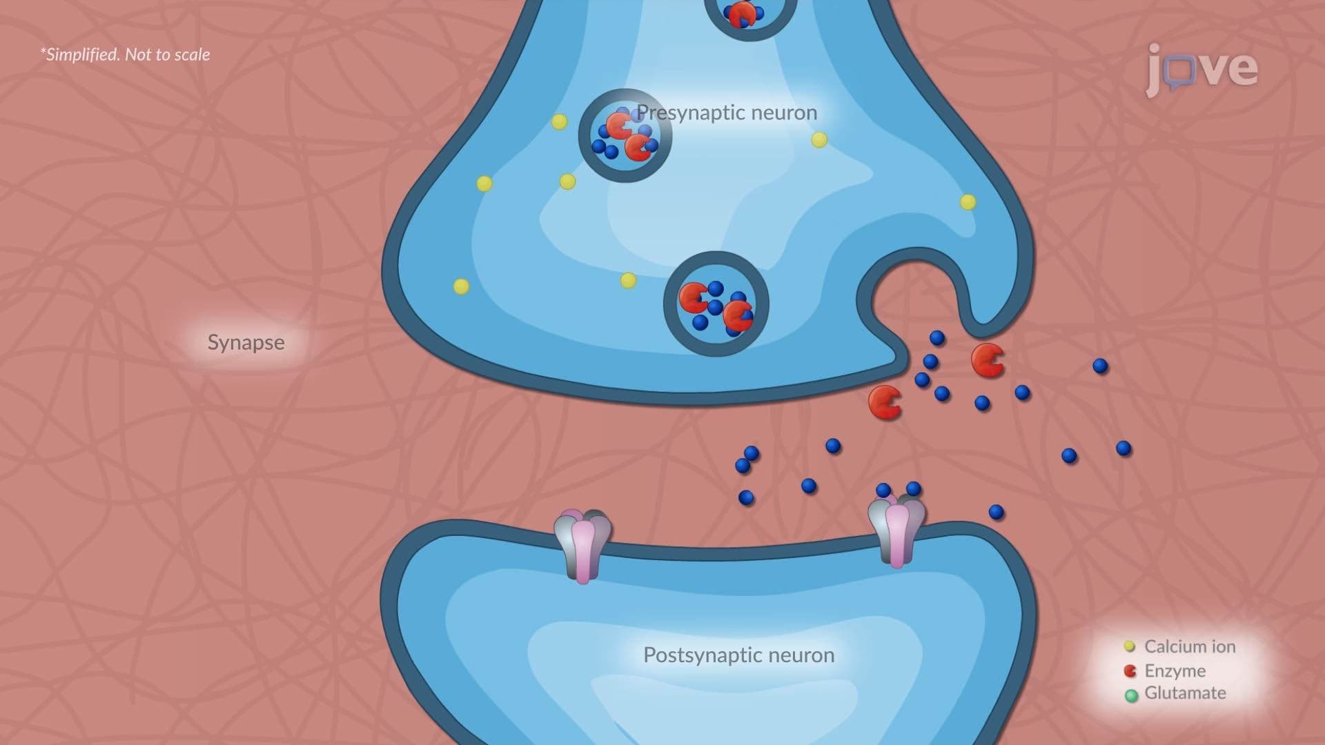
3. Ischemic Stroke ll: Pathophysiology
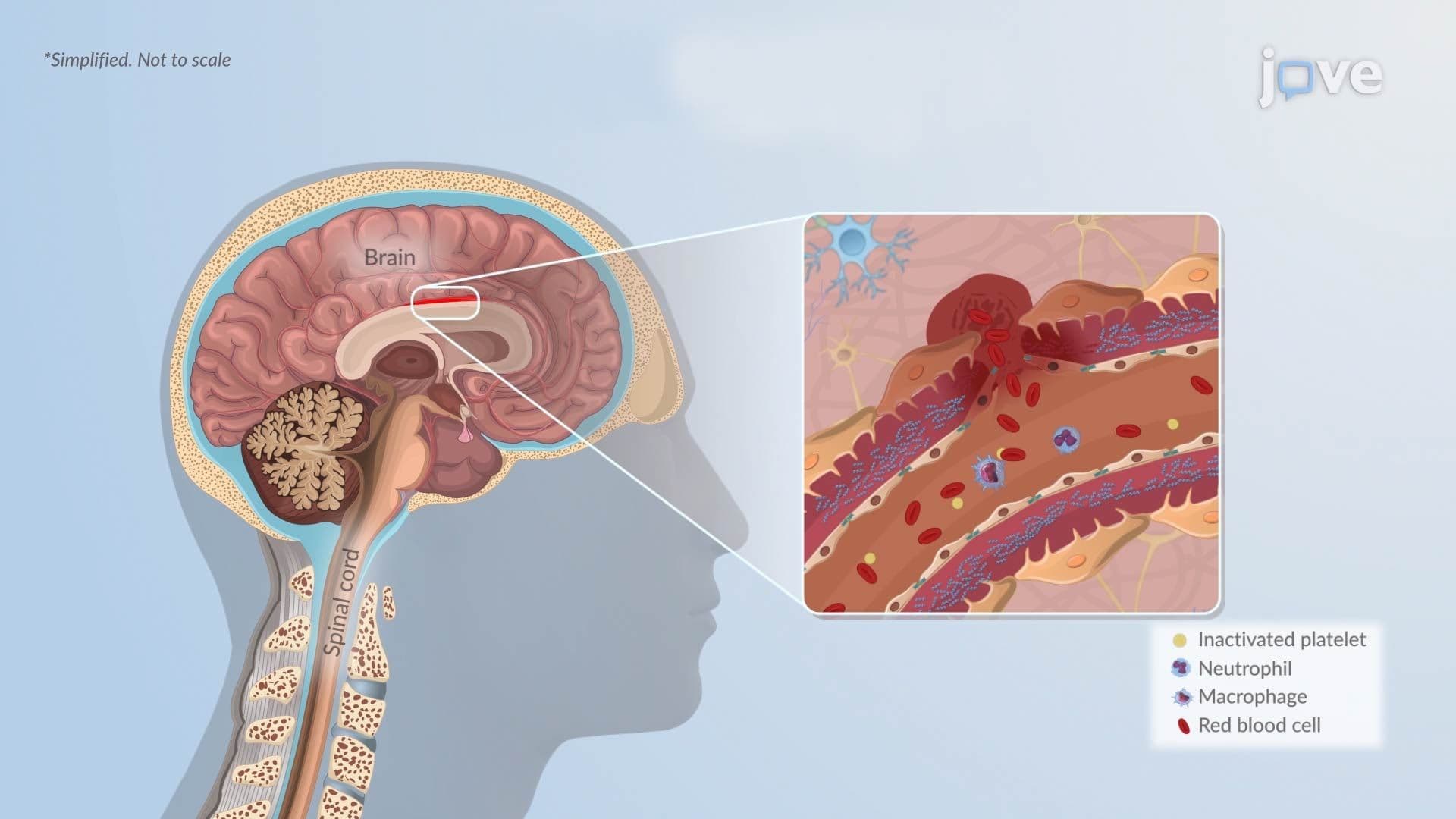
4. Hemorrhagic Stroke l: Introduction
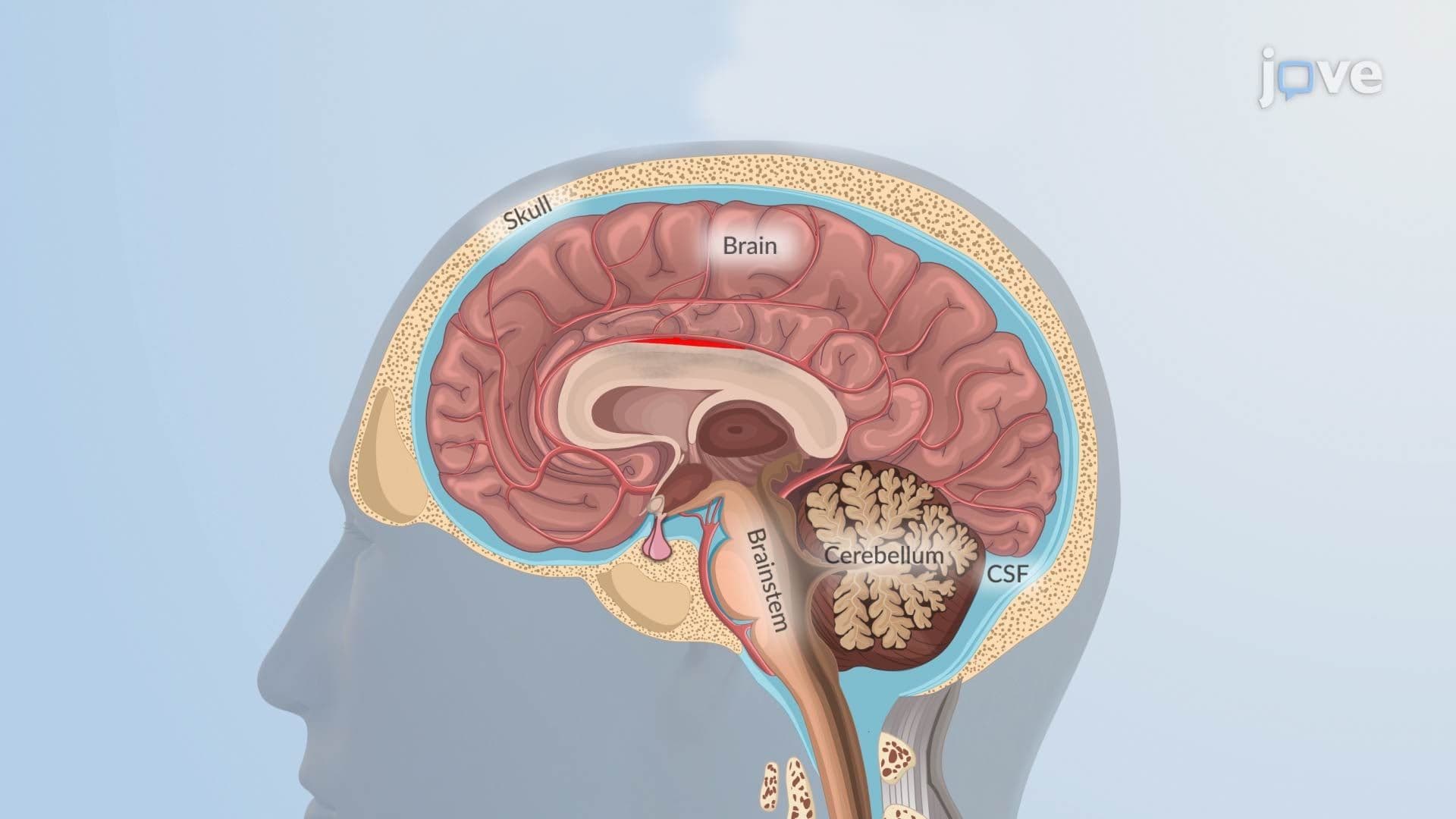
5. Hemorrhagic Stroke ll: Pathophysiology
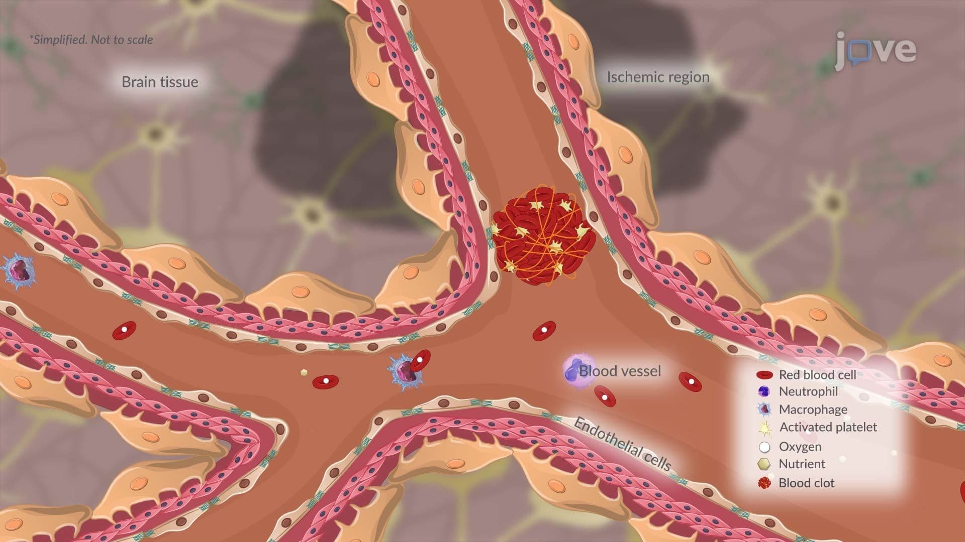
6. Transient Ischemic Attack l: Introduction
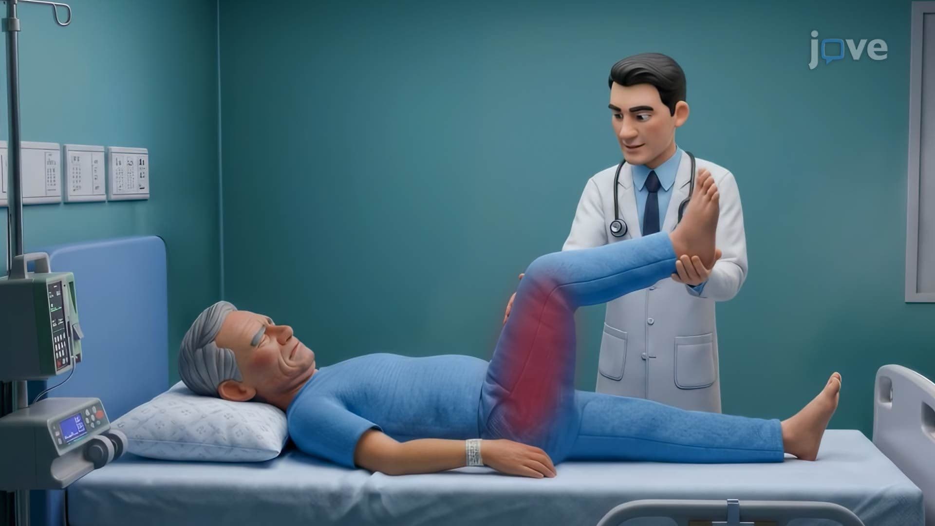
7. Bacterial Meningitis I: Introduction
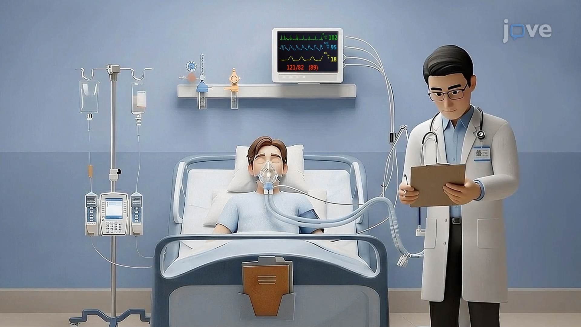
8. Bacterial Meningitis II: Pathophysiology
9. Encephalitis l: Introduction
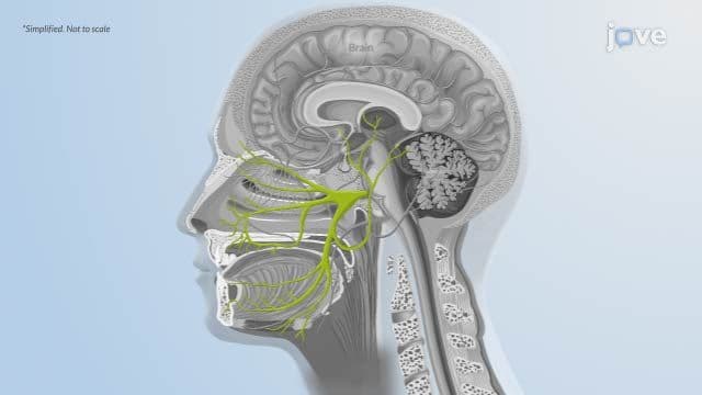
10. Encephalitis ll: Pathophysiology

11. Brain Abscess l: Introduction
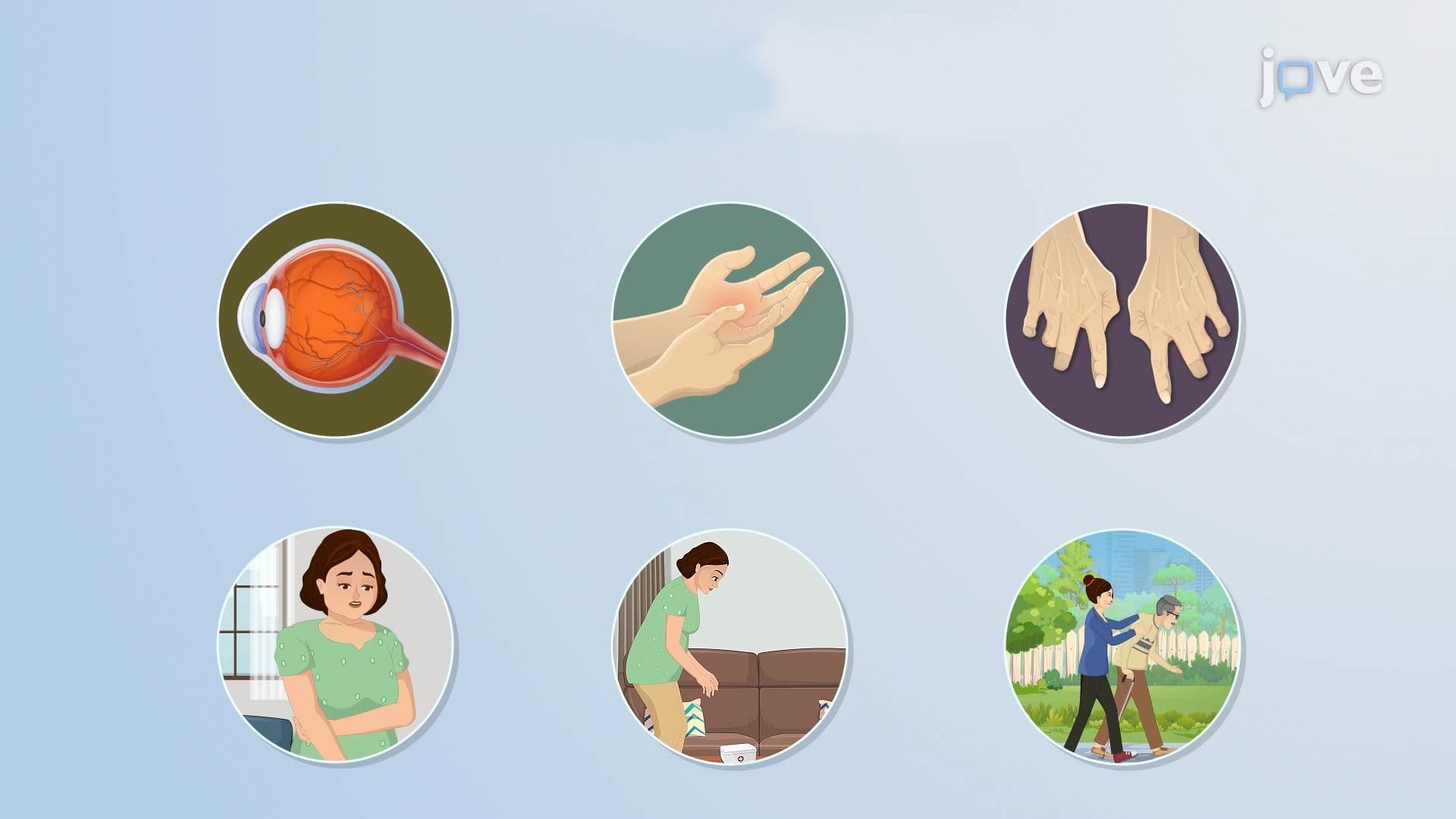
12. Multiple Sclerosis l: Introduction
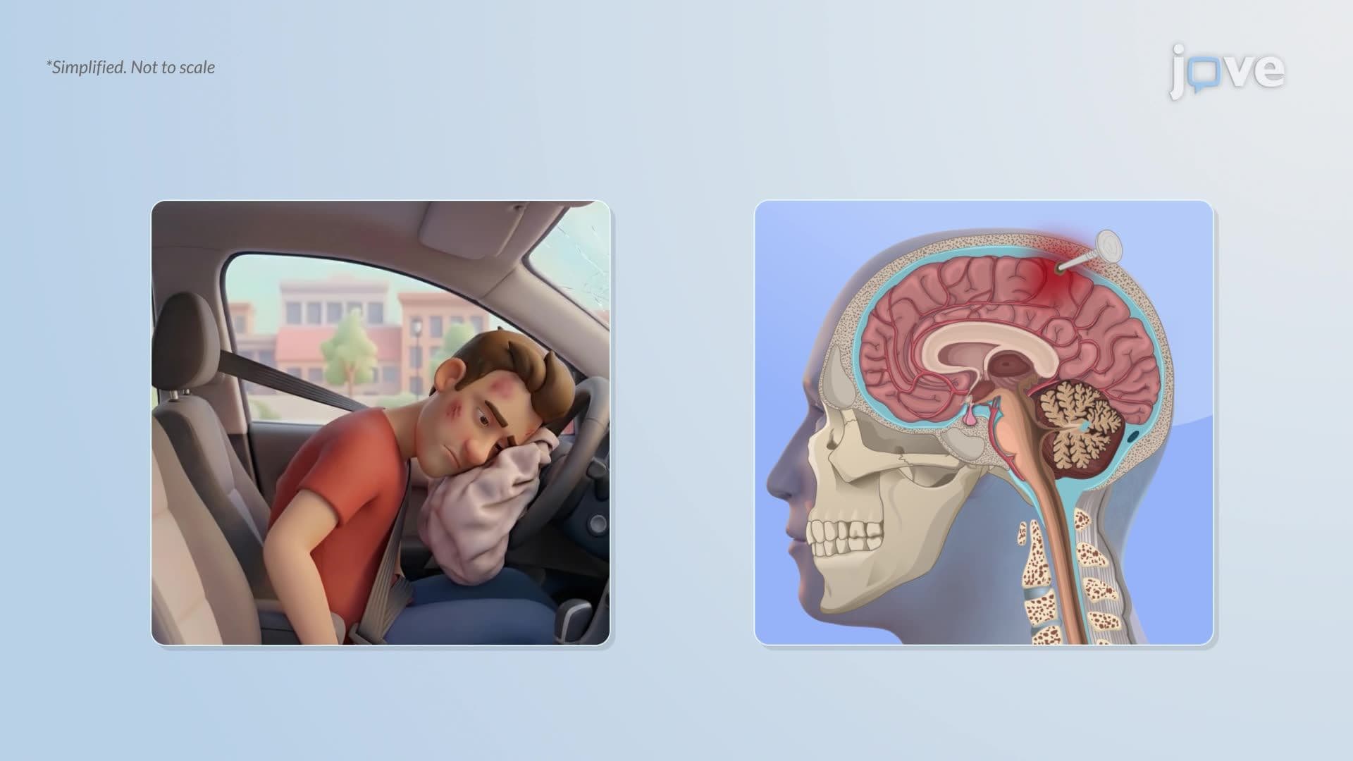
13. Traumatic Brain Injury l: Introduction
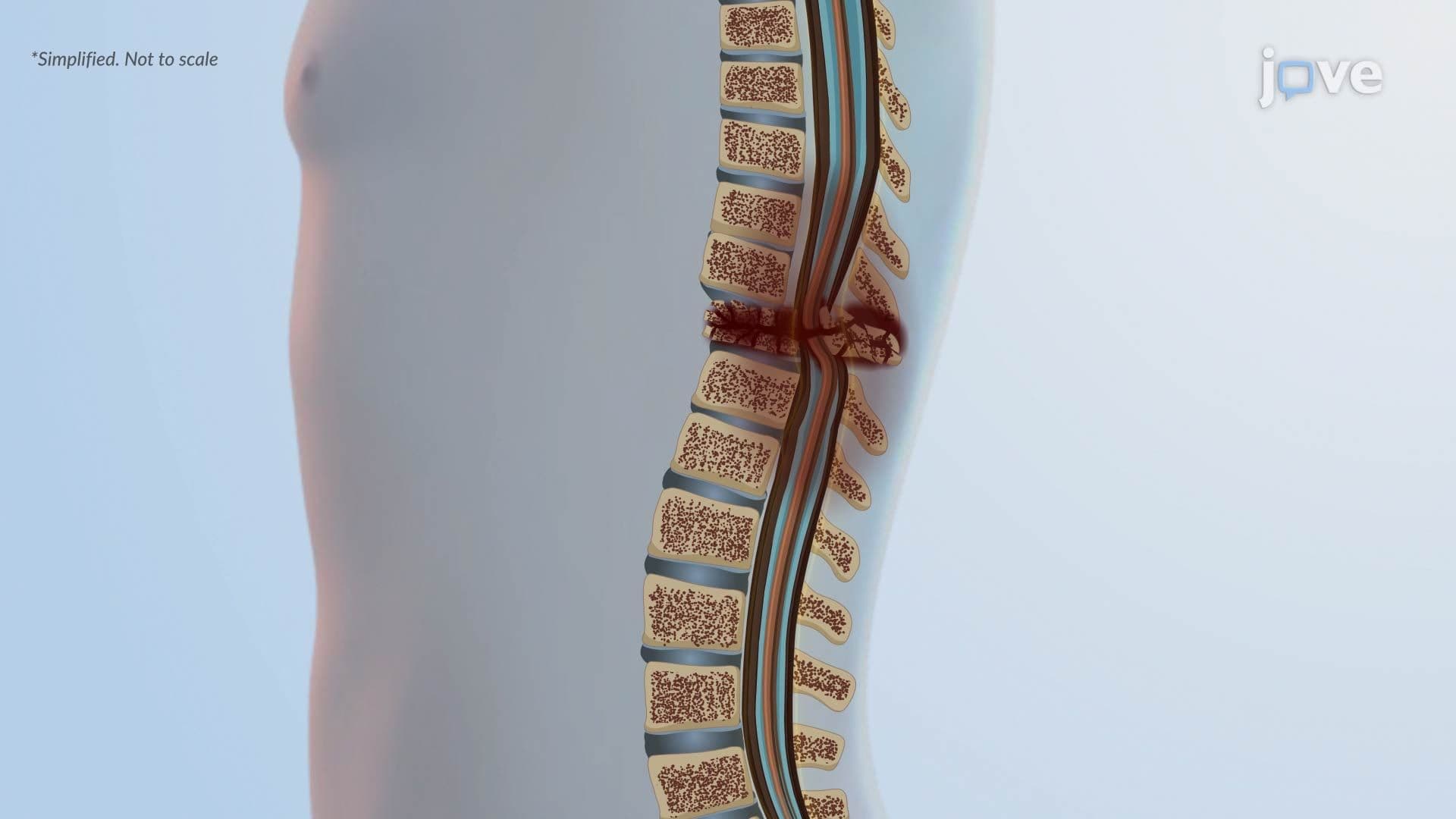
14. Spinal Cord Injury ll: Pathophysiology
15. Secondary Spinal Cord Injury llI: Pathophysiology
16. Increased Intracranial Pressure l: Introduction
17. Increased Intracranial Pressure ll: Pathophysiology
18. Cerebral Edema l: Introduction
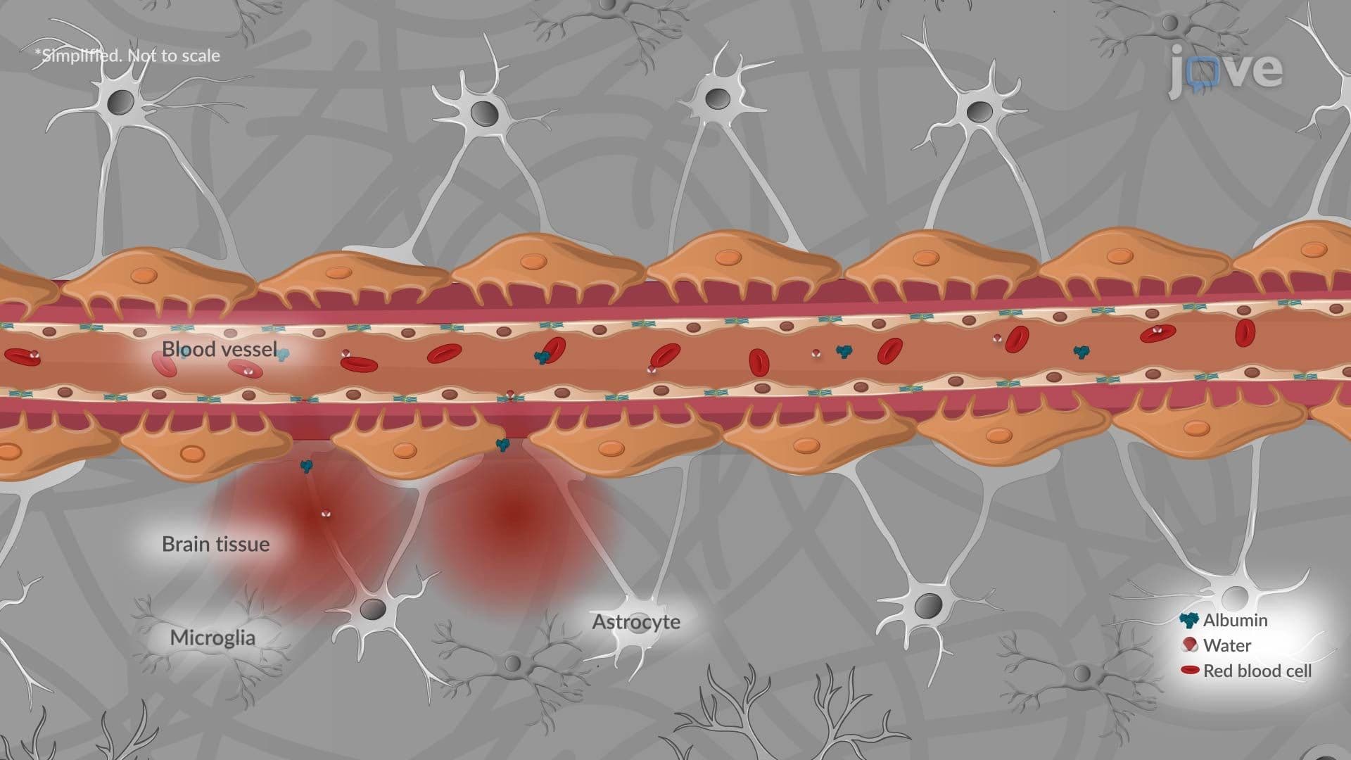
19. Cerebral Edema ll: Pathophysiology
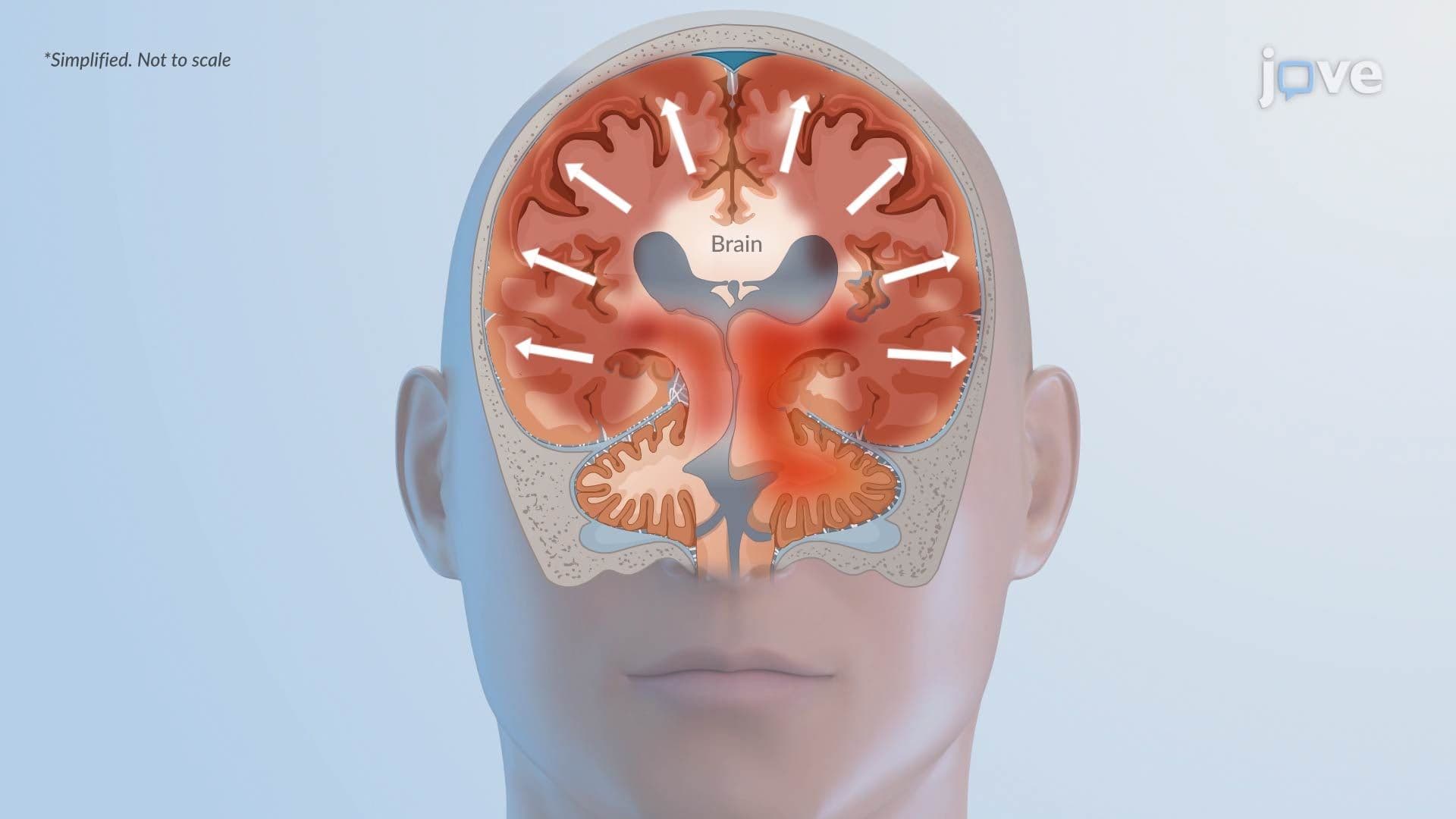
20. Cytotoxic Edema: Pathophysiology

21. Alzheimer Disease l: Introduction
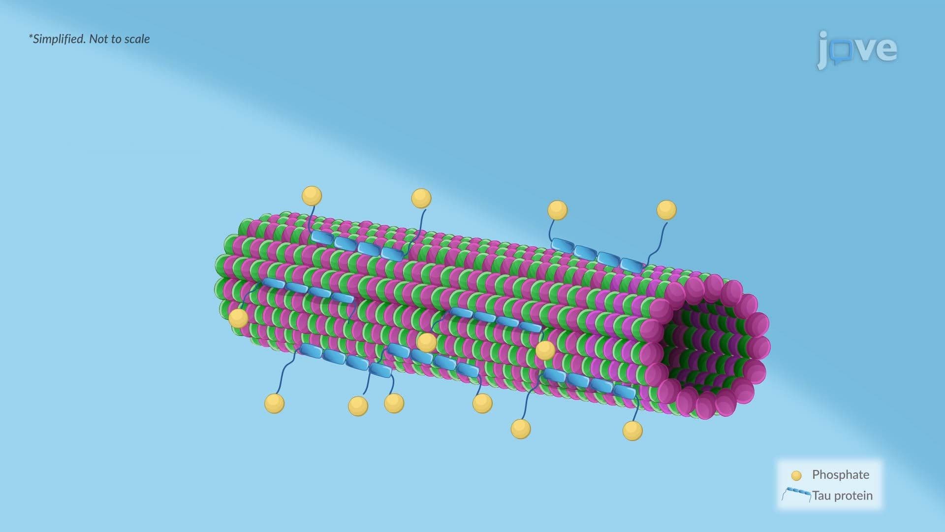
22. Alzheimer Disease ll: Pathophysiology
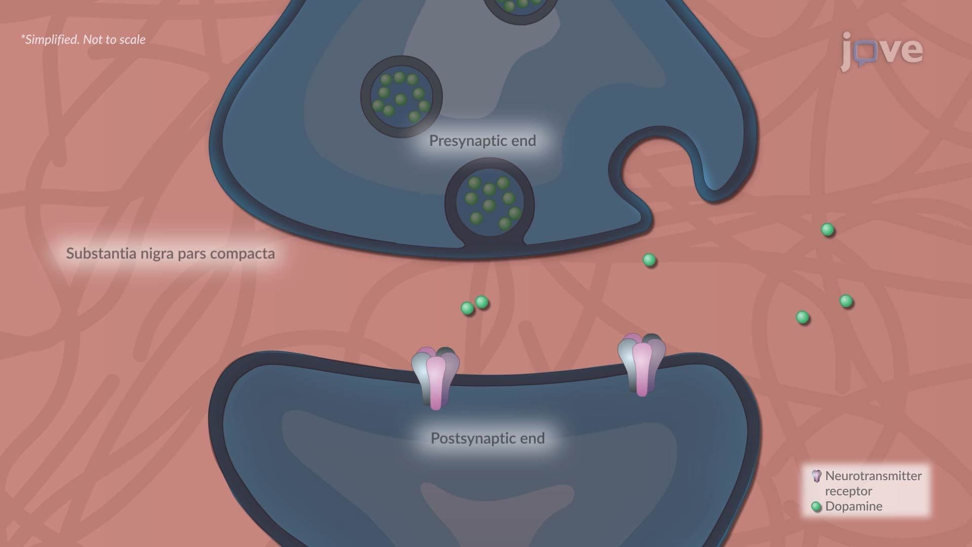
23. Parkinson Disease l: Introduction
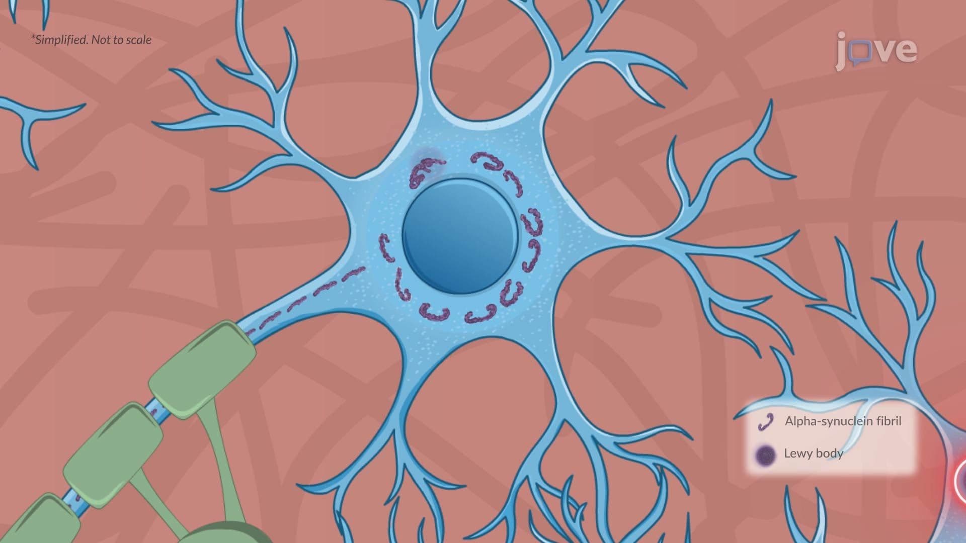
24. Parkinson Disease ll: Pathophysiology
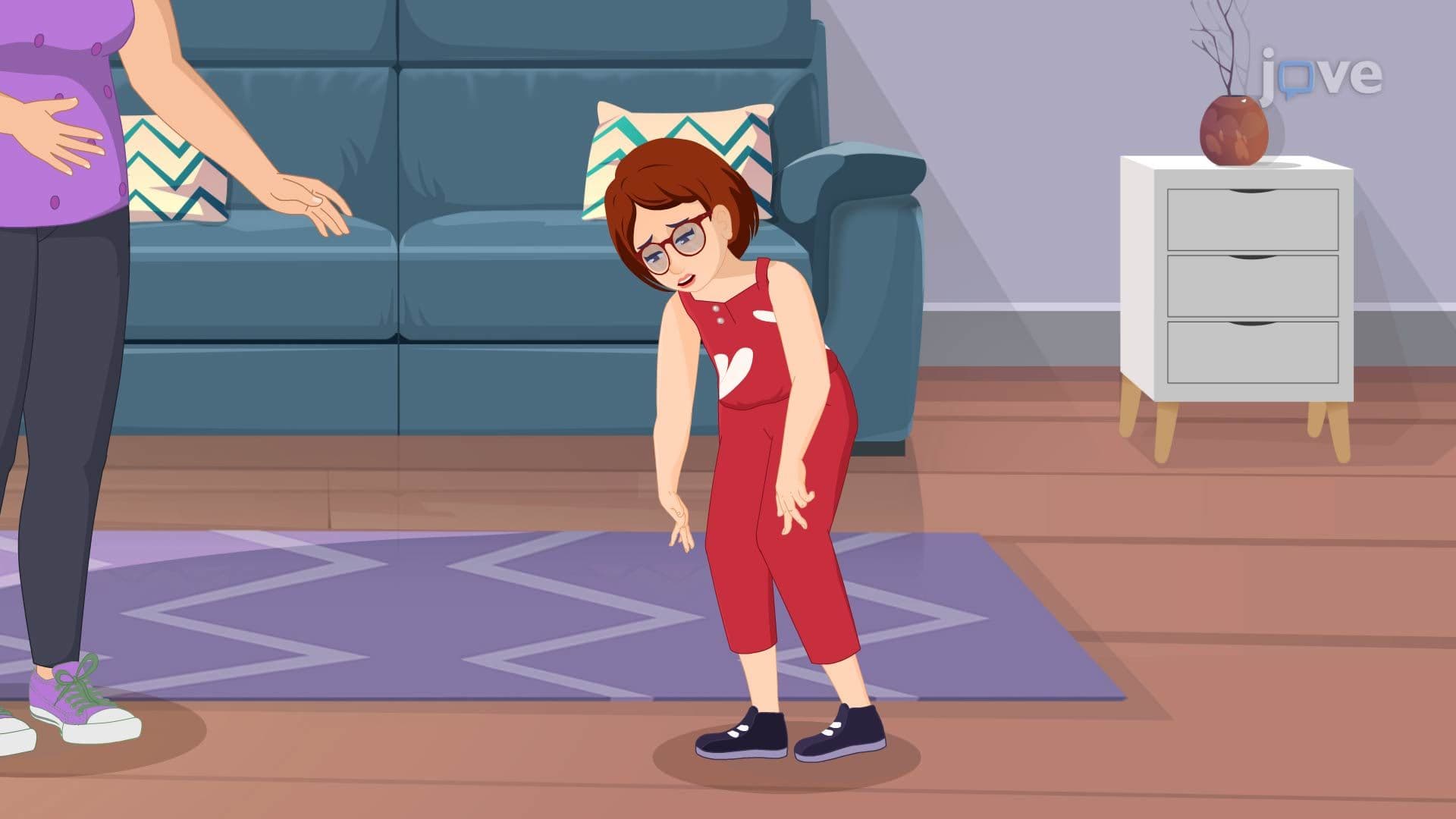
25. Huntington Disease l: Introduction
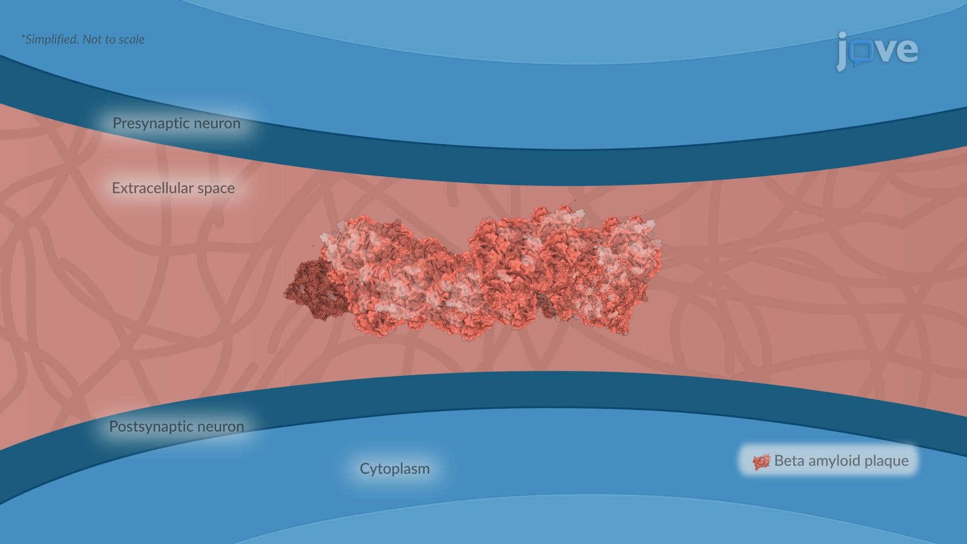
26. Dementia l: Introduction
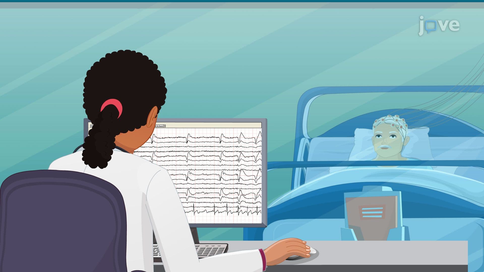
27. Seizures l: Introduction
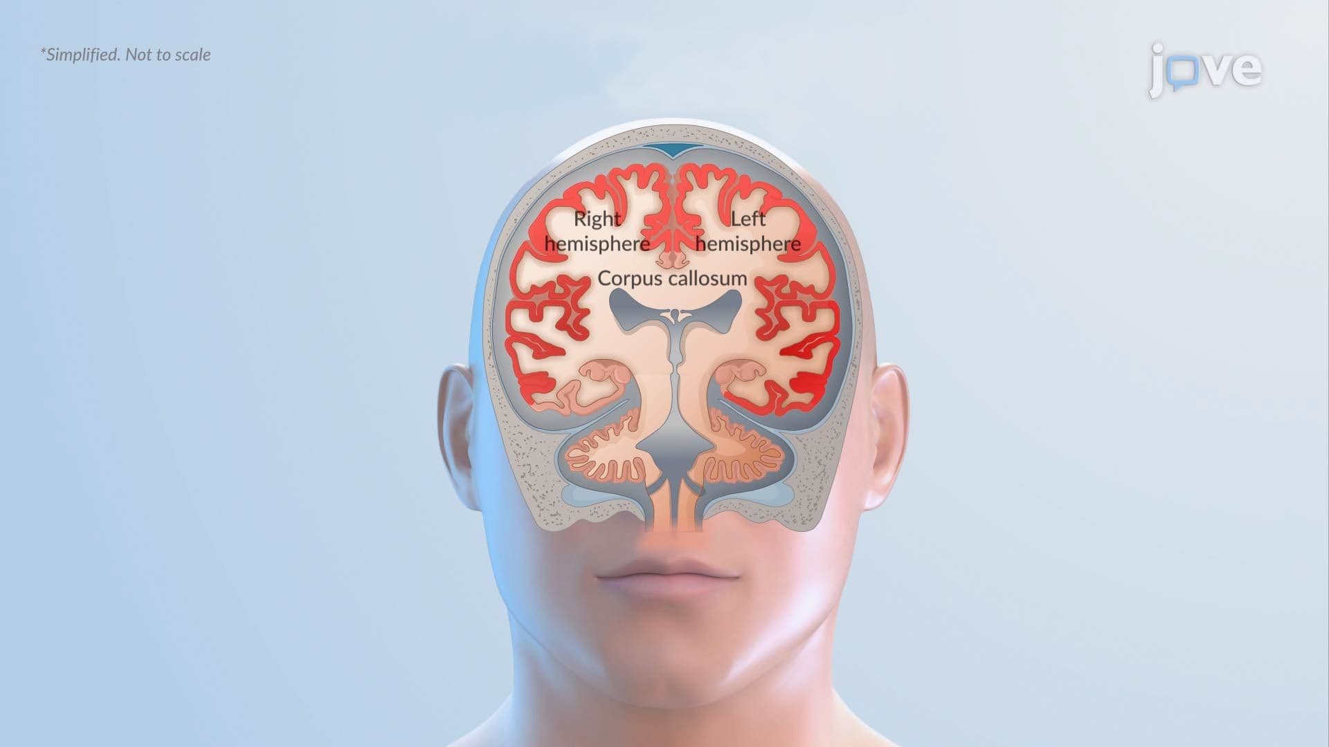
28. Seizures ll: Types
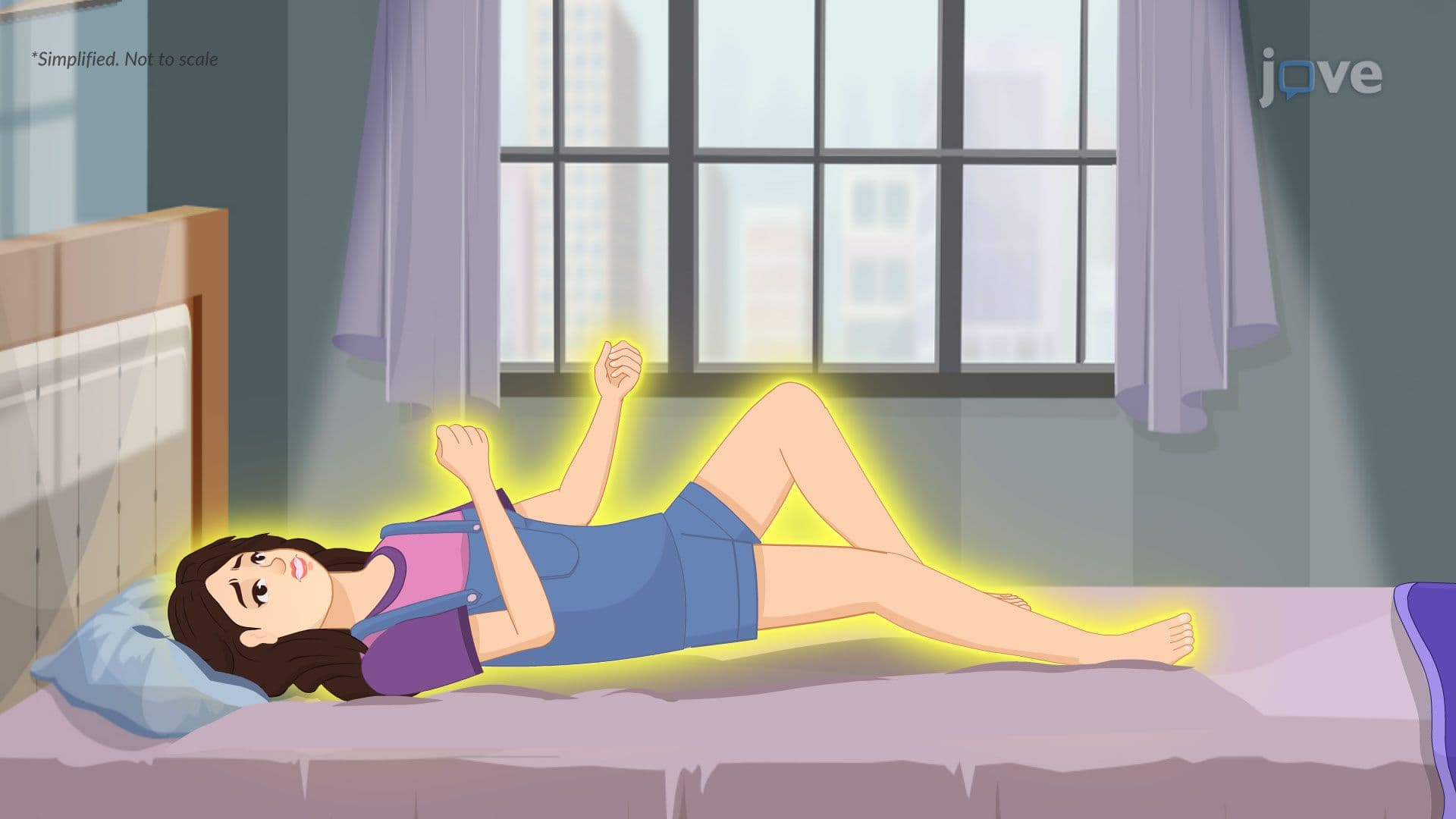
29. Epilepsy ll: Types

30. Myasthenia Gravis ll: Pathophysiology
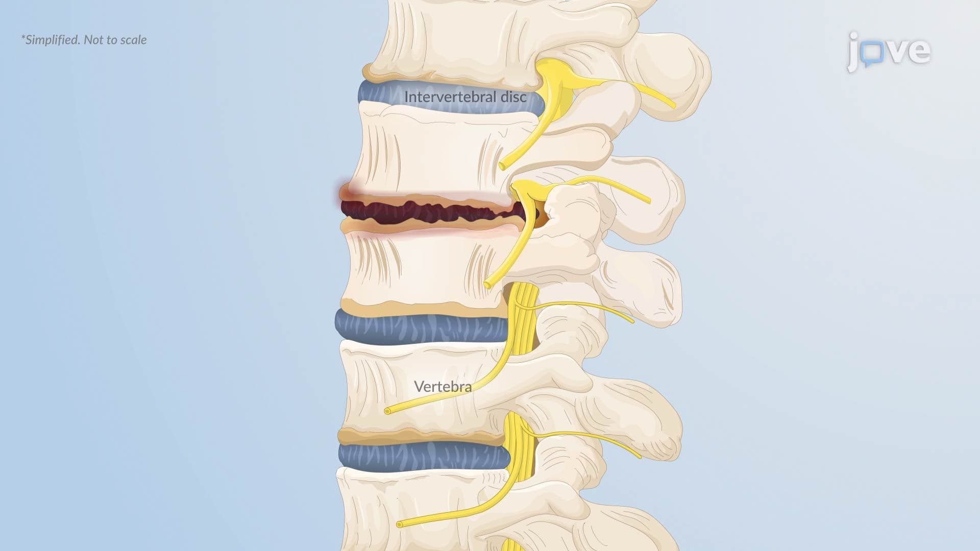
31. Herniated Intervertebral Disc l: Introduction

32. Degenerative Disc Disease I: Introduction
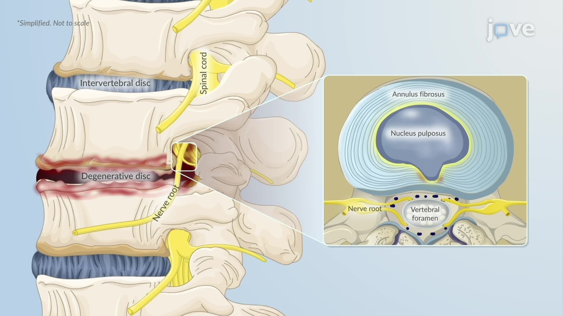
33. Degenerative Disc Disease ll: Pathophysiology
34. Alterations in Muscle Tone ll
35. Alterations in Muscle Tone lll
Nervous System Disorders Fundamentals covers the essential mechanisms, classifications, and clinical consequences of major CNS and PNS conditions—from stroke and traumatic brain injury to Alzheimer's disease and seizure disorders. JoVE Coach guides students through vascular, inflammatory, degenerative, and traumatic pathologies, connecting cellular-level dysfunction to real-world neurological outcomes critical for US healthcare and science education.
- Understand the two major stroke classifications—ischemic and hemorrhagic—and how each disrupts cerebral blood flow at the cellular and vascular level
- Identify the pathophysiological mechanisms behind increased intracranial pressure, cerebral edema subtypes, and herniation syndromes
- Analyze how neurodegenerative diseases such as Alzheimer's, Parkinson's, and Huntington's disease progressively damage specific brain regions and circuits
- Explore the inflammatory and immune-mediated processes driving bacterial meningitis, encephalitis, brain abscess, and multiple sclerosis
- Learn how traumatic brain injury and spinal cord injury produce both immediate primary damage and delayed secondary injury cascades
- Understand the mechanisms underlying seizure generation, epilepsy classification, and neuromuscular disorders like myasthenia gravis
- Apply knowledge of disc degeneration, muscle tone alterations, and dementia to interpret clinical presentations and pathophysiology questions
- Identify key risk factors, diagnostic indicators, and US-relevant clinical signs associated with each nervous system disorder
1. Stroke: Ischemic and Hemorrhagic Types Stroke is a sudden disruption of cerebral blood flow resulting in neurological dysfunction. Ischemic strokes—the most common type—occur when a cerebral artery is blocked by a thrombus (often from atherosclerotic plaque rupture) or an embolus (frequently from atrial fibrillation). Hemorrhagic strokes result from vessel rupture, causing either intracerebral hemorrhage directly into brain tissue or subarachnoid hemorrhage into the space around the brain, often from a ruptured berry aneurysm. Both types can be life-threatening, and distinguishing them is essential for clinical management in US emergency settings.
2. Ischemic Stroke Pathophysiology and the Ischemic Penumbra When cerebral blood flow is blocked, oxygen and glucose delivery fail within minutes. An infarct core of irreversibly dead cells forms at the center, surrounded by the penumbra—metabolically stressed tissue that may still be salvageable. Ion pump failure allows sodium and calcium to flood cells, triggering cytotoxic edema. Excess calcium drives glutamate release and excitotoxicity, damaging neurons further. Inflammatory cells worsen injury and contribute to vasogenic edema. The longer the blockage persists, the larger the infarct core grows, reinforcing why rapid intervention—such as tPA administration in US stroke centers—is time-critical.
3. Hemorrhagic Stroke Pathophysiology After a vessel ruptures, blood accumulates as a hematoma, compressing surrounding brain tissue and raising intracranial pressure. Secondary injury follows as hemoglobin and iron leak into tissue, triggering oxidative stress and microglial-mediated inflammation that compromises the blood-brain barrier. In subarachnoid hemorrhage, vasospasm can cause delayed cerebral ischemia days after the initial bleed, while blood in the cerebrospinal fluid can obstruct drainage, causing hydrocephalus. These cascading secondary events often determine long-term neurological outcomes and are the focus of intensive care management in US neurocritical care units.
4. Transient Ischemic Attack (TIA) A transient ischemic attack—commonly called a "mini-stroke"—involves a temporary focal loss of blood flow that causes stroke-like symptoms resolving within hours without permanent tissue infarction. Mechanisms include large-artery atherosclerosis causing transient flow restriction, emboli that temporarily block then dissolve from vessels, and reduced perfusion distal to a high-grade carotid stenosis. TIA is a major warning sign: roughly 10–15% of patients in the US experience a full stroke within 90 days of a TIA, making rapid evaluation with tools like the ABCD2 score clinically essential.
5. Bacterial Meningitis: Introduction and Pathophysiology Bacterial meningitis is an acute, life-threatening infection of the leptomeninges and cerebrospinal fluid. Common pathogens in US adults include *Streptococcus pneumoniae* and *Neisseria meningitidis*, while *Listeria monocytogenes* affects immunocompromised individuals and older adults. Bacteria colonizing the nasopharynx invade the bloodstream, penetrate the blood-CSF barrier, and multiply in the relatively immune-privileged subarachnoid space. This triggers intense inflammation—cytokine release, neutrophil recruitment, BBB disruption, and edema—raising intracranial pressure and reducing cerebral perfusion. Classic signs include fever, severe headache, neck stiffness, and positive Kernig and Brudzinski signs.
6. Encephalitis and Brain Abscess Encephalitis is inflammation of the brain parenchyma itself, most commonly caused by viruses such as herpes simplex virus type 1 (HSV-1), which has a tropism for the temporal lobes, or arboviruses like West Nile virus. Autoimmune encephalitis—such as anti-NMDA receptor encephalitis—involves antibody-mediated disruption of neural signaling. In contrast, a brain abscess is a localized pus-filled mass arising from bacterial invasion—often from sinus, dental, or cardiac sources—that acts as a space-occupying lesion, elevating intracranial pressure and causing focal neurological deficits, seizures, and papilledema. Both conditions require urgent diagnosis and treatment in US hospital settings.
7. Multiple Sclerosis (MS) Multiple sclerosis is a chronic autoimmune CNS disease in which the immune system attacks myelin sheaths, demyelinating axons in the brain and spinal cord. Over time, axonal damage accumulates, producing irreversible neurological disability. MS typically affects US adults between ages 20 and 40, occurring twice as often in women. Symptoms are diverse—optic neuritis, spasticity, paresthesias, bladder dysfunction, and cognitive impairment—and often follow a relapsing-remitting pattern where episodes of neurological worsening alternate with partial or full recovery. It is one of the most common causes of non-traumatic disability in young American adults.
8. Traumatic Brain Injury (TBI) TBI results from external mechanical forces disrupting normal brain function, classified by severity using the Glasgow Coma Scale (GCS), loss of consciousness duration, and post-traumatic amnesia. Mild TBI (GCS 13–15), commonly called a concussion, frequently occurs in US contact sports. Moderate TBI (GCS 9–12) often results from motor vehicle crashes, while severe TBI (GCS 3–8) can follow major falls or high-impact collisions. Primary injury is immediate and mechanical; secondary injury unfolds over hours to days involving inflammation, excitotoxicity, and ischemia—processes that are the targets of US neurointensive care interventions.
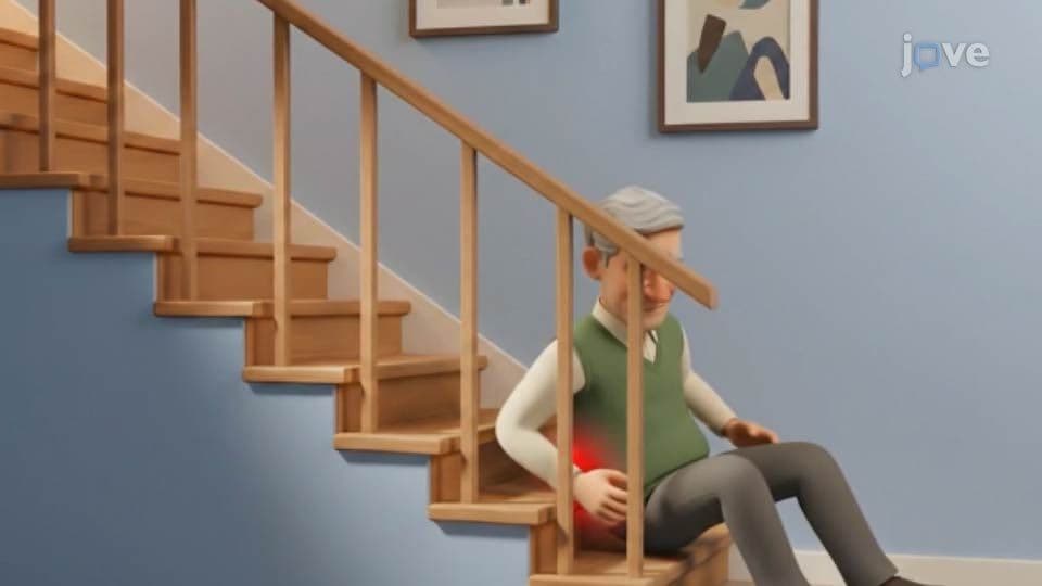
9. Spinal Cord Injury: Primary and Secondary Mechanisms Spinal cord injury involves an immediate primary phase—compression, contusion, laceration, or distraction of cord tissue—that disrupts neurons, supporting cells, and blood vessels. A secondary injury phase then unfolds over minutes to weeks, driven by ATP depletion, glutamate-mediated excitotoxicity, calcium overload, inflammatory cell infiltration, and reactive oxygen species. Oligodendrocyte loss strips axons of myelin, impairing signal conduction. In later stages, reactive astrocytes form a glial scar that limits but does not reverse injury. Understanding this two-phase model is foundational for rehabilitation medicine and spinal cord research programs at US institutions like the NIH.
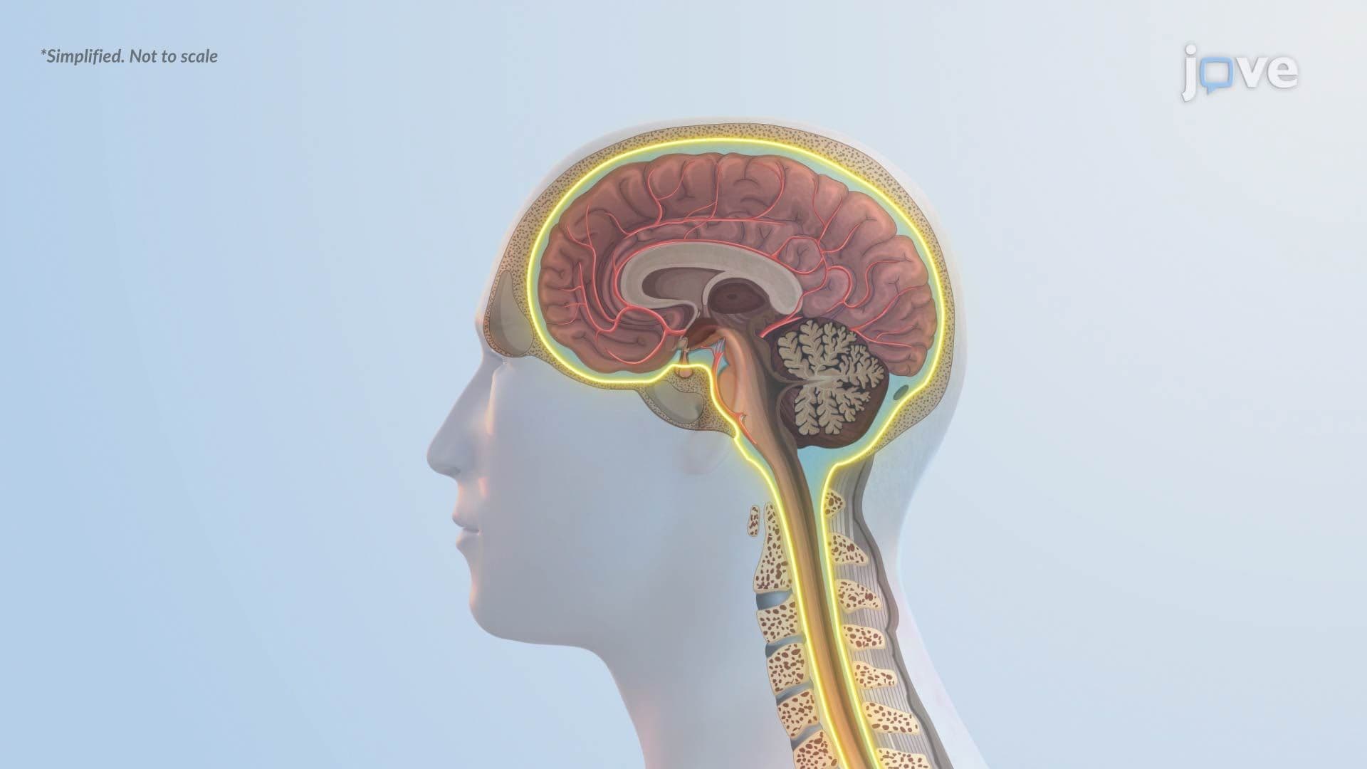
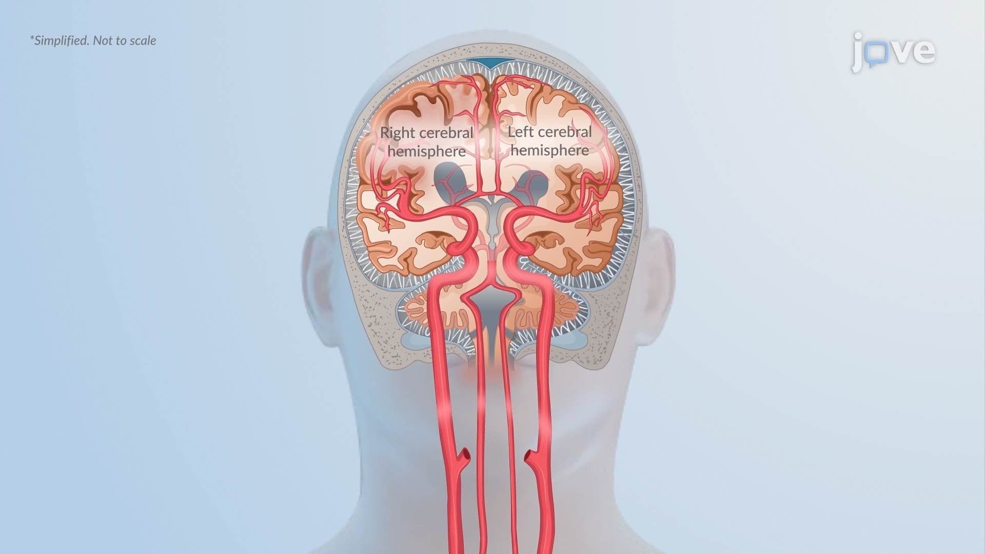
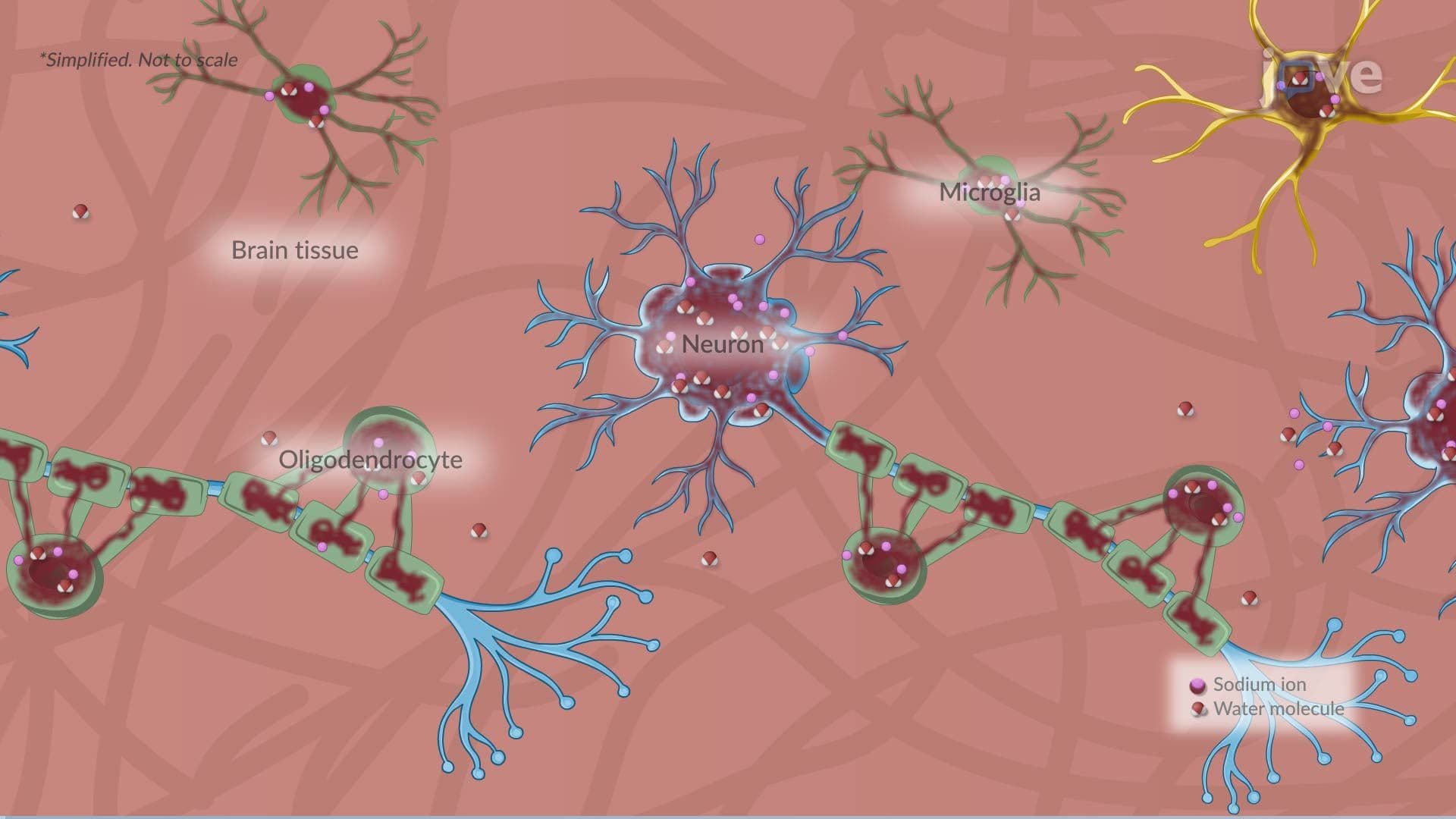
10. Increased Intracranial Pressure and Cerebral Edema The Monro-Kellie doctrine states that total intracranial volume is fixed; any increase in one component—brain tissue, CSF, or blood—must be offset by another or ICP rises. Causes include hemorrhage, large infarcts, tumors, and infections. Cerebral edema is a major driver: vasogenic edema arises from blood-brain barrier breakdown allowing protein-rich fluid into extracellular space; cytotoxic edema results from cellular energy failure and intracellular water accumulation; interstitial edema occurs with hydrocephalus; and ionic edema develops via osmotic gradients without protein leakage. Uncontrolled ICP elevation leads to herniation syndromes, brainstem compression, and death.
11. Neurodegenerative Disorders: Alzheimer's, Parkinson's, and Huntington's Disease Alzheimer's disease, the leading cause of US dementia, features extracellular beta-amyloid plaques and intracellular neurofibrillary tangles from hyperphosphorylated tau, causing progressive hippocampal and cortical atrophy. Parkinson's disease involves degeneration of dopaminergic neurons in the substantia nigra, producing the classic triad of resting tremor, rigidity, and bradykinesia, alongside Lewy body accumulation of misfolded alpha-synuclein. Huntington's disease is caused by a CAG repeat expansion in the HTT gene, leading to striatal neuronal death. Together, these disorders represent the major focus of neurodegeneration research and clinical neurology across US academic medical centers.
12. Seizures, Epilepsy, and Myasthenia Gravis Seizures result from abnormal, excessive, or synchronous neuronal activity in the brain and are classified as focal (originating in one hemisphere) or generalized (involving both hemispheres simultaneously). Epilepsy is a chronic disorder defined by recurrent unprovoked seizures. Myasthenia gravis is an autoimmune neuromuscular junction disorder in which antibodies target acetylcholine receptors at the neuromuscular junction, causing fluctuating muscle weakness that worsens with activity—particularly affecting extraocular and bulbar muscles. All three conditions are heavily represented on US licensing exams including the USMLE and NCLEX, and require a strong mechanistic understanding for clinical reasoning.
13. Disc Disease and Alterations in Muscle Tone Herniated intervertebral disc occurs when the nucleus pulposus protrudes through the annulus fibrosus, compressing spinal nerve roots and causing radiculopathy—pain, weakness, or numbness in the distribution of the affected nerve. Degenerative disc disease involves age-related dehydration and structural breakdown of disc tissue, reducing shock absorption and narrowing the spinal canal. Alterations in muscle tone—including spasticity (increased tone from upper motor neuron lesions) and flaccidity (decreased tone from lower motor neuron damage)—reflect the level and extent of nervous system injury and guide clinical assessment in US physical and occupational therapy settings.
Frequently Asked Questions
The infarct core is the zone of brain tissue that has already died due to complete loss of blood flow—cells here are irreversibly damaged within minutes. The ischemic penumbra surrounds the core and consists of metabolically stressed but still-viable tissue. It receives minimal residual blood flow, keeping cells alive temporarily but at serious risk of dying if circulation is not restored quickly. This is why treatments like tissue plasminogen activator (tPA), given within 4.5 hours at certified US stroke centers, aim to salvage penumbral tissue before it progresses to irreversible infarction.
Cerebral edema refers specifically to the abnormal accumulation of fluid within brain tissue, which can occur by four distinct mechanisms: vasogenic, cytotoxic, interstitial, and ionic. Increased intracranial pressure (ICP) is the consequence—or result—of edema (or other volume-expanding processes like hemorrhage or tumor) exceeding the skull's ability to compensate, as described by the Monro-Kellie doctrine. Think of edema as the cause and elevated ICP as the downstream effect. Both concepts are frequently tested together on the USMLE and NCLEX because managing one requires understanding the other.
On the USMLE Step 1, expect questions that require you to work through a clinical vignette and identify the underlying cellular or molecular mechanism—for example, explaining why cytotoxic edema occurs in ischemia or why Alzheimer's disease causes cholinergic deficits. Step 2 CK tests clinical decision-making, such as differentiating ischemic from hemorrhagic stroke before treatment. The NCLEX-RN frequently tests neurological assessment skills: recognizing signs of increased ICP, identifying Kernig and Brudzinski signs in meningitis, or prioritizing interventions for a patient with a TBI. Pathophysiology-focused mastery of these conditions is essential for both exams.
No—these terms are related but distinct. A seizure is a single event of abnormal, excessive neuronal firing that can have many causes, including fever (febrile seizure in children), low blood sugar, or head trauma. Epilepsy is a chronic neurological disorder diagnosed when a person has had two or more unprovoked seizures, or one seizure with a high likelihood of recurrence based on EEG or brain imaging findings. Not everyone who has a seizure has epilepsy. This distinction matters both clinically and on US exams like the MCAT and USMLE, where understanding the classification of focal versus generalized seizures is also expected.
Both conditions present with fever and headache, which can make early differentiation challenging. Bacterial meningitis primarily infects the meninges—the membranes surrounding the brain—and is strongly associated with neck stiffness (meningismus), positive Kernig and Brudzinski signs, and rapid systemic deterioration. Encephalitis, in contrast, involves direct inflammation of brain parenchyma, so it more prominently features altered mental status, cognitive changes, behavioral disturbances, and seizures as early findings. In practice, both conditions may coexist as meningoencephalitis, especially with pathogens like Cryptococcus neoformans in immunocompromised patients. Lumbar puncture with CSF analysis is key to differentiating them in US clinical settings.
The United States sees approximately 17,000 new traumatic spinal cord injuries each year, most commonly from motor vehicle crashes, falls, and sports injuries. The two-phase injury model covered in this course—immediate primary mechanical damage followed by a prolonged secondary cascade of excitotoxicity, inflammation, and demyelination—directly explains why patients with spinal cord injuries often worsen neurologically in the hours and days after trauma. This model also informs current US clinical practices, such as avoiding hypotension and hypoxia in spinal cord injury patients in the ICU, since these factors worsen the secondary injury phase. Rehabilitation centers like the Shepherd Center in Atlanta or the Kessler Foundation in New Jersey apply these principles daily.
Most students find the cerebral edema subtypes and the pathophysiology of increased ICP the most conceptually difficult, largely because the four edema types (vasogenic, cytotoxic, interstitial, ionic) are easy to confuse. The key is anchoring each type to its mechanism: vasogenic = BBB breakdown with protein leakage; cytotoxic = cellular energy failure and pump dysfunction; interstitial = CSF forced across the ependyma in hydrocephalus; ionic = osmotic gradients without protein leakage. The secondary injury cascades in stroke and spinal cord injury are also challenging because they involve multiple simultaneous molecular events. Drawing out each pathway step-by-step is a proven strategy.
The most effective approach is to organize these disorders into mechanistic categories rather than memorizing each condition in isolation. Group vascular disorders (stroke, TIA, ICP) together, inflammatory/infectious disorders (meningitis, encephalitis, brain abscess) together, and neurodegenerative diseases (Alzheimer's, Parkinson's, Huntington's) as a cluster. For each condition, map out: the cause → the cellular/molecular mechanism → the resulting structural change → the clinical signs. Use active recall techniques such as flashcards for key pathways (e.g., the excitotoxicity cascade, Monro-Kellie doctrine, tau hyperphosphorylation) and practice applying these mechanisms to clinical vignettes, which mirrors the format of the USMLE, NCLEX, and MCAT.
Parkinson's disease is caused by the loss of dopamine-producing neurons in the substantia nigra, which reduces dopamine signaling in the striatum and disrupts the basal ganglia motor circuit. This mechanistic understanding directly explains why the mainstay treatment in the US is levodopa (L-DOPA)—a dopamine precursor that can cross the blood-brain barrier and be converted into dopamine, partially restoring the depleted signaling. Similarly, understanding that misfolded alpha-synuclein (Lewy bodies) propagates in a prion-like manner between neurons explains why Parkinson's disease progresses over time and why researchers at US institutions are exploring therapies that target alpha-synuclein aggregation to slow disease progression.
This microcourse includes 35 concept videos that walk you through the building blocks of Pathophysiology. Each video is short, about 1 minute, so you can cover a full topic during a coffee break or between classes. The full sequence starts with Stroke: Introduction and Types and ends with Alterations in Muscle Tone lll.
The playlist moves from big-picture ideas to the precise vocabulary used in Pathophysiology. Early videos introduce Stroke: Introduction and Types, Ischemic Stroke l: Introduction, and Ischemic Stroke ll: Pathophysiology. The middle of the series focuses on Hemorrhagic Stroke ll: Pathophysiology, Transient Ischemic Attack l: Introduction, and Bacterial Meningitis I: Introduction. The final stretch covers Bacterial Meningitis II: Pathophysiology, Encephalitis l: Introduction, Encephalitis ll: Pathophysiology, Brain Abscess l: Introduction, Multiple Sclerosis l: Introduction, Traumatic Brain Injury l: Introduction, and Alterations in Muscle Tone lll.
The natural next step is Respiratory System Disorders. From there, you can move to Gastrointestinal System Disorders. Once you finish those, the full Pathophysiology curriculum of 5 microcourses on JoVE Coach opens up, taking you from foundational concepts to advanced systems.
Related Subjects