Loading subject page…


15,262 views


Video Summary: Insulin the Receptor and Signaling Pathways Explained
Did you know that your body processes glucose from a single meal through over 50 million insulin receptors in just your muscle tissue alone? Insulin the receptor and signaling pathways form the foundation of how cells throughout your body respond to blood sugar changes after eating. When a diabetic patient at Johns Hopkins Hospital receives insulin therapy, this intricate molecular machinery determines whether glucose successfully enters their cells or remains dangerously elevated in their bloodstream. Watch the full video on JoVE Coach to master this concept with expert-led visuals and step-by-step explanations.
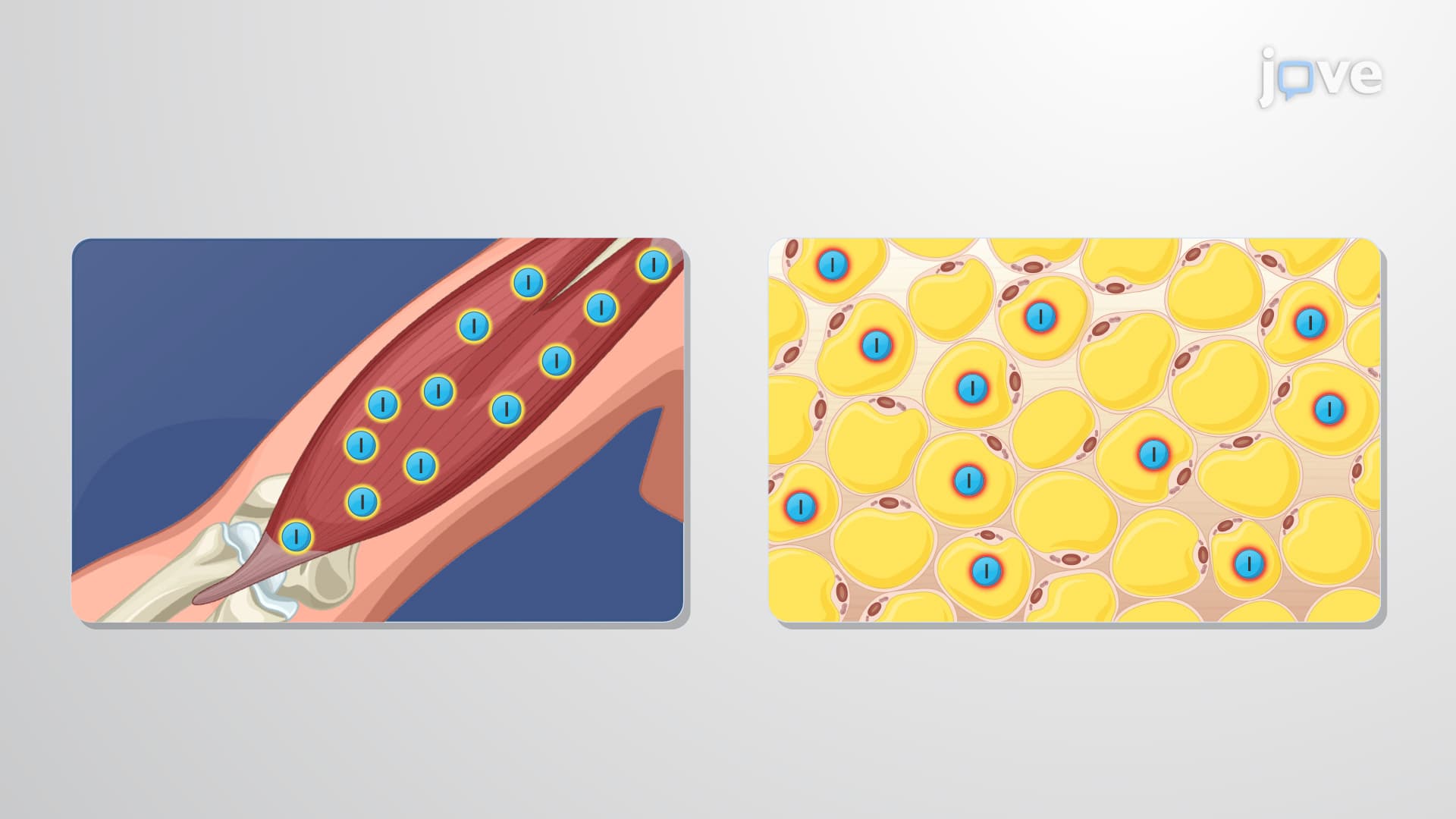
The insulin receptor represents one of medicine's most clinically relevant examples of receptor tyrosine kinase signaling. This sophisticated molecular machine consists of two α-subunits and two β-subunits, held together by disulfide bonds to form a stable heterotetramer that spans the cell membrane. The receptor's abundance varies dramatically across tissues-adipocytes contain roughly 100,000 receptors per cell, while skeletal muscle fibers house millions, reflecting their critical roles in glucose homeostasis.
When insulin binds to the extracellular α-subunits, it triggers a conformational change that activates the intracellular tyrosine kinase domains of the β-subunits. This activation initiates autophosphorylation, where each β-subunit phosphorylates specific tyrosine residues on its partner. Students preparing for the MCAT should understand that this creates docking sites for adapter proteins like Shc and insulin receptor substrates (IRS-1 through IRS-4), which serve as scaffolds for downstream signaling complexes.
The activated receptor launches two major signaling branches. The Shc-MAP kinase pathway primarily drives cell growth and differentiation-crucial for understanding cancer biology in advanced coursework. Meanwhile, the IRS-PI3-kinase axis controls metabolic responses. PI3-kinase converts PIP2 to PIP3, creating a lipid messenger that recruits and activates Akt (also called PKB). This mechanism frequently appears on AP Biology exams when discussing signal transduction.
Akt activation represents the critical junction where insulin signaling meets glucose metabolism. In muscle and fat cells, Akt phosphorylates proteins that normally sequester GLUT4 transporters in intracellular vesicles. Upon phosphorylation, these vesicles fuse with the plasma membrane, increasing glucose uptake capacity by up to 20-fold. Once inside, glucose-6-phosphate can enter glycolysis for immediate energy, the pentose phosphate pathway for NADPH production, or convert to glucose-1-phosphate for glycogen storage-metabolic flexibility essential for maintaining blood sugar between meals.
This intricate system explains why insulin resistance, common in type 2 diabetes affecting 37 million Americans, creates such devastating metabolic consequences. Understanding these pathways provides the foundation for comprehending diabetes pathophysiology, a topic heavily emphasized in pre-med curricula and clinical practice.
Related Micro-courses