Loading video…


 16 Concepts
 15.3K views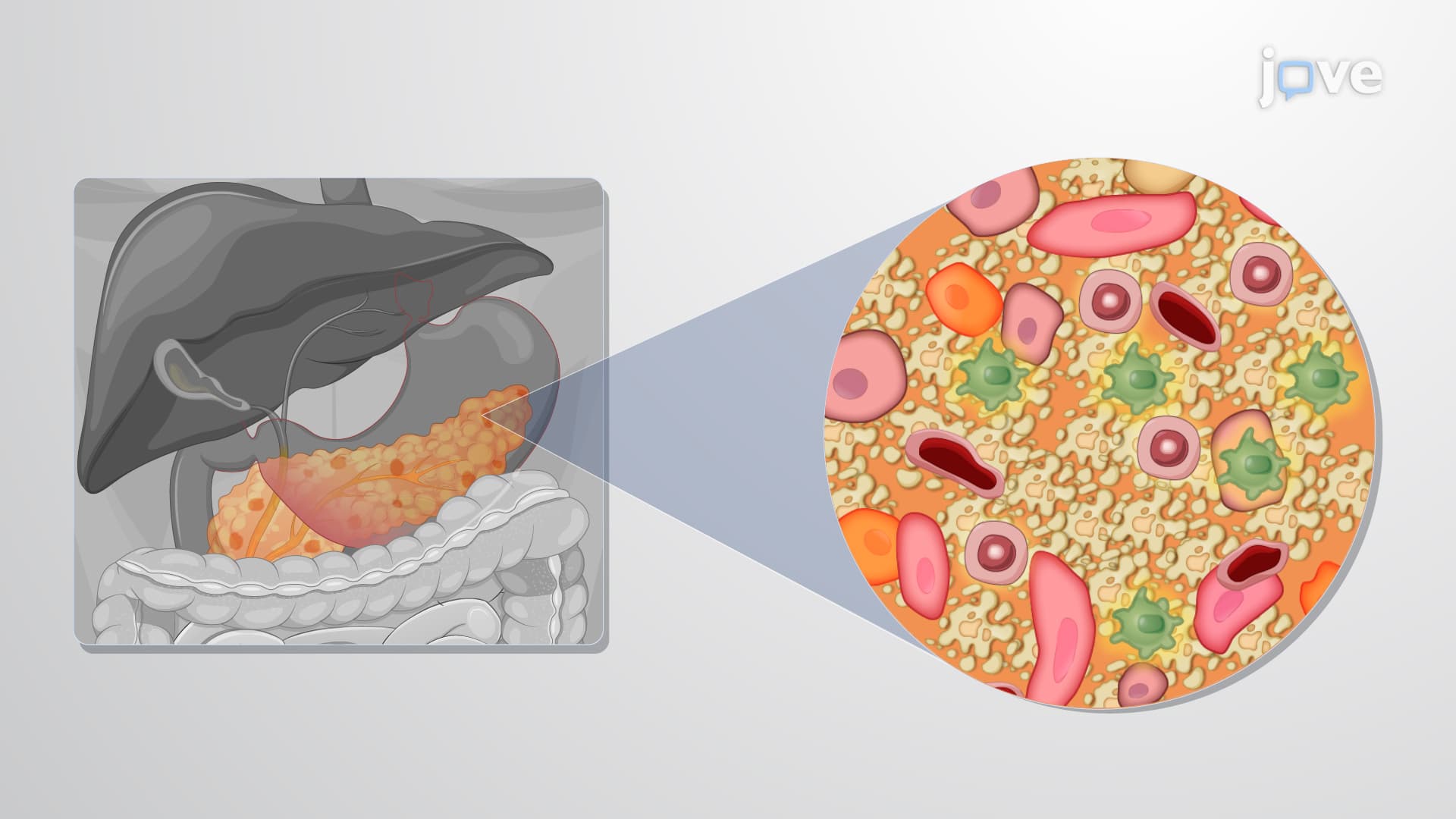

Video Summary: What Is Pathophysiology of Diabetes
Did you know that nearly 37 million Americans live with diabetes, yet many don't understand the underlying cellular breakdown causing their condition? The pathophysiology of diabetes reveals how this chronic metabolic disorder develops through distinct mechanisms that disrupt normal blood sugar regulation. In Type 1 diabetes, autoimmune attacks destroy insulin-producing cells, while Type 2 involves multiple system failures affecting insulin production and cellular response. Watch the full video on JoVE Coach to master this concept with expert-led visuals and step-by-step explanations.
The pathophysiology of diabetes encompasses the complex biological processes that lead to chronic hyperglycemia and its associated complications. Unlike simply memorizing symptoms, understanding these mechanisms reveals why diabetes affects multiple organ systems and requires targeted therapeutic approaches.
Type 1 diabetes represents a classic autoimmune disorder where the body's immune system mistakenly targets pancreatic beta cells. This process begins when environmental triggers-such as viral infections like Coxsackievirus or Epstein-Barr virus-activate immune responses in genetically susceptible individuals. T-lymphocytes infiltrate pancreatic islets, releasing inflammatory cytokines including interleukin-1 and tumor necrosis factor-alpha.
The destruction follows a predictable timeline. Initially, patients maintain normal glucose levels despite ongoing cellular damage. However, once approximately 80% of beta cells are destroyed, insulin production drops below critical thresholds, leading to clinical diabetes. This explains why Type 1 patients often present with rapid symptom onset-their remaining beta cells can no longer compensate for the massive cellular loss.
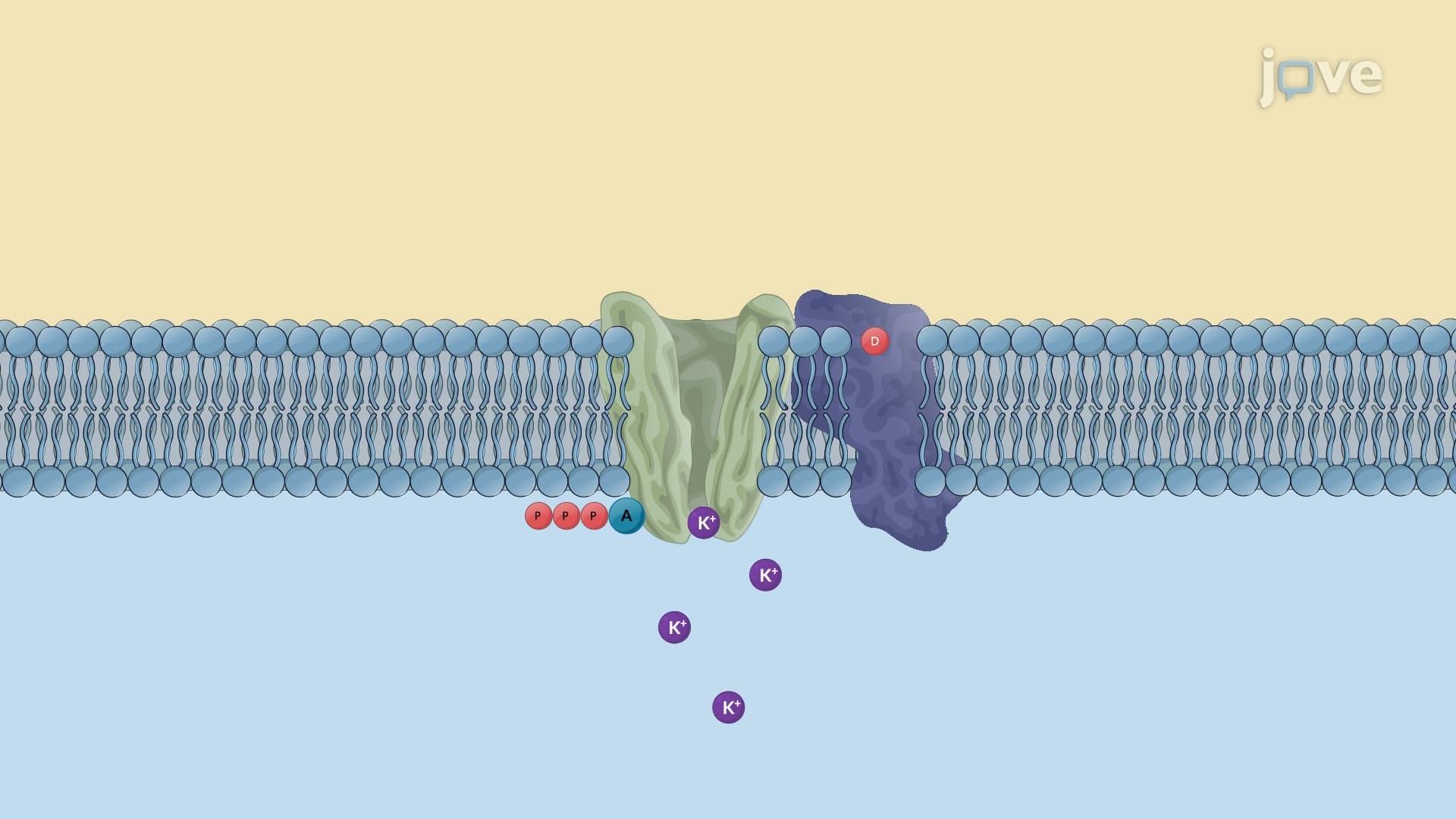
Type 2 diabetes involves three interconnected pathophysiological defects. First, pancreatic beta cells develop secretory dysfunction, producing insufficient insulin relative to glucose loads. Unlike Type 1's complete destruction, these cells remain present but function poorly, explaining why some Type 2 patients initially respond to medications that stimulate insulin release.
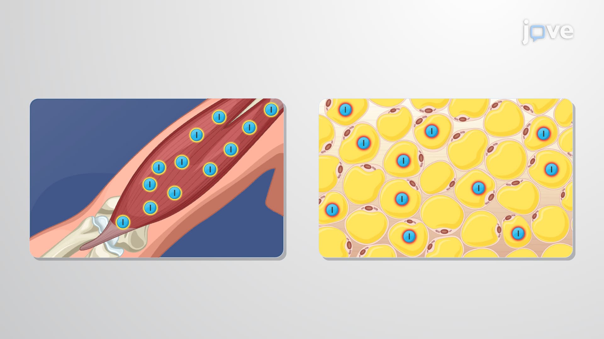
Second, peripheral insulin resistance develops in target tissues. Skeletal muscle cells, which normally consume 80% of glucose during fed states, become less responsive to insulin signals. Similarly, adipose tissue fails to suppress lipolysis appropriately, releasing free fatty acids that worsen insulin resistance. Hepatic insulin resistance allows continued glucose production even when blood sugars are elevated.
Understanding these mechanisms proves essential for MCAT passages on endocrine physiology and AP Biology questions about cellular signaling. Students preparing for health professions should recognize how pathophysiology knowledge guides treatment decisions-Type 1 patients require insulin replacement, while Type 2 patients may benefit from medications targeting specific defects like insulin resistance or beta cell dysfunction.
Related Micro-courses