Loading subject page…


111,914 views


Video Summary: Neuron Structure the Cell Body and Dendrites
Your brain processes over 100 billion electrical signals every second-but how do individual neurons manage this incredible feat? The neuron cell body dendrite structure forms the foundation of neural communication, with specialized components like the soma containing vital organelles and dendrites branching out to receive chemical messages. Consider how neurologists at Johns Hopkins use detailed knowledge of perikaryon anatomy to diagnose neurodegenerative diseases by examining cellular changes in patient brain tissue. Watch the full video on JoVE Coach to master this concept with expert-led visuals and step-by-step explanations.
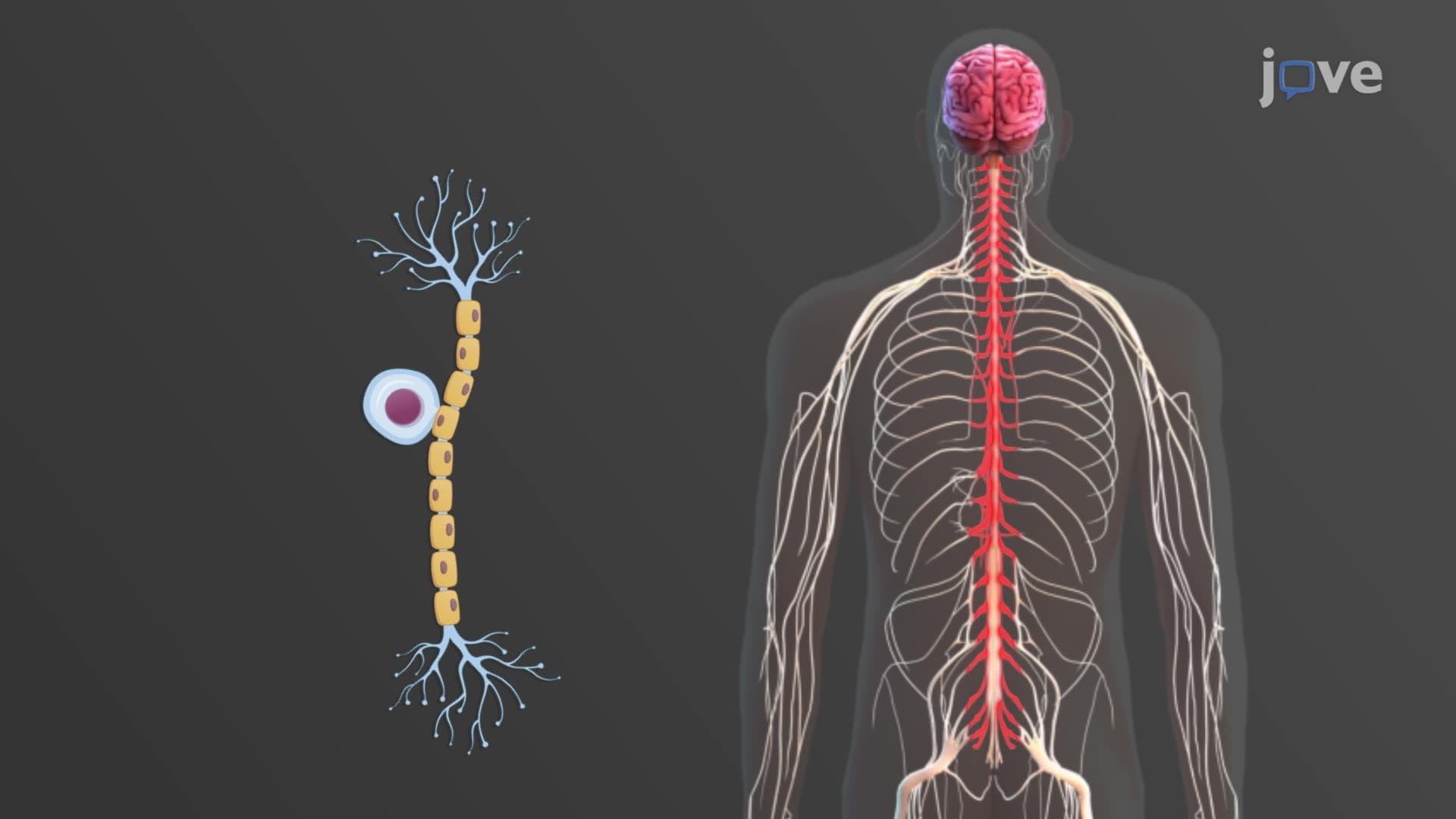
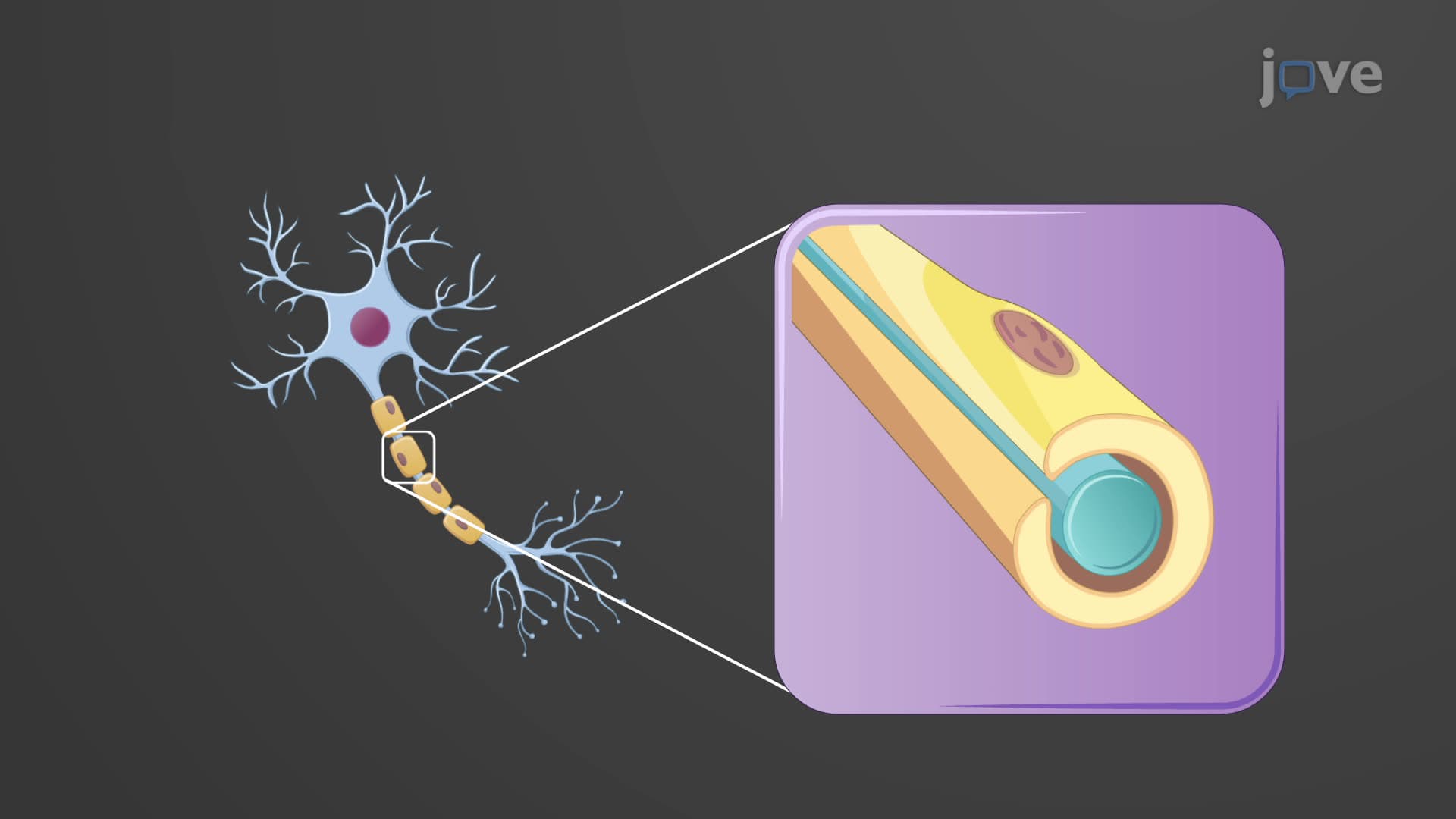
The architectural design of neurons reflects their specialized function as the brain's communication network. Unlike typical cells that simply maintain themselves, neurons must constantly receive, process, and transmit electrical and chemical signals across vast networks. The soma and dendrite neuron anatomy represents a sophisticated solution to this challenge, with each component optimized for specific aspects of neural communication.
The cell body neuron function extends far beyond basic cellular maintenance. As the perikaryon, this structure houses the nucleus and coordinates all metabolic activities essential for neuronal survival and signal processing. The presence of Nissl bodies-dense clusters of ribosomes and rough endoplasmic reticulum-distinguishes neurons from other cell types and enables the massive protein synthesis required for neurotransmitter production and membrane maintenance.
Medical students studying for the MCAT encounter detailed questions about these organelles, particularly how Nissl body distribution changes in diseased states. For instance, researchers at the Mayo Clinic have documented how Nissl body dissolution occurs early in Alzheimer's disease, making soma structure analysis crucial for diagnostic pathology. The cytoskeletal elements, including neurofibrils and neurotubules, form highways for transporting materials between the cell body and distant axon terminals-a journey that can span over three feet in motor neurons controlling your toes.
Dendrite spine branching represents one of nature's most elegant solutions for maximizing surface area within confined space. These tree-like extensions can contain thousands of synaptic contacts, each equipped with specific neurotransmitter receptors. The strategic placement of dendritic spines creates microenvironments where chemical signals convert to electrical graded potentials, allowing neurons to integrate multiple inputs simultaneously.
AP Biology students often struggle with understanding how dendrite receptor input determines neural response patterns. Consider how a single pyramidal neuron in your cerebral cortex receives approximately 10,000 synaptic inputs-some excitatory, others inhibitory. The dendrite's ability to spatially and temporally integrate these signals determines whether the neuron will fire an action potential, making dendritic architecture crucial for complex behaviors like decision-making and memory formation.
The distinction between nuclei and ganglia reflects fundamental differences in central versus peripheral nervous system organization. Neurologists at Cleveland Clinic regularly use this knowledge when diagnosing conditions affecting specific brain nuclei, such as Parkinson's disease (substantia nigra) or Huntington's disease (caudate nucleus). Understanding these organizational principles proves essential for medical professionals interpreting brain imaging studies and planning therapeutic interventions.
Related Micro-courses