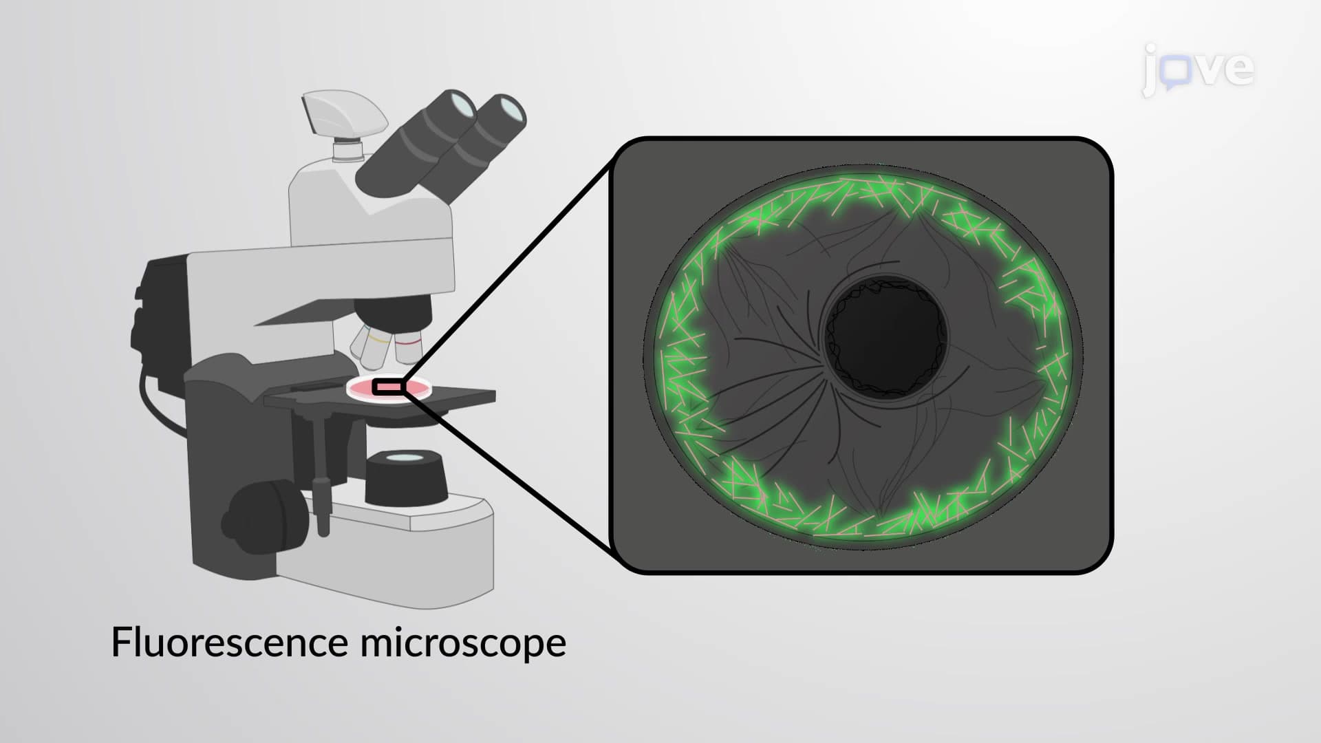
The cytoskeleton I: actin and microfilaments forms the dynamic structural framework essential for cellular function. This comprehensive course through JoVE Coach explores how actin cytoskeleton function drives cell movement, division, and structural support across human physiology, from muscle contraction in cardiac cells to white blood cell migration during immune responses.
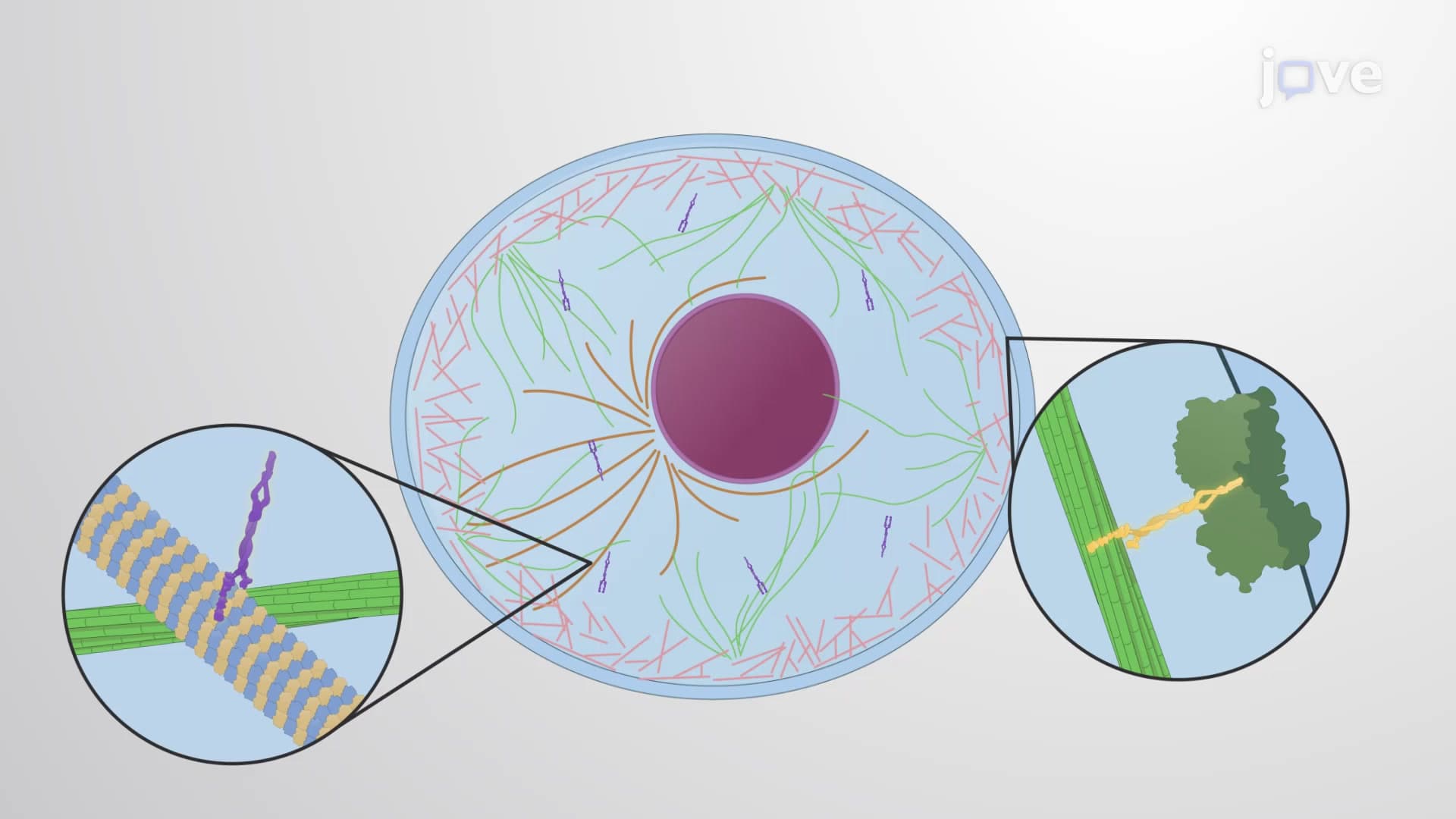
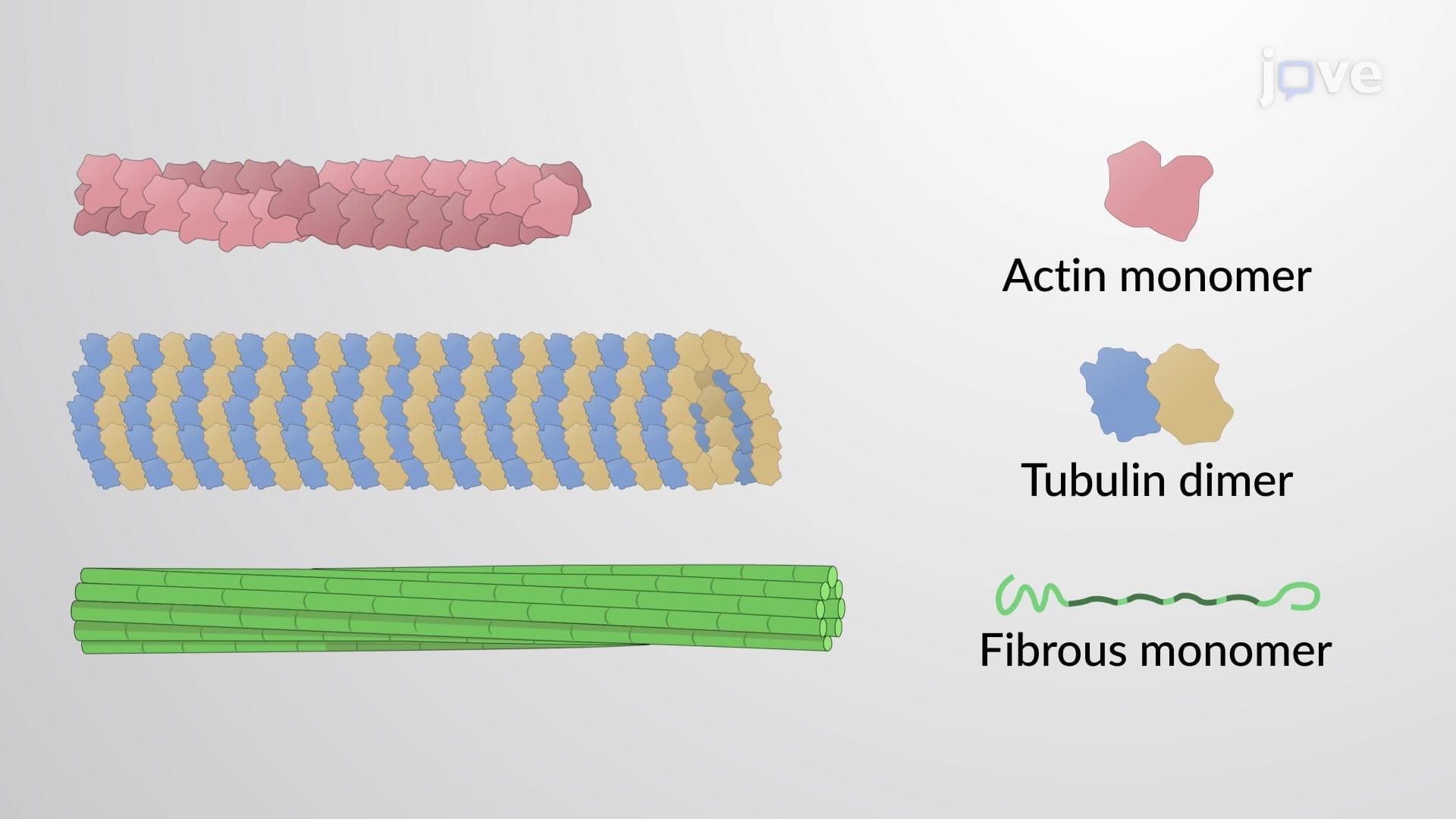
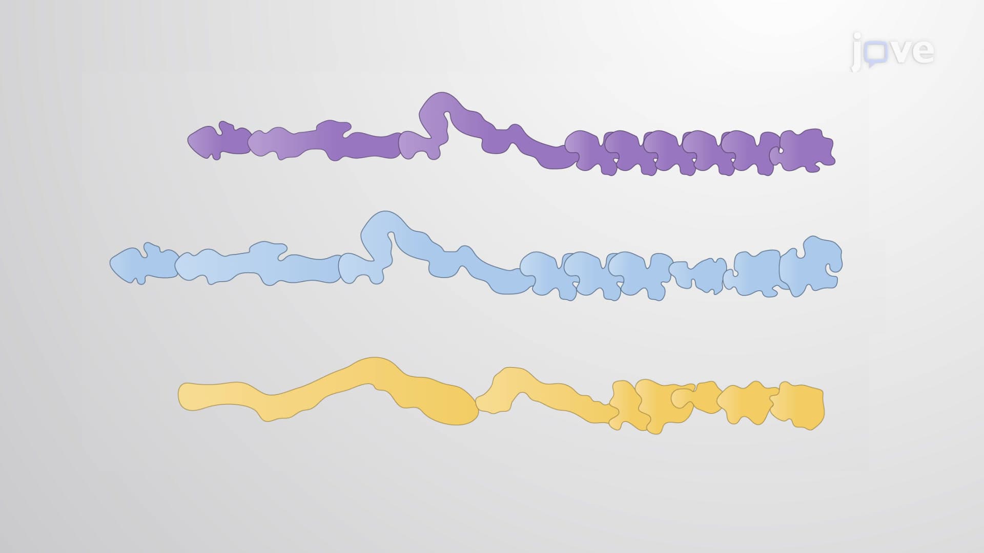
1. Cytoskeletal Architecture and Components The cytoskeleton comprises three distinct filament types: microfilaments (actin), microtubules (tubulin), and intermediate filaments. Each serves specialized functions in American healthcare contexts—actin microfilaments enable immune cell migration to infection sites, microtubules form mitotic spindles during cancer cell division studies at institutions like MD Anderson, and intermediate filaments provide mechanical strength to skin cells, relevant for dermatological treatments. These protein networks interconnect through accessory proteins called plakins, creating integrated cellular scaffolding that maintains cell shape while allowing dynamic reorganization for processes like wound healing in burn units across US hospitals.
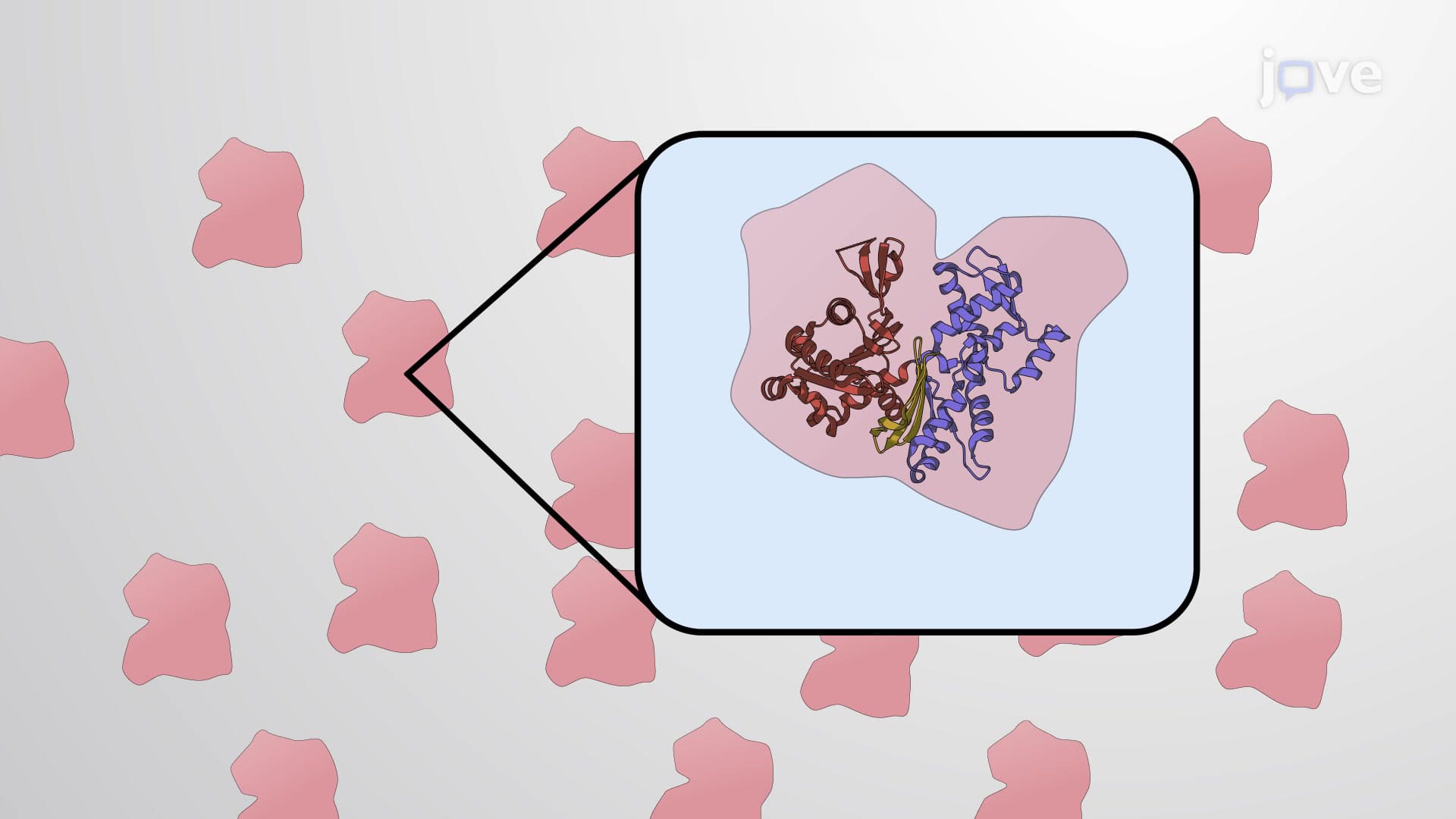
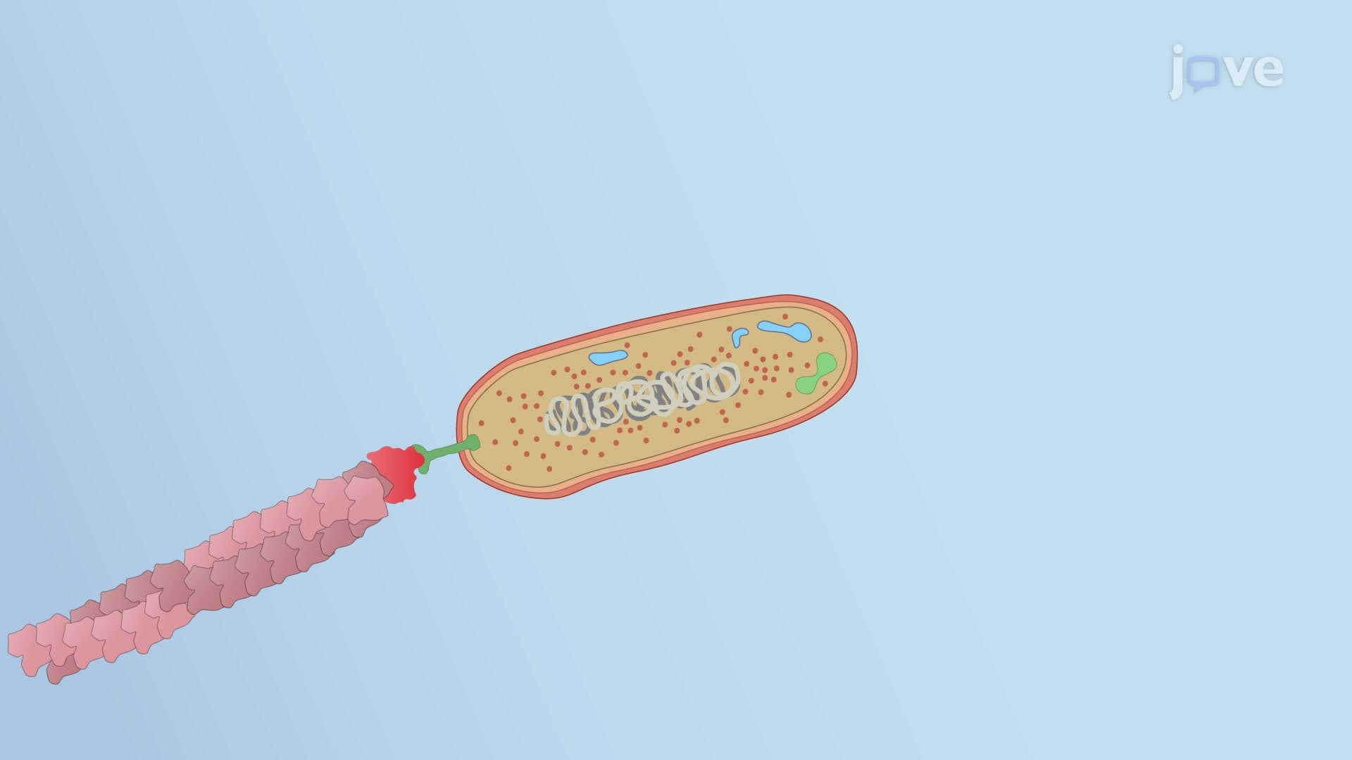
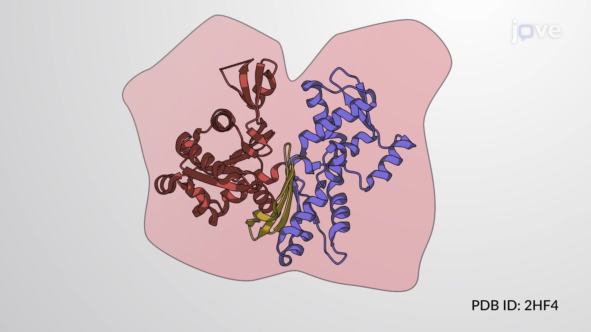
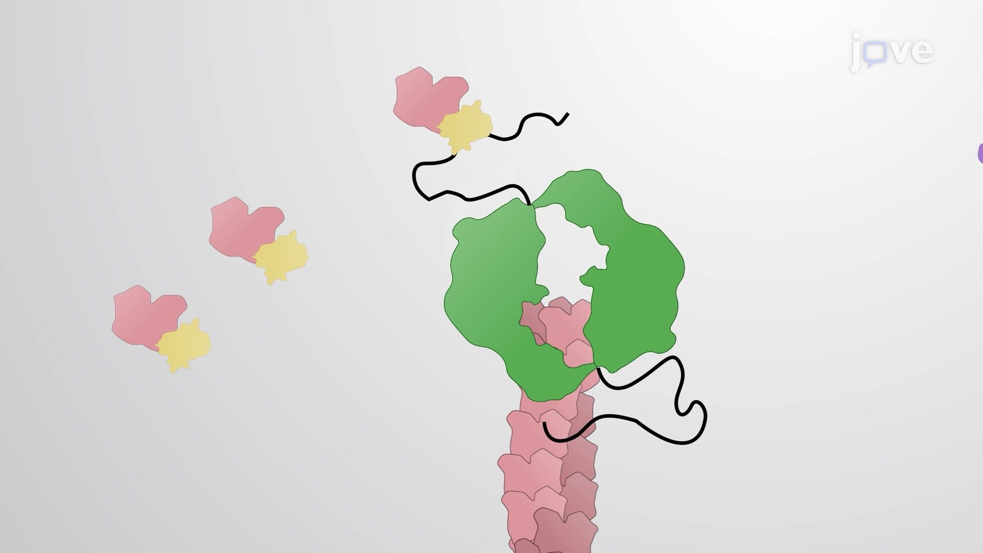
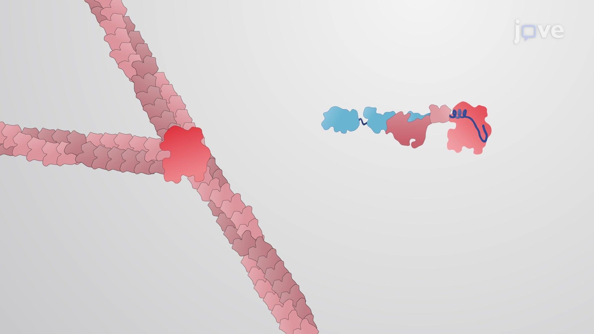
2. Actin Structure and Polymerization Dynamics G-actin monomers contain ATP-binding sites and polymerize into helical F-actin filaments through nucleation, elongation, and steady-state phases. This process resembles assembly lines in American manufacturing—individual components (G-actin) join systematically to create functional products (F-actin). The plus-end grows faster than the minus-end, creating polarity essential for directional processes. In US medical research, understanding actin polymerization helps explain how cancer cells metastasize through blood vessels, as malignant cells exploit actin dynamics to invade healthy tissues. Formin proteins guide straight filament formation while Arp2/3 complexes create branched networks, similar to interstate highway systems versus city street grids.
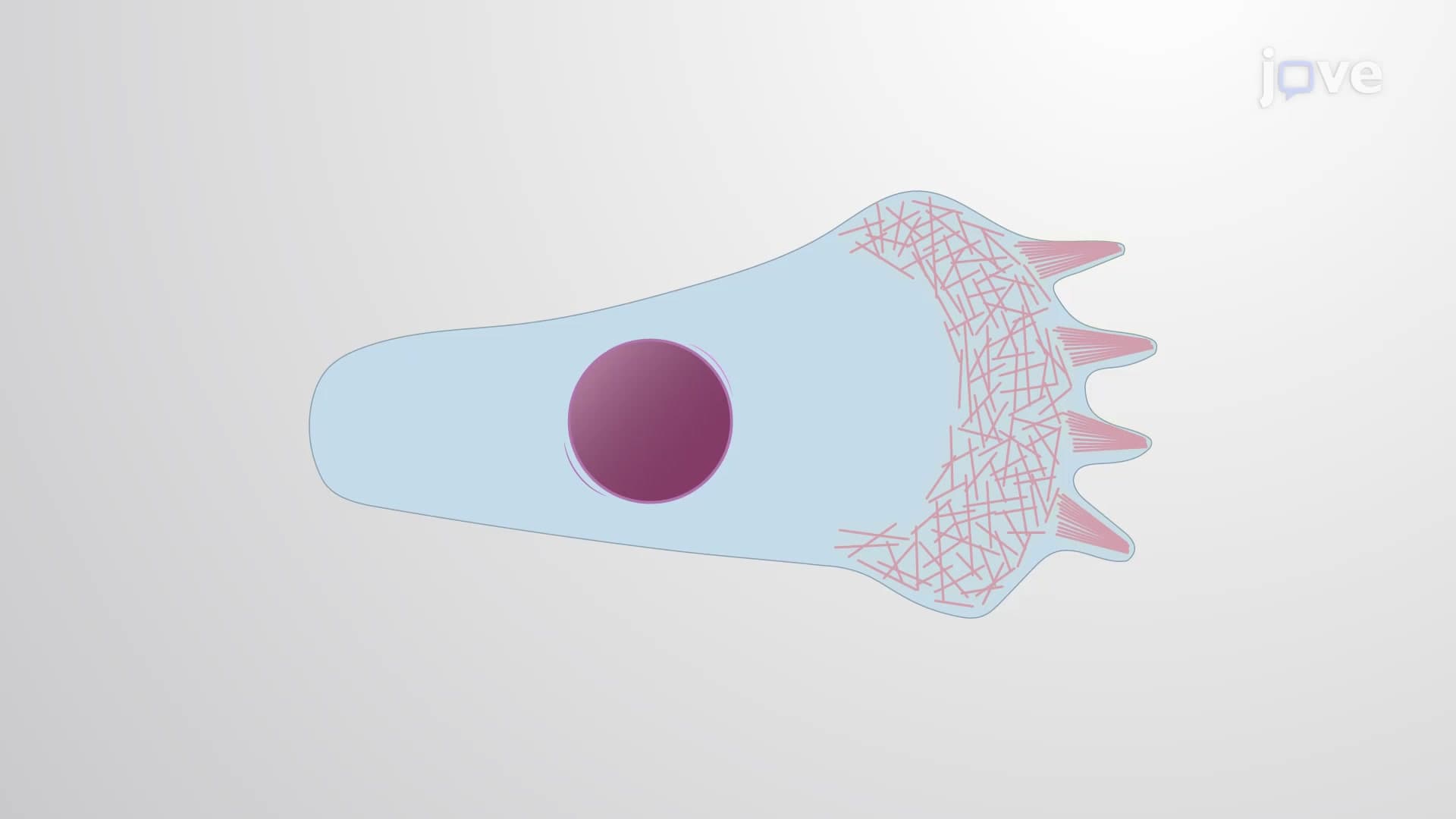
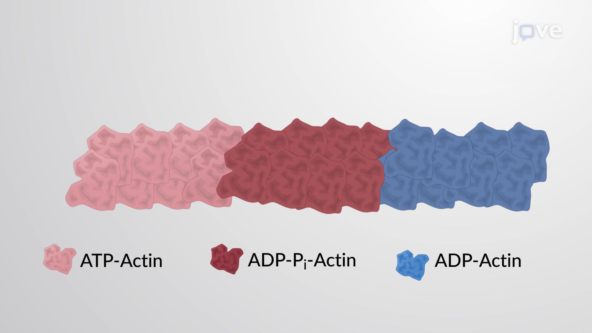
3. Actin Treadmilling and Critical Concentration Treadmilling maintains constant filament length while allowing continuous monomer flow from minus to plus ends. This phenomenon occurs when free actin concentration falls between critical concentrations of both filament ends. In American physiology, this process enables continuous cell migration—like white blood cells patrolling bloodstream for infections. The critical concentration concept parallels economic equilibrium points taught in US business schools, where supply and demand balance. ATP hydrolysis drives this process, with ATP-actin at growing ends gradually converting to ADP-actin at shrinking ends, creating the energy gradient necessary for sustained cellular movement during immune responses or tissue repair.
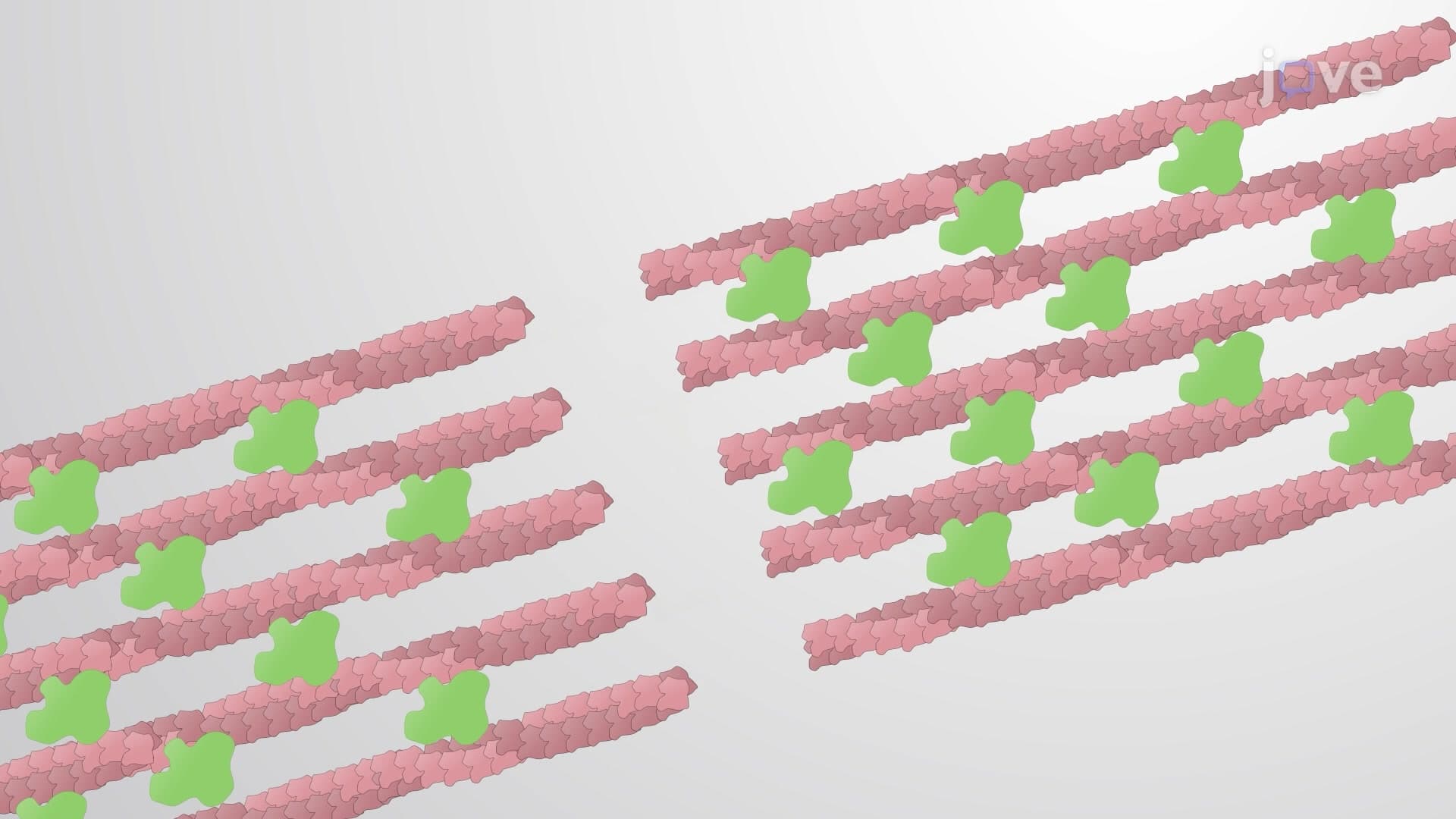
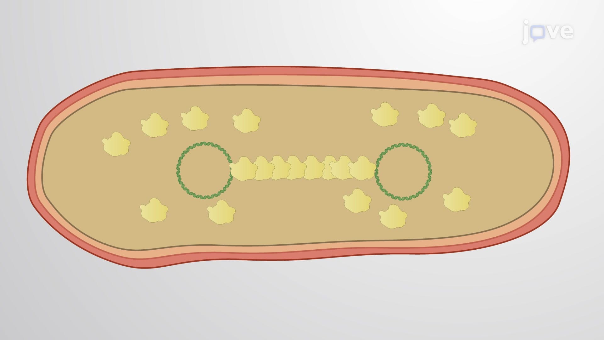
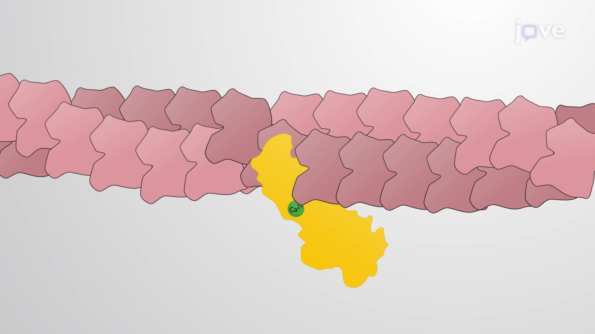
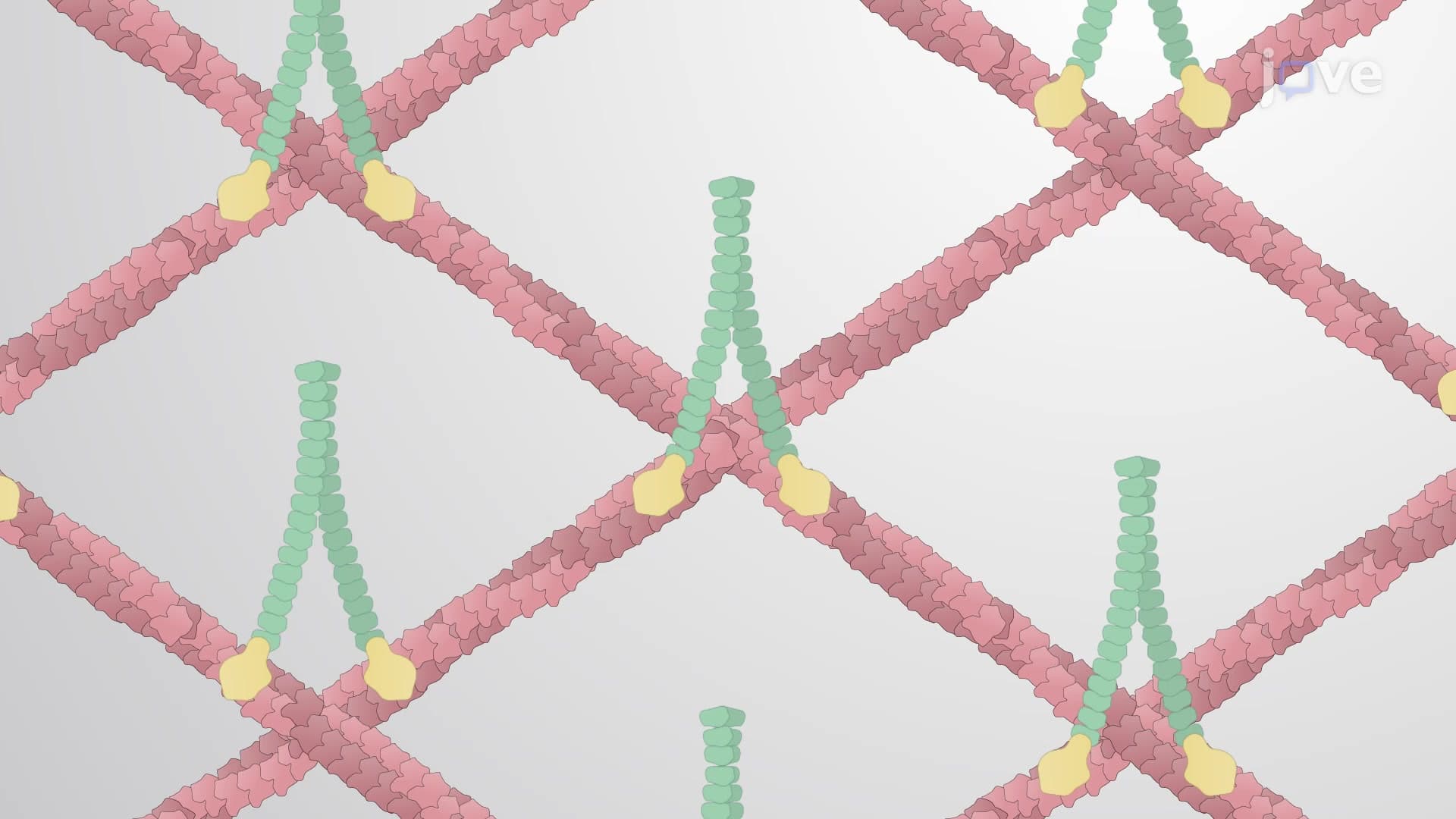
4. Actin-Binding Proteins and Filament Organization Diverse actin-binding proteins create distinct cellular structures—alpha-actinin forms loose bundles allowing myosin insertion (crucial for heart muscle function), while fimbrin creates tight parallel bundles in intestinal microvilli for nutrient absorption. Cofilin and gelsolin regulate filament disassembly, acting like cellular demolition crews during tissue remodeling. These proteins enable specialized functions across human organ systems studied in US medical schools: stress fibers in smooth muscle cells control blood vessel diameter, contractile rings enable cell division in rapidly growing tissues like bone marrow, and dendritic networks in immune cells facilitate antigen capture and presentation to T-cells.
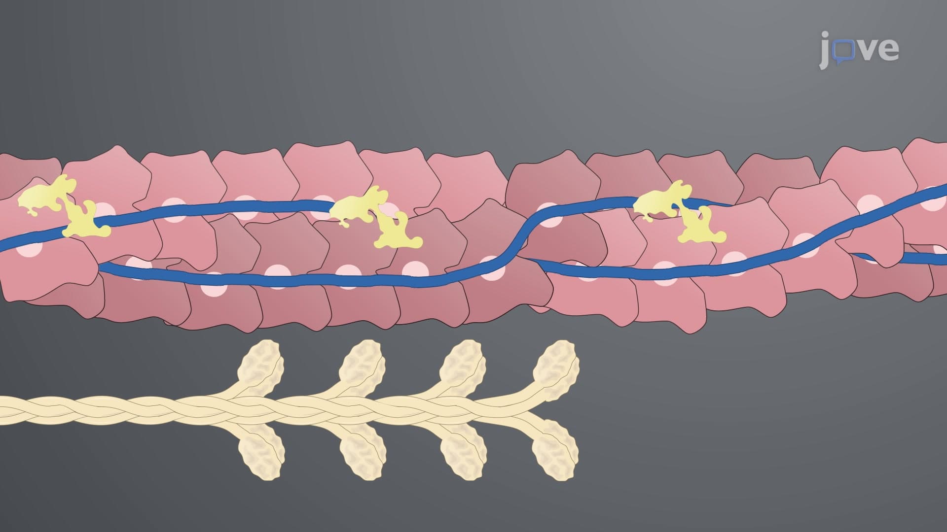
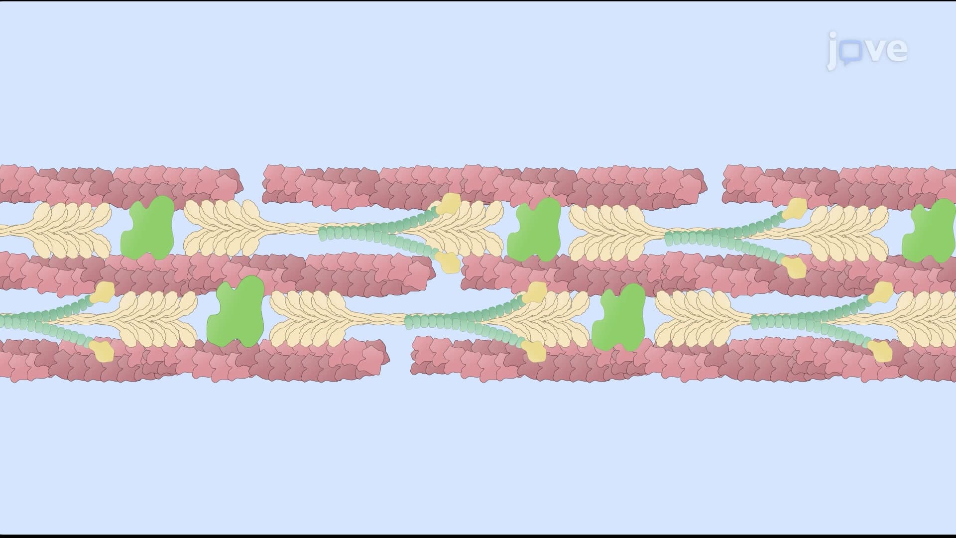
5. Myosin Motor Proteins and Cellular Contractility Myosin superfamily proteins convert chemical energy (ATP) into mechanical work through cyclic binding to actin filaments. Myosin II in skeletal muscle creates the contractile force enabling movement—from marathon runners in Boston to weightlifters in American gyms. The cross-bridge cycle involves ATP binding, hydrolysis, and release, generating force strokes that slide actin filaments past myosin. In non-muscle cells, myosin II forms stress fibers and contractile rings, enabling processes like blood clot retraction in hospital emergency rooms and cell division in growing tissues. Understanding myosin function proves essential for treating muscle diseases like muscular dystrophy, extensively researched at American institutions including Johns Hopkins and Mayo Clinic.