Loading subject page…


61,312 views
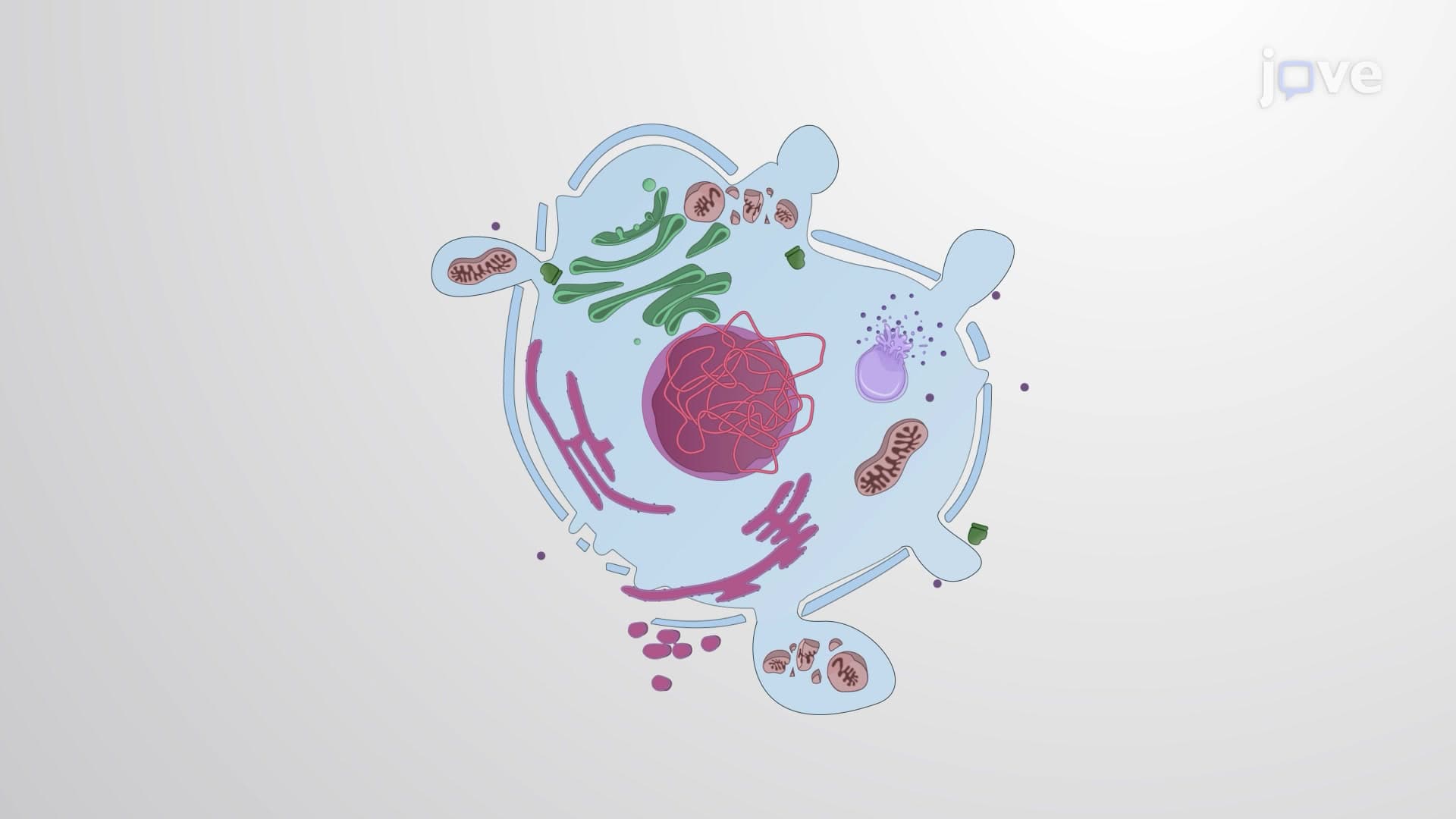

Video Summary: What Is Necrosis
Ever wondered why tissue damage from frostbite or severe burns leads to blackened, dead tissue that must be surgically removed? Necrosis is the process where cells die and their membranes rupture, releasing contents that trigger dangerous inflammation in surrounding healthy tissue. This cellular death mechanism differs dramatically from the body's controlled cell death pathways and plays a critical role in conditions like heart attacks, strokes, and gangrene seen in US emergency departments daily. Watch the full video on JoVE Coach to master this concept with expert-led visuals and step-by-step explanations.
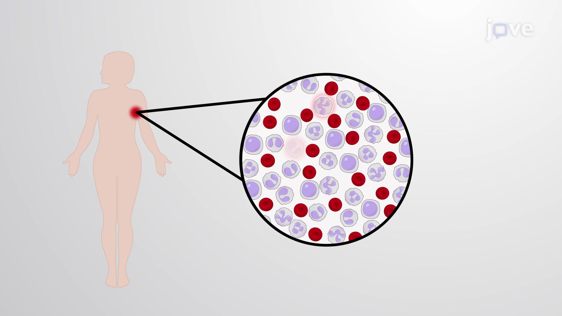
Necrosis represents one of the most dramatic forms of cell death, characterized by the complete breakdown of cellular integrity. Unlike the orderly process of apoptosis where cells quietly self-destruct, necrosis involves violent cell membrane rupture that releases toxic cellular contents into surrounding tissues. This inflammatory cascade makes necrosis particularly dangerous in medical settings, as seen in conditions like myocardial infarction (heart attacks) where necrotic heart muscle releases enzymes that doctors measure to confirm tissue death.
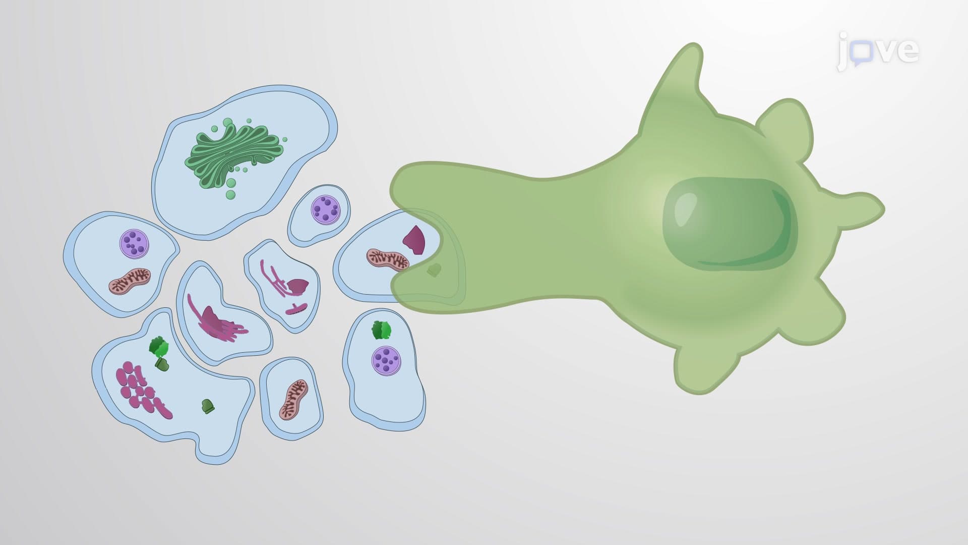
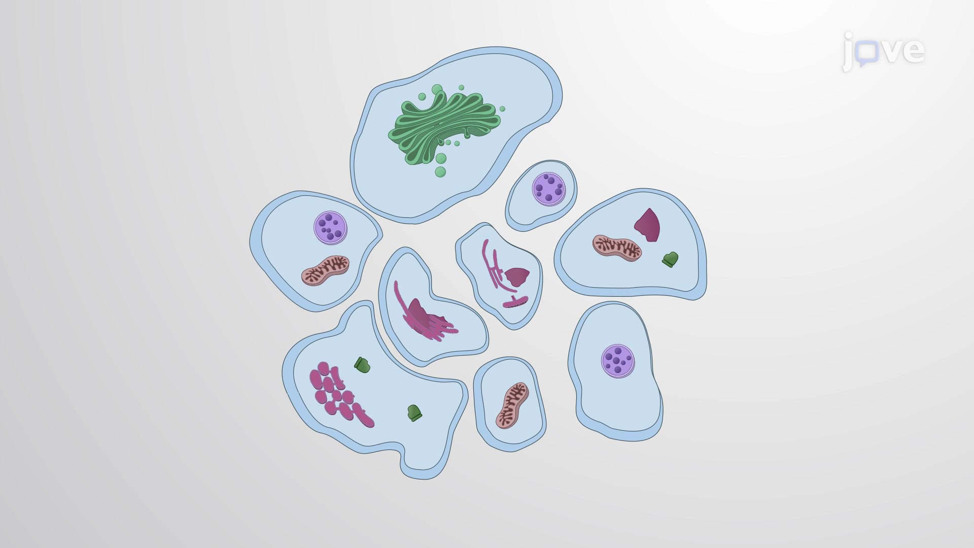
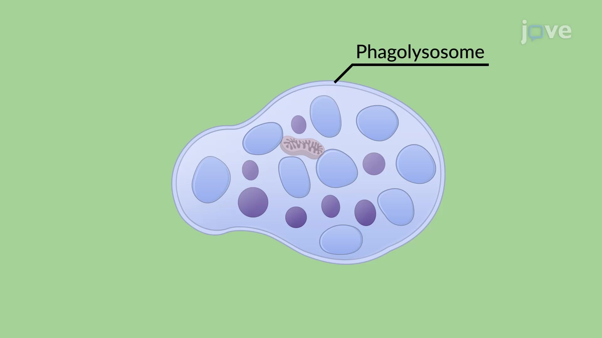
The most common form occurs when cells face overwhelming stress beyond their survival capacity. Physical trauma from car accidents, extreme temperatures causing frostbite, or toxic chemical exposure can trigger this pathway. The process begins with mitochondrial damage-these cellular powerhouses can no longer produce adequate ATP (cellular energy currency). Simultaneously, calcium regulation fails, causing massive calcium influx that makes cells swell like water balloons until they burst. This accidental necrosis explains why burn victims require immediate medical attention to prevent widespread tissue death and infection.
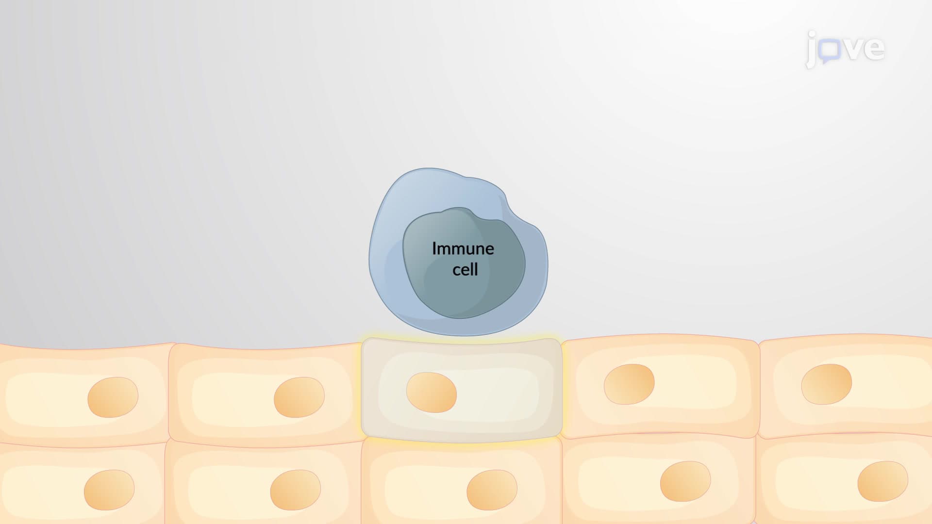
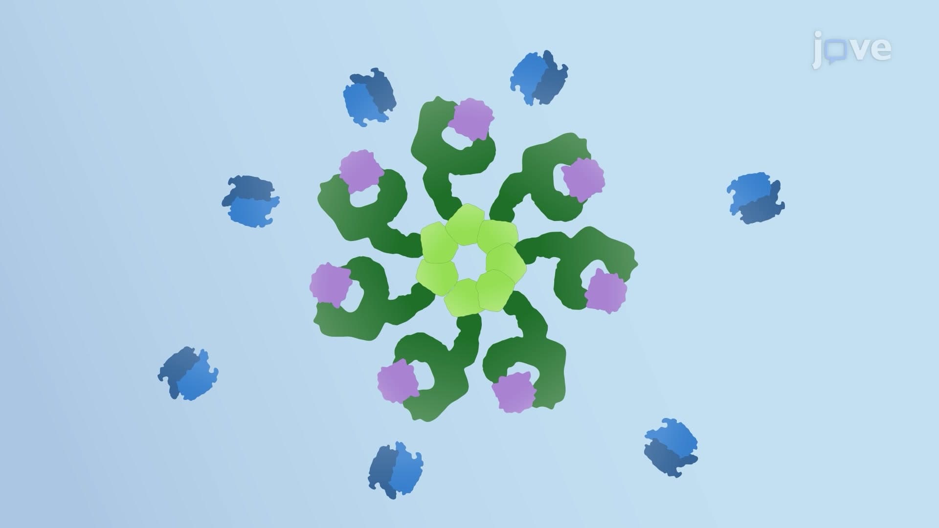
Surprisingly, cells also possess a controlled form of necrosis called necroptosis, discovered relatively recently by researchers at major US institutions like Harvard Medical School and Memorial Sloan Kettering. This pathway activates when the normal apoptosis machinery fails-imagine it as a cellular "Plan B" for death. Key proteins RIP1 and RIP3 form molecular complexes that ultimately activate MLKL, a protein that punches holes in cell membranes. This controlled destruction still causes inflammation but serves important biological functions, such as eliminating virus-infected cells that have disabled their apoptosis mechanisms.
Understanding necrosis proves crucial for students preparing for healthcare careers and advanced placement exams. The MCAT frequently tests necrosis concepts in biological sciences sections, while nursing students encounter necrotic tissue assessment on the NCLEX-RN. In AP Biology courses, necrosis appears in cell biology units alongside apoptosis comparisons. Medical students studying pathology learn to identify necrotic tissue patterns that guide treatment decisions-from the gray-black appearance of gangrenous tissue to the characteristic enzyme release patterns following heart attacks in US cardiac care units.
Related Micro-courses