Loading subject page…


40,213 views

Video Summary: Actin Polymerization and Cell Motility Explained
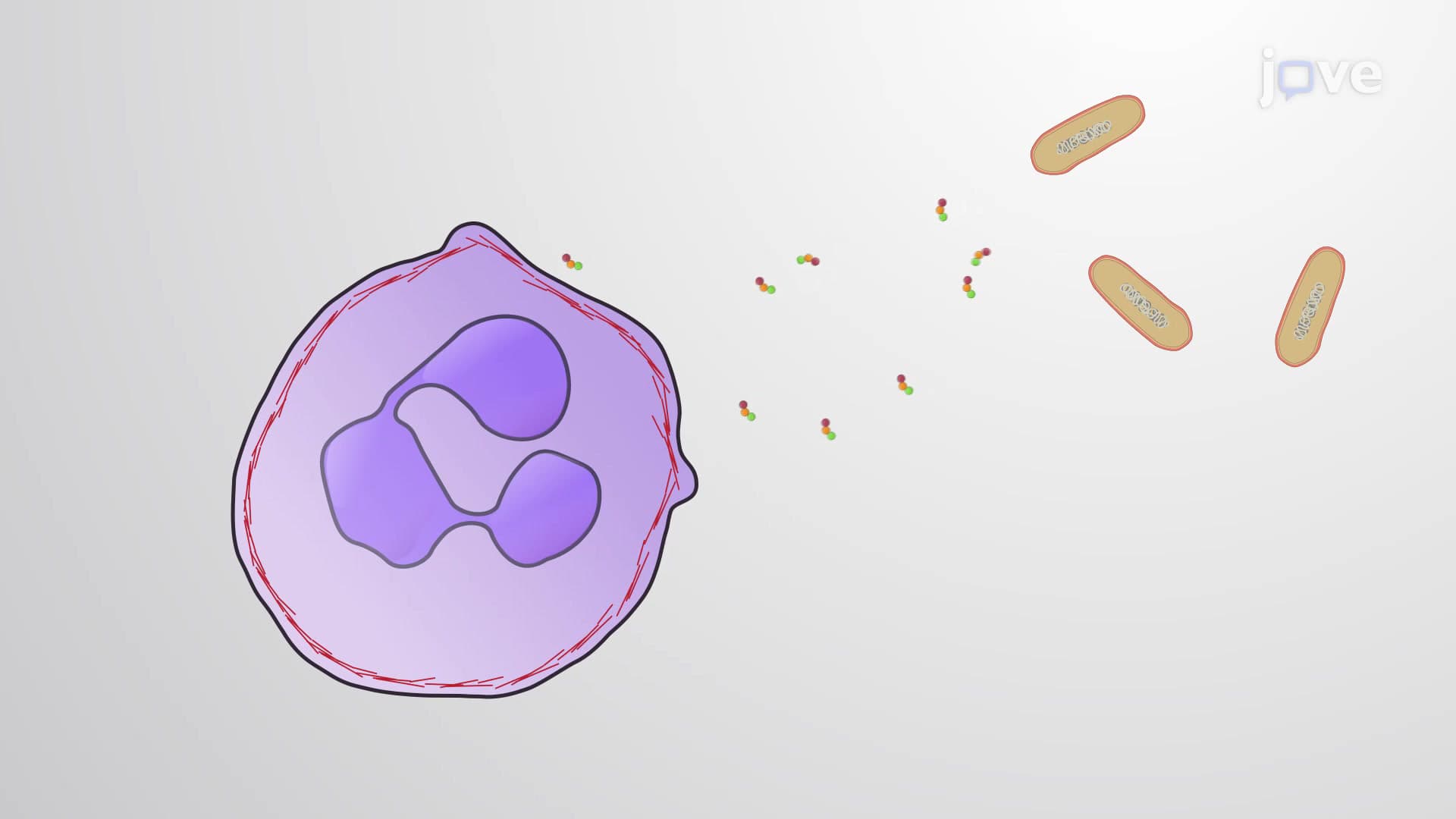
Did you know that cancer cells hijack the same actin polymerization and cell motility mechanisms that allow immune cells to chase down infections? When a white blood cell in your bloodstream detects a bacterial invasion at a wound site, it must rapidly reorganize its internal actin filaments to squeeze through blood vessel walls and migrate to the infection. This process, known as Actin Polymerization And Cell Motility Explained, involves the continuous assembly and disassembly of protein filaments that literally push the cell membrane forward, creating the protrusions needed for movement. Watch the full video on JoVE Coach to master this concept with expert-led visuals and step-by-step explanations.
Cell migration represents one of biology's most elegant examples of molecular machinery in action. At its core lies the actin cytoskeleton-a dynamic network of protein filaments that undergoes constant remodeling to power cellular movement. This process becomes critically important in numerous physiological contexts, from wound healing to immune responses, making it a cornerstone topic in AP Biology, college cell biology courses, and pre-medical curricula.
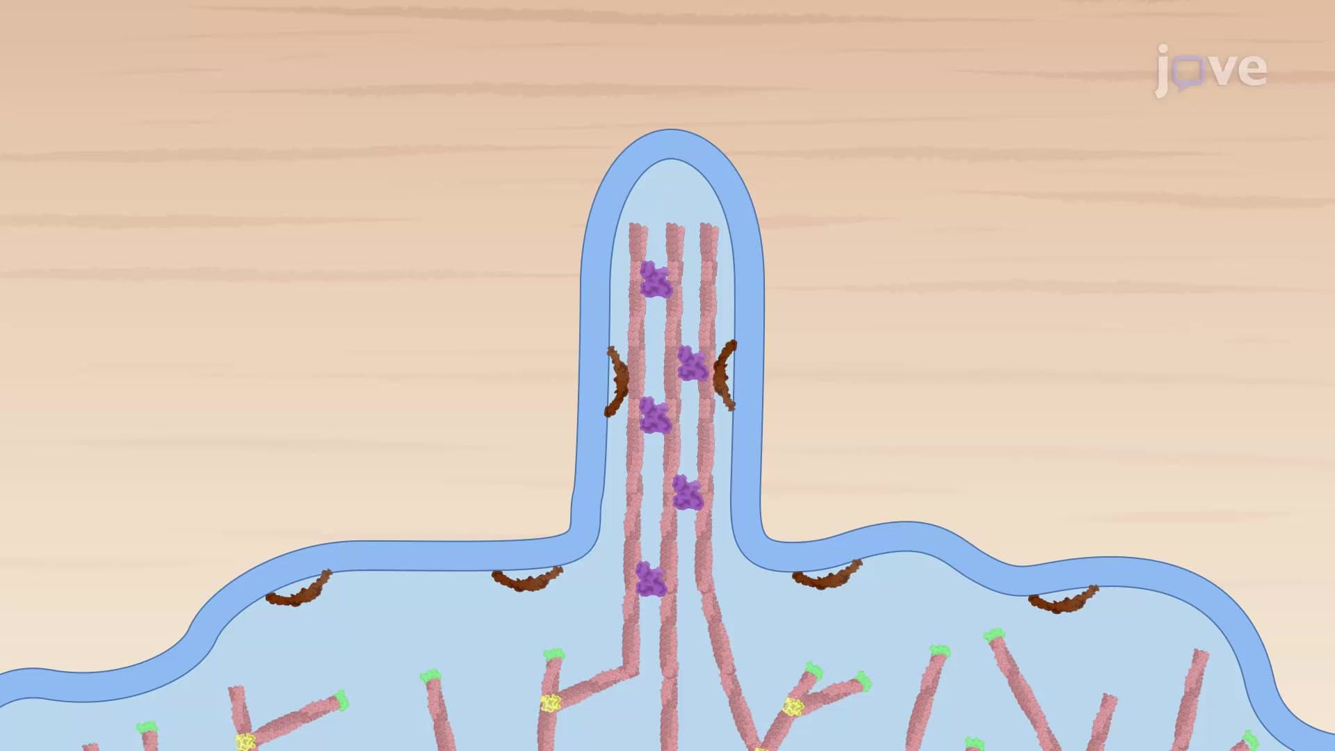
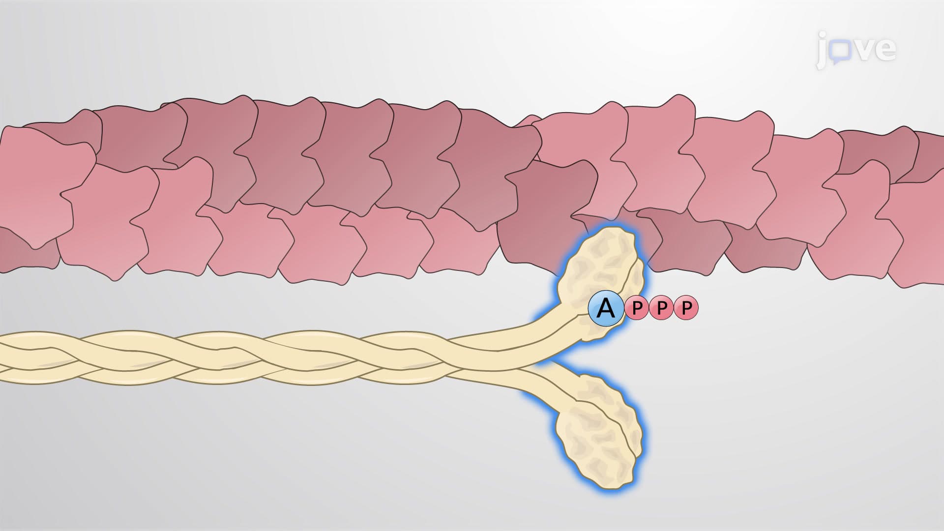
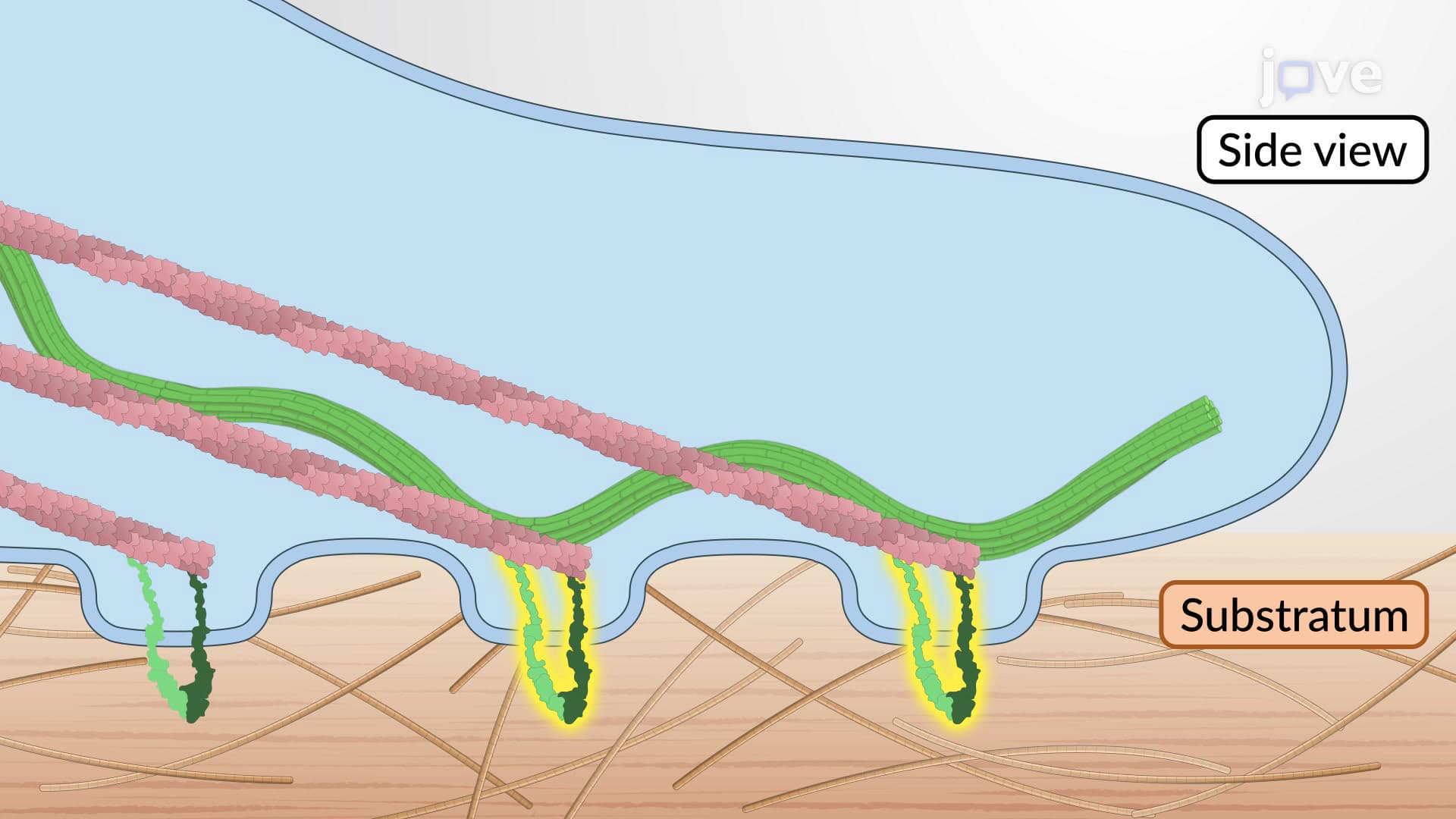
Actin exists as a 42-kilodalton globular protein that serves as the building block for cellular architecture. In its monomeric form (G-actin), this protein remains relatively inactive. However, under appropriate cellular conditions, these monomers polymerize into long, helical filaments called F-actin (filamentous actin). Each filament exhibits distinct polarity, with a fast-growing plus end (also called the barbed end) and a slower-growing minus end (pointed end). This structural asymmetry proves crucial for directional cell movement.
The polymerization process follows precise biochemical rules that students encounter in advanced biology exams. ATP-bound actin monomers preferentially add to the plus end, while older ADP-bound subunits at the minus end undergo depolymerization. This creates a phenomenon called "treadmilling," where the filament maintains constant length while individual subunits flow from the plus to minus end-a concept frequently tested on the MCAT and college biochemistry exams.
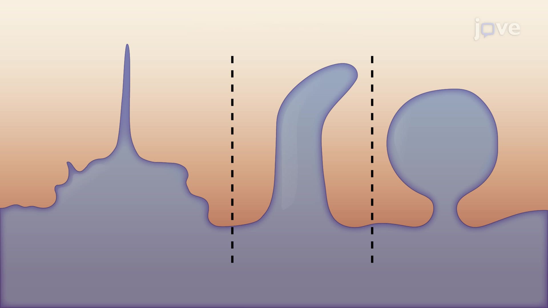
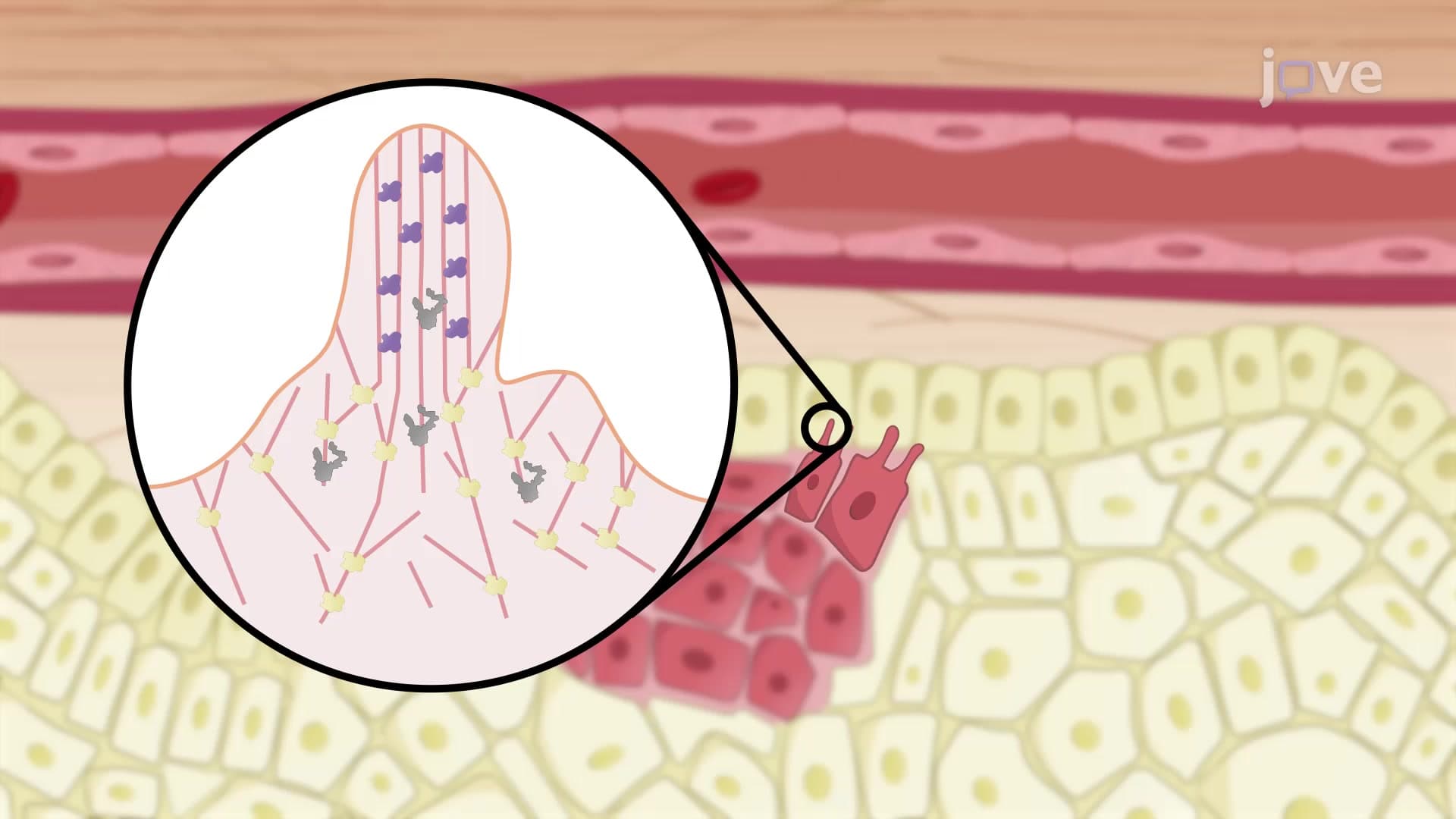
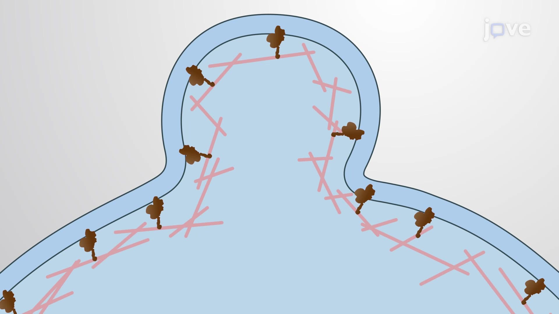
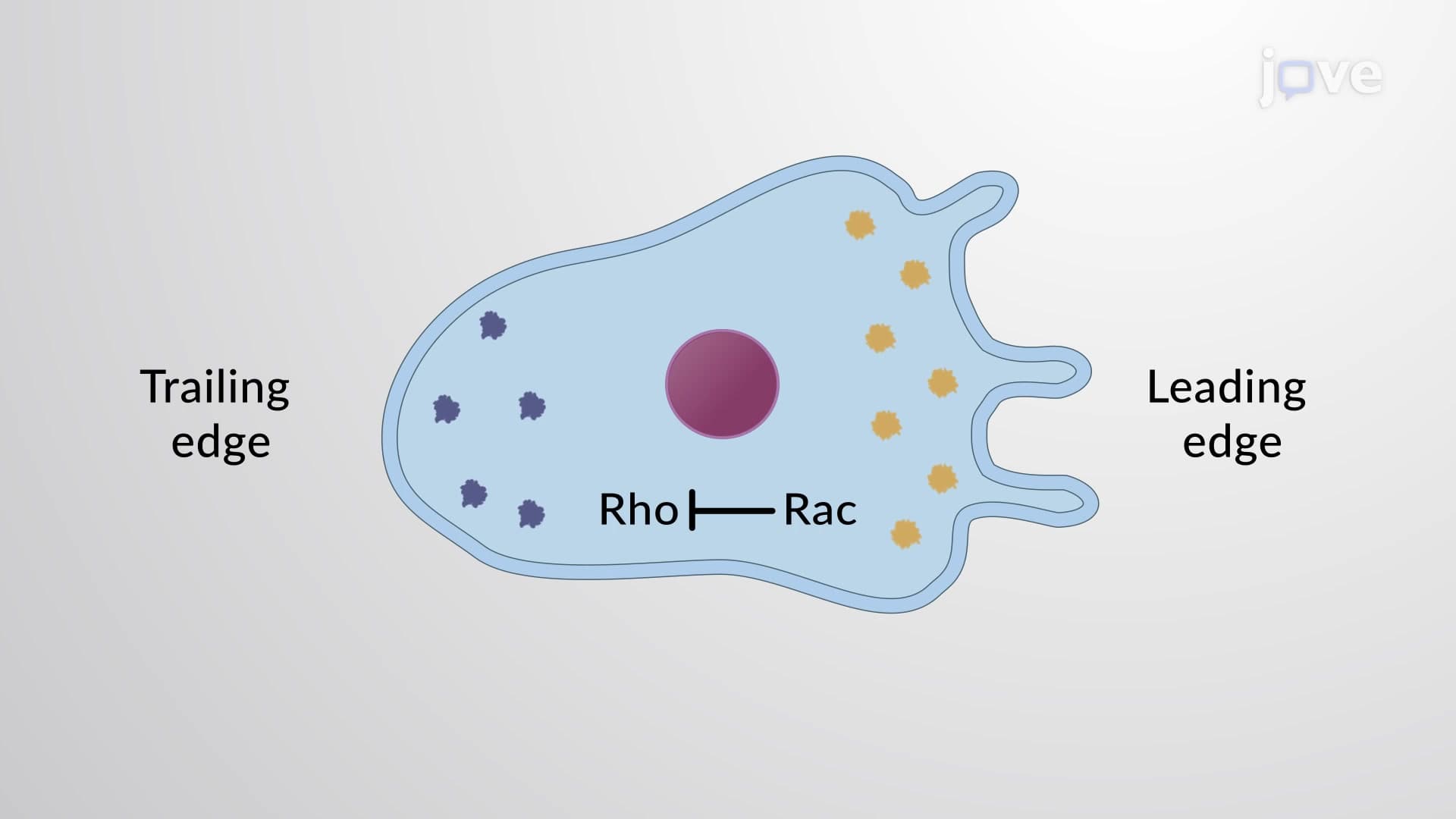
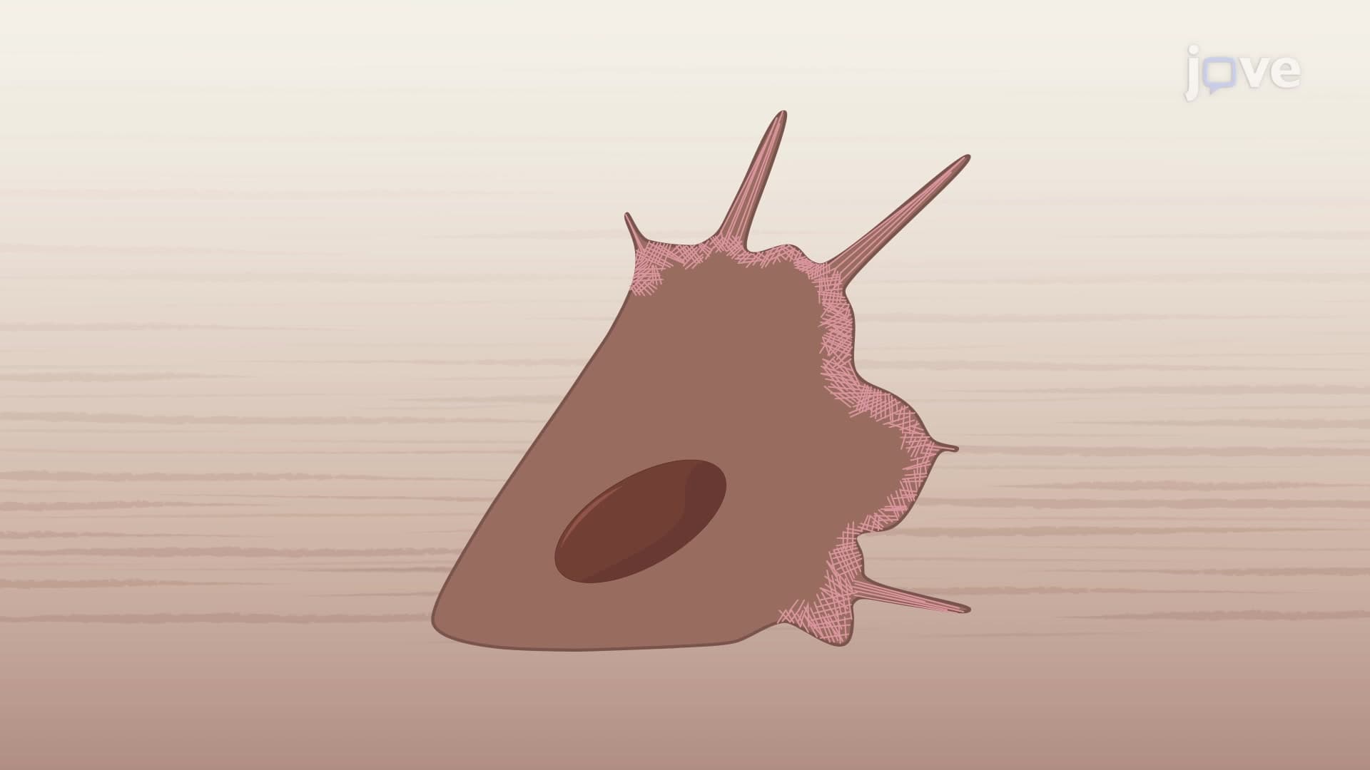
The mechanical basis of cell motility centers on actin's ability to generate pushing forces against the cell membrane. When cells receive migratory signals-such as chemokine gradients during immune cell recruitment to infection sites-actin polymerization becomes highly localized at the cell's leading edge. The growing filaments literally push against the membrane, creating finger-like projections called filopodia or sheet-like extensions called lamellipodia.
Consider how neutrophils respond to bacterial infections in tissue. These immune cells must rapidly extend membrane protrusions to navigate through tight spaces between cells, a process entirely dependent on coordinated actin dynamics. The force generated by just a few hundred polymerizing filaments can exceed the membrane's resistance, demonstrating the remarkable efficiency of this molecular motor system.
Understanding actin polymerization and cell motility has profound implications for medical research and treatment. Cancer metastasis relies heavily on altered actin dynamics that allow malignant cells to invade surrounding tissues and enter the bloodstream. Pharmaceutical researchers at institutions like Johns Hopkins and Stanford University actively develop drugs targeting actin regulators as potential cancer therapies. This connection between basic cell biology and clinical applications frequently appears in USMLE questions and advanced placement biology essays, emphasizing the importance of mastering these fundamental concepts.
Related Micro-courses