Loading subject page…


40,213 views
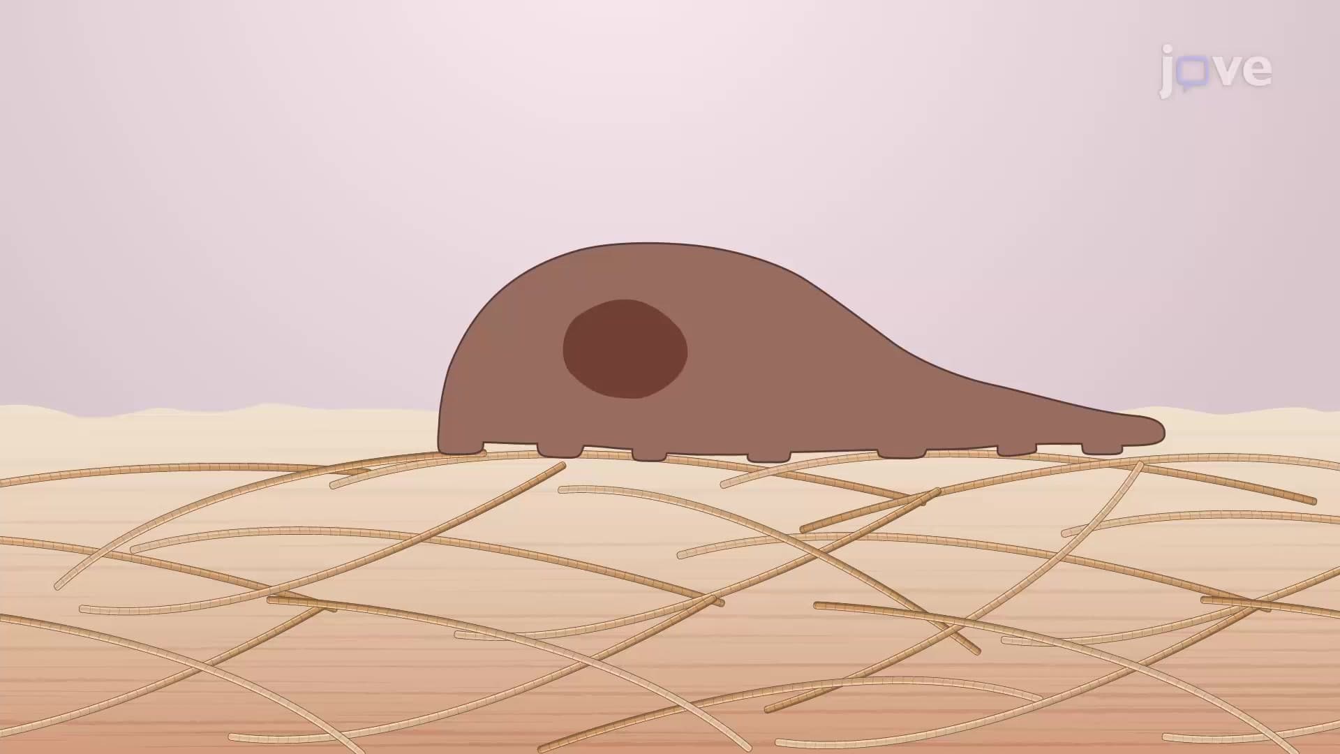

Video Summary: What Is Cell Migration
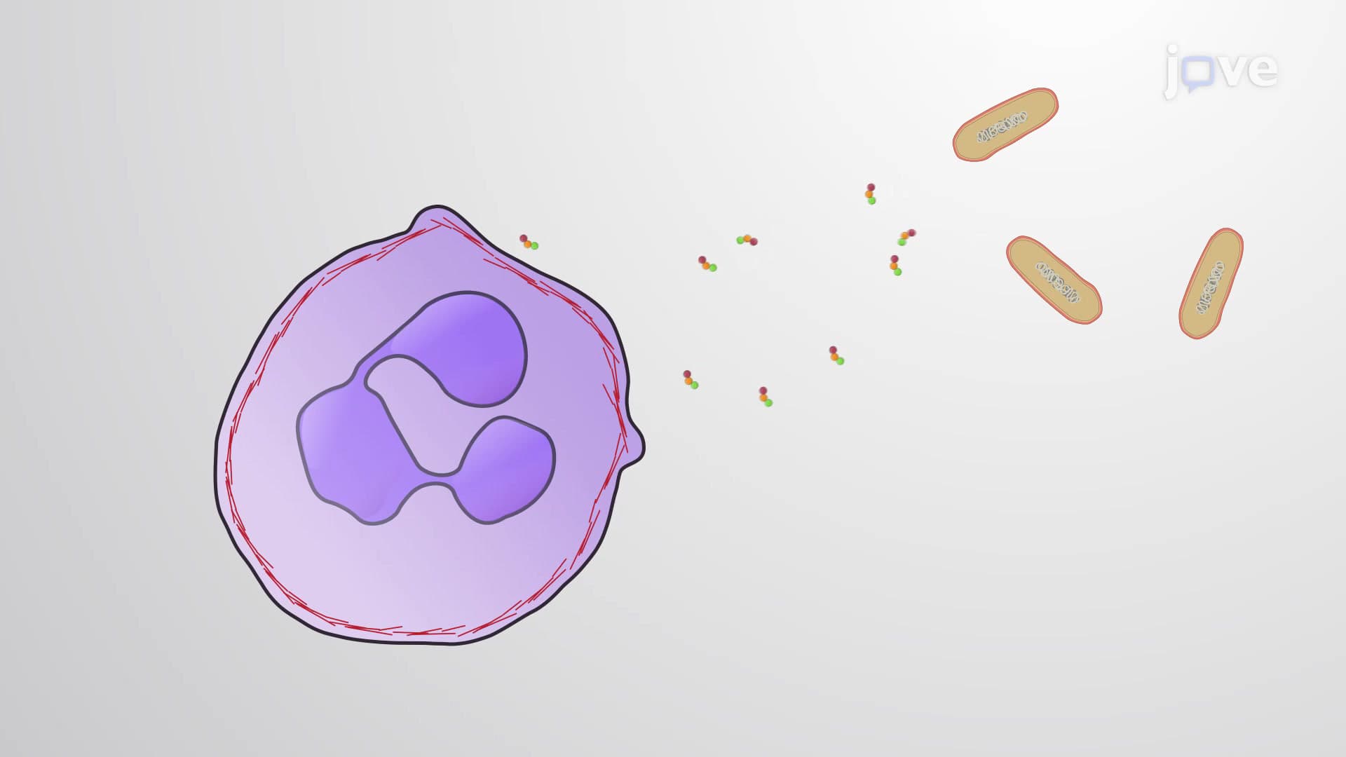
Ever wonder how your scraped knee heals itself? Cell migration, the coordinated movement of cells from one location to another, drives this remarkable repair process throughout your body. When you cut your finger in the kitchen, specialized cells called fibroblasts immediately begin moving toward the wound site, guided by chemical signals released from damaged tissue. This fundamental biological process enables everything from wound healing to embryonic development. Watch the full video on JoVE Coach to master this concept with expert-led visuals and step-by-step explanations.
Cell migration represents one of biology's most sophisticated transportation systems, enabling cells to navigate through complex tissue environments with remarkable precision. This process involves coordinated changes in cell shape, adhesion properties, and internal force generation that allow cells to move purposefully through three-dimensional spaces.
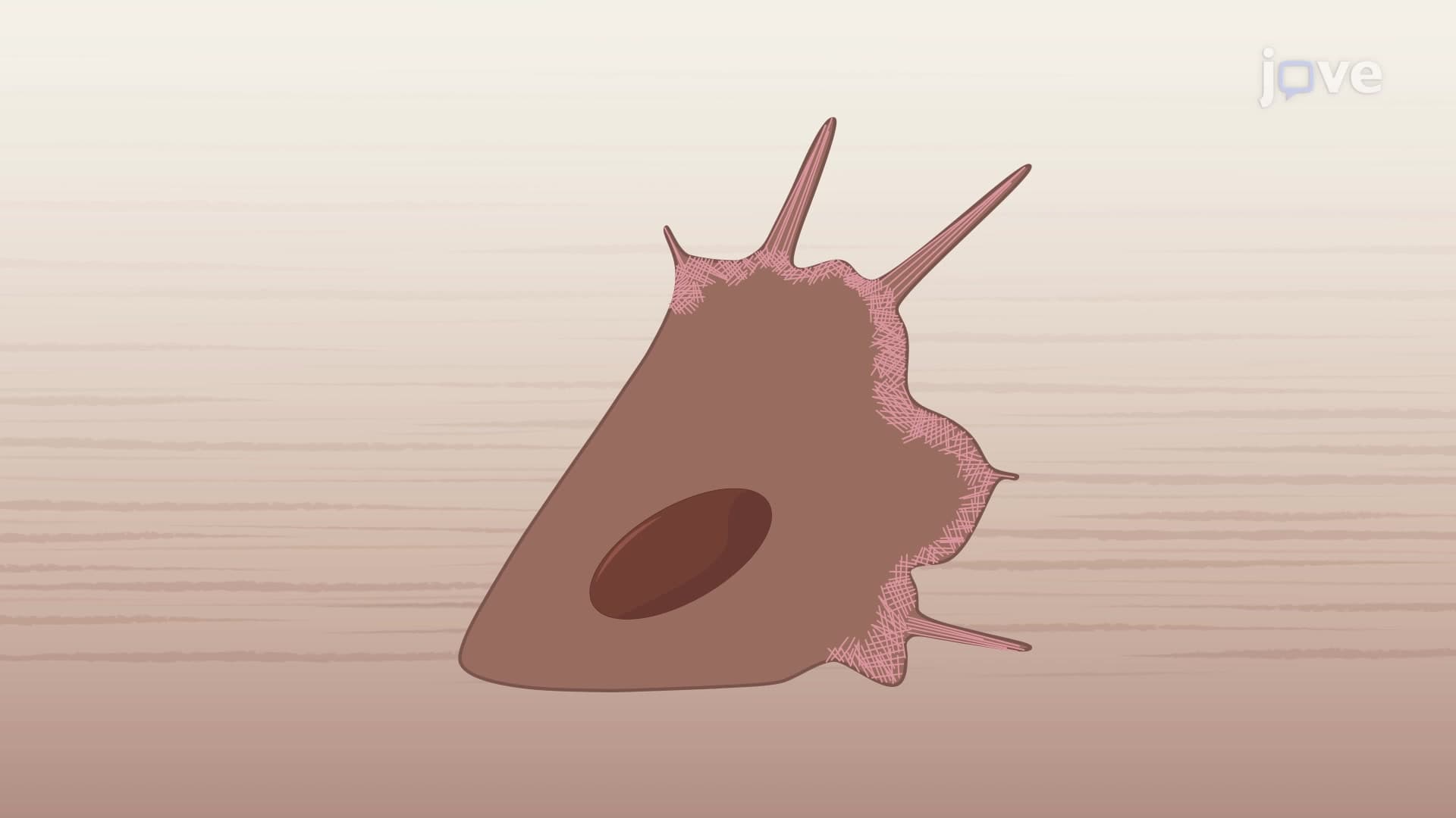
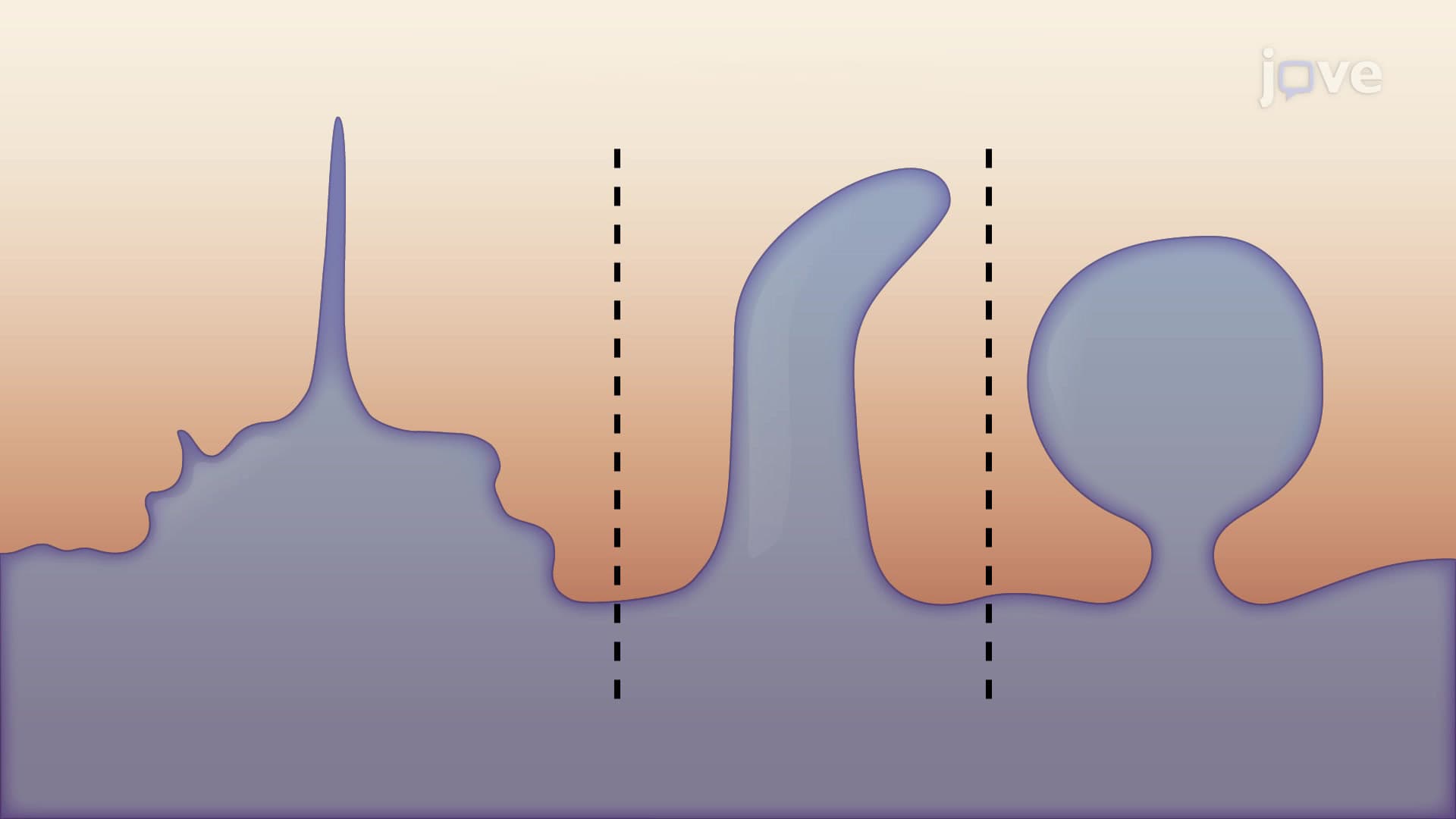
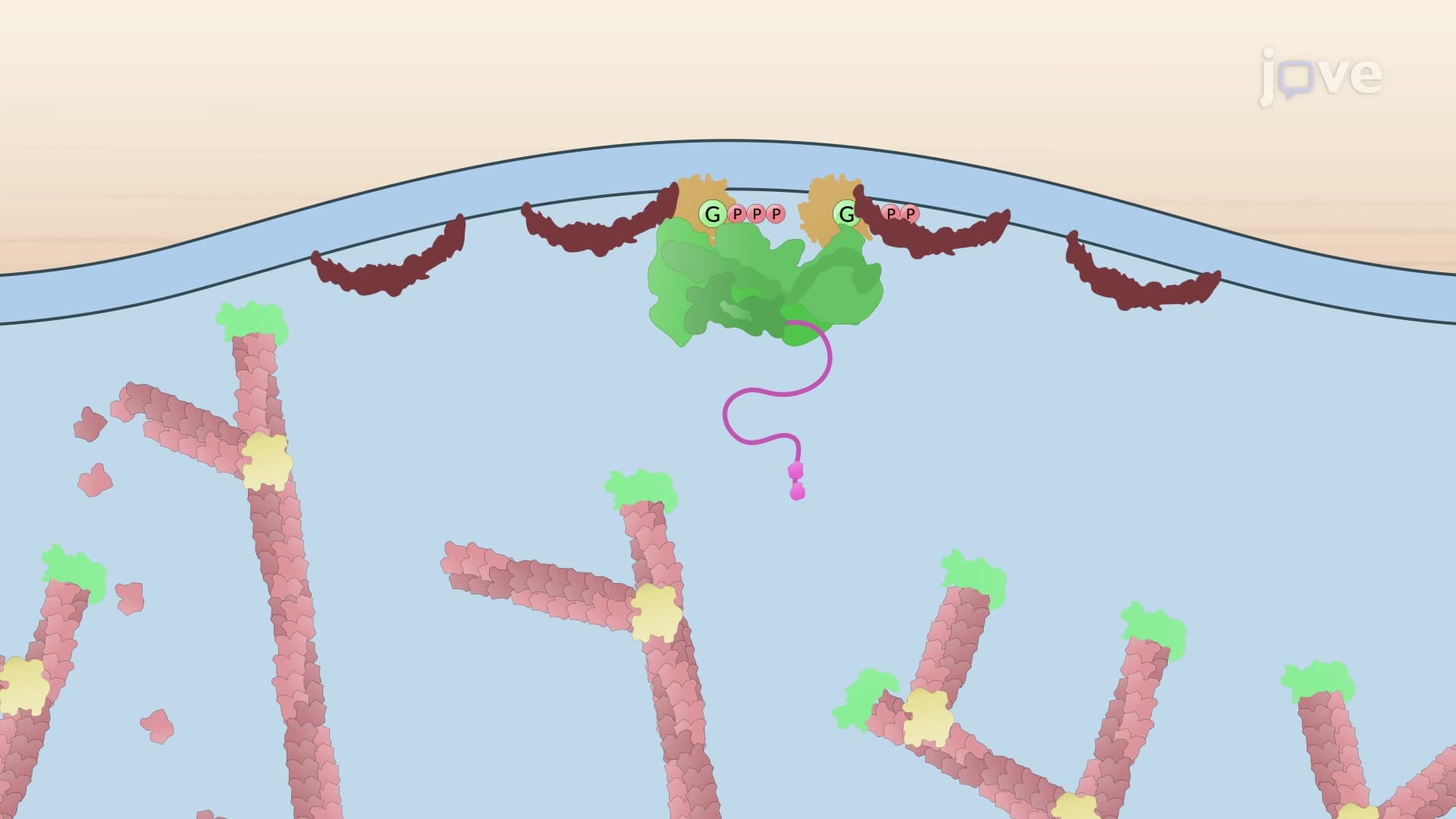
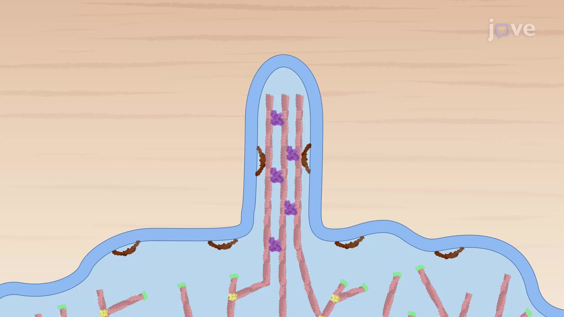
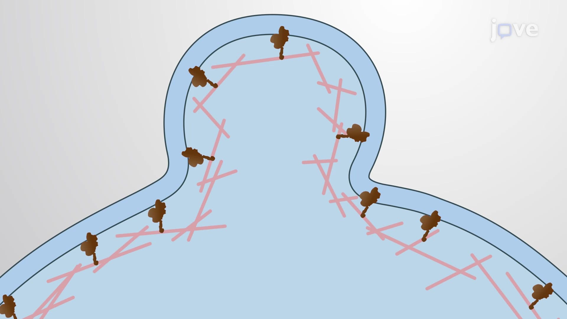
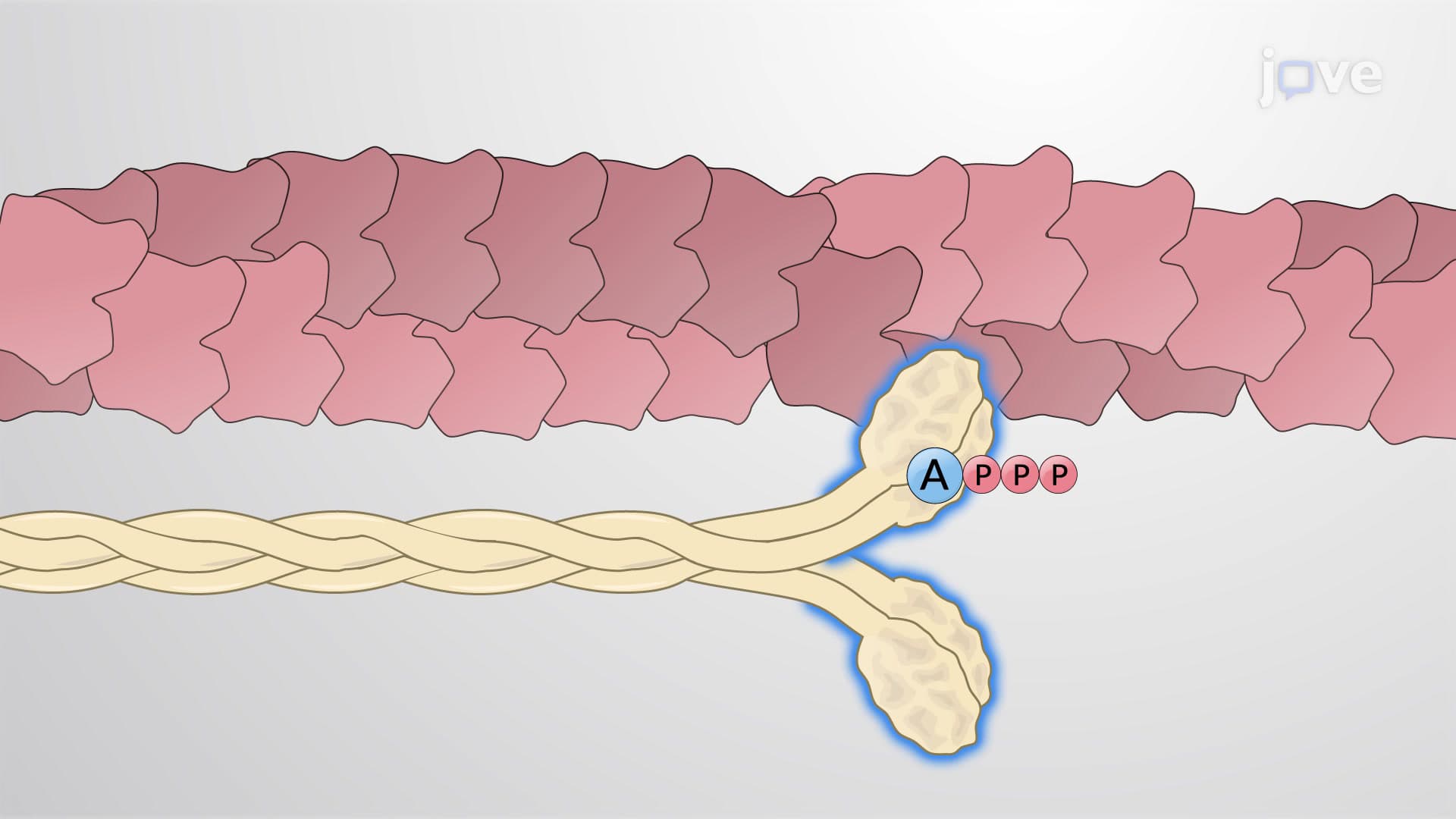
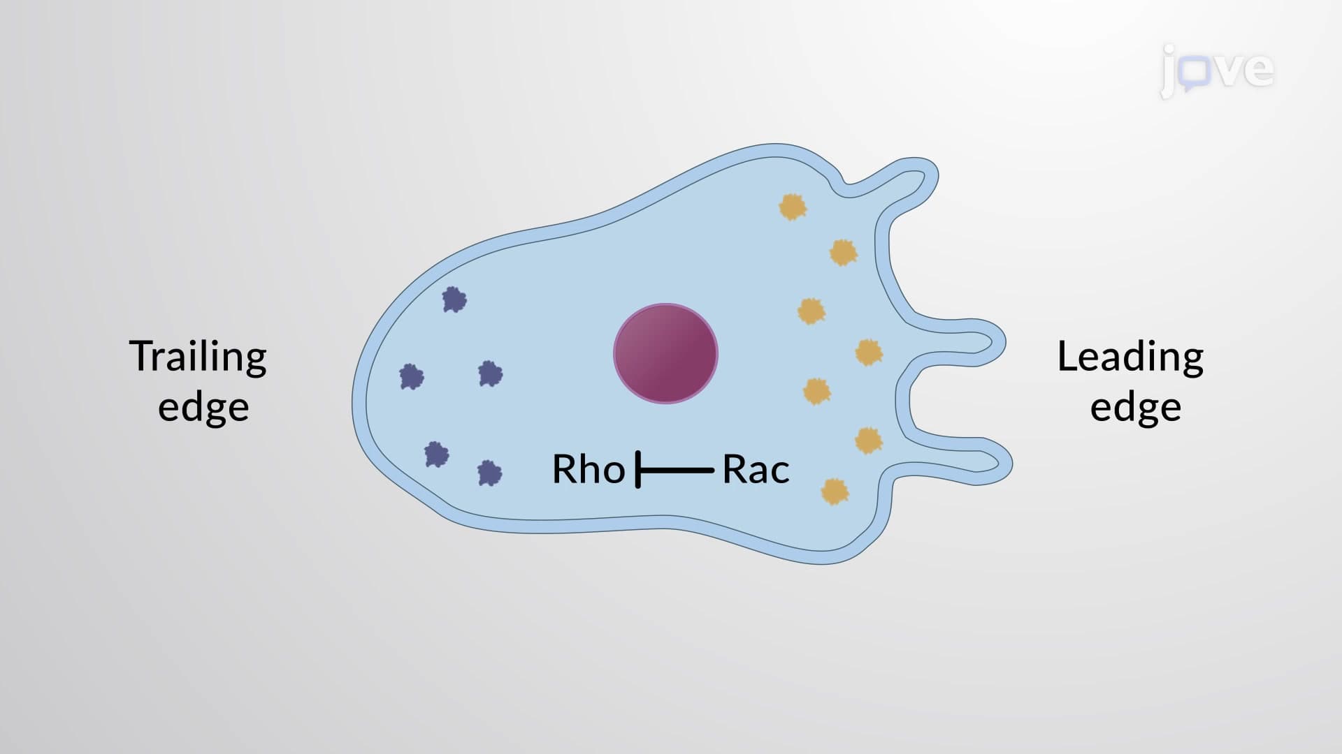
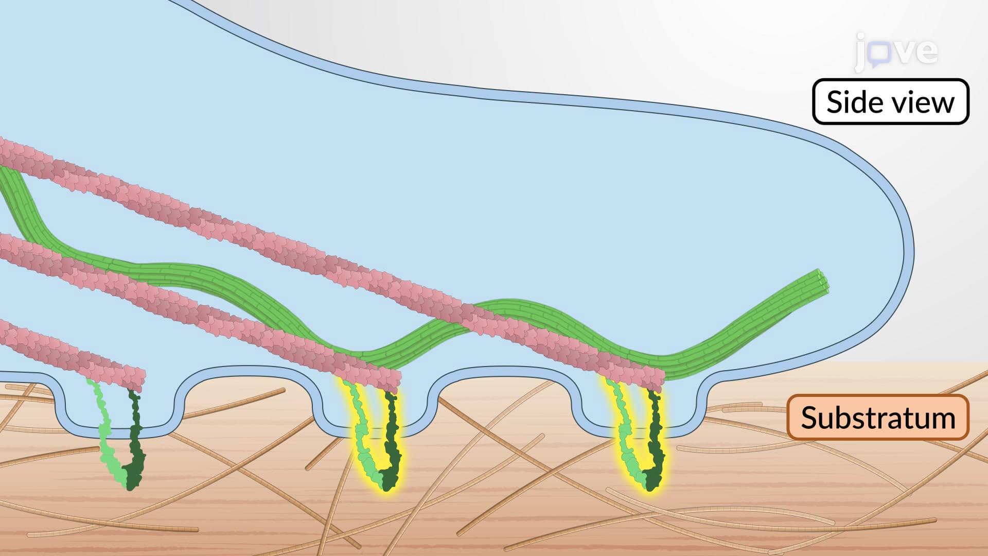
The migration process relies on dynamic interactions between the cell's internal skeleton (cytoskeleton) and external environment. At the leading edge, cells extend finger-like projections called filopodia and sheet-like structures called lamellipodia. These extensions contain dense networks of actin filaments that polymerize rapidly, literally pushing the cell membrane forward. Meanwhile, specialized protein complexes called focal adhesions anchor these protrusions to the surrounding extracellular matrix, creating temporary "molecular anchors" that cells use to pull themselves forward.
This concept frequently appears on AP Biology exams, particularly in questions about tissue repair and embryonic development. Students should understand that focal adhesions aren't permanent connections-they form and dissolve in cycles, allowing cells to maintain forward momentum while releasing their "grip" at the trailing edge.
Cell migration dysfunction underlies numerous medical conditions treated in US hospitals daily. Cancer metastasis occurs when tumor cells gain abnormal migratory abilities, allowing them to invade distant organs. Conversely, chronic wounds in diabetic patients often result from impaired cell migration, preventing proper healing. Understanding these mechanisms helps explain why treatments like growth factor therapy and stem cell therapies show promise in clinical trials at institutions like the Mayo Clinic and Johns Hopkins.
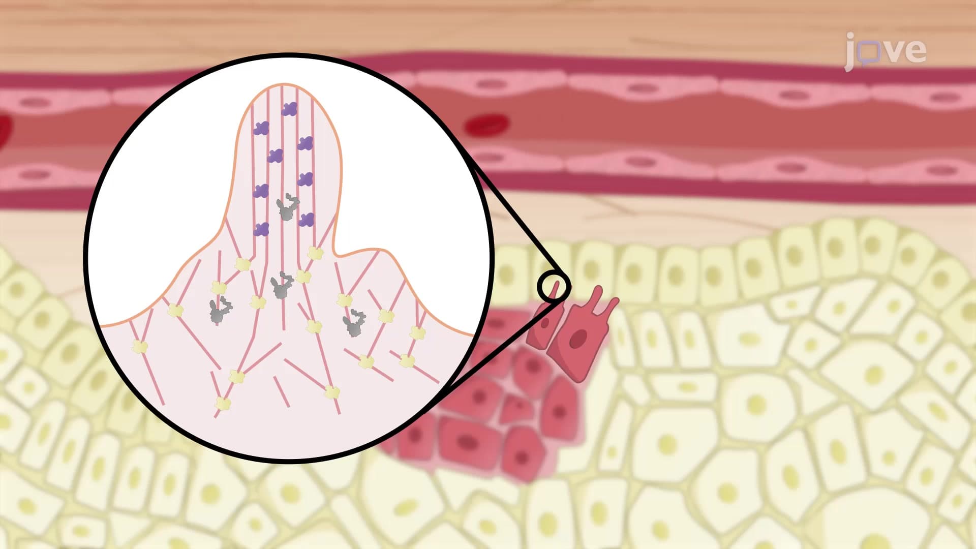
While individual cells like fibroblasts migrate independently toward chemical signals, epithelial cells demonstrate collective migration patterns. During wound healing, sheets of epithelial cells move together while maintaining connections through cell-cell junctions, resembling a coordinated "cellular convoy" advancing across the wound surface. This collective behavior ensures complete coverage and prevents gaps that could lead to infection-a concept commonly tested on MCAT Cell Biology sections and college-level physiology exams.
Related Micro-courses