Loading video…


 7 Concepts
 5.4K views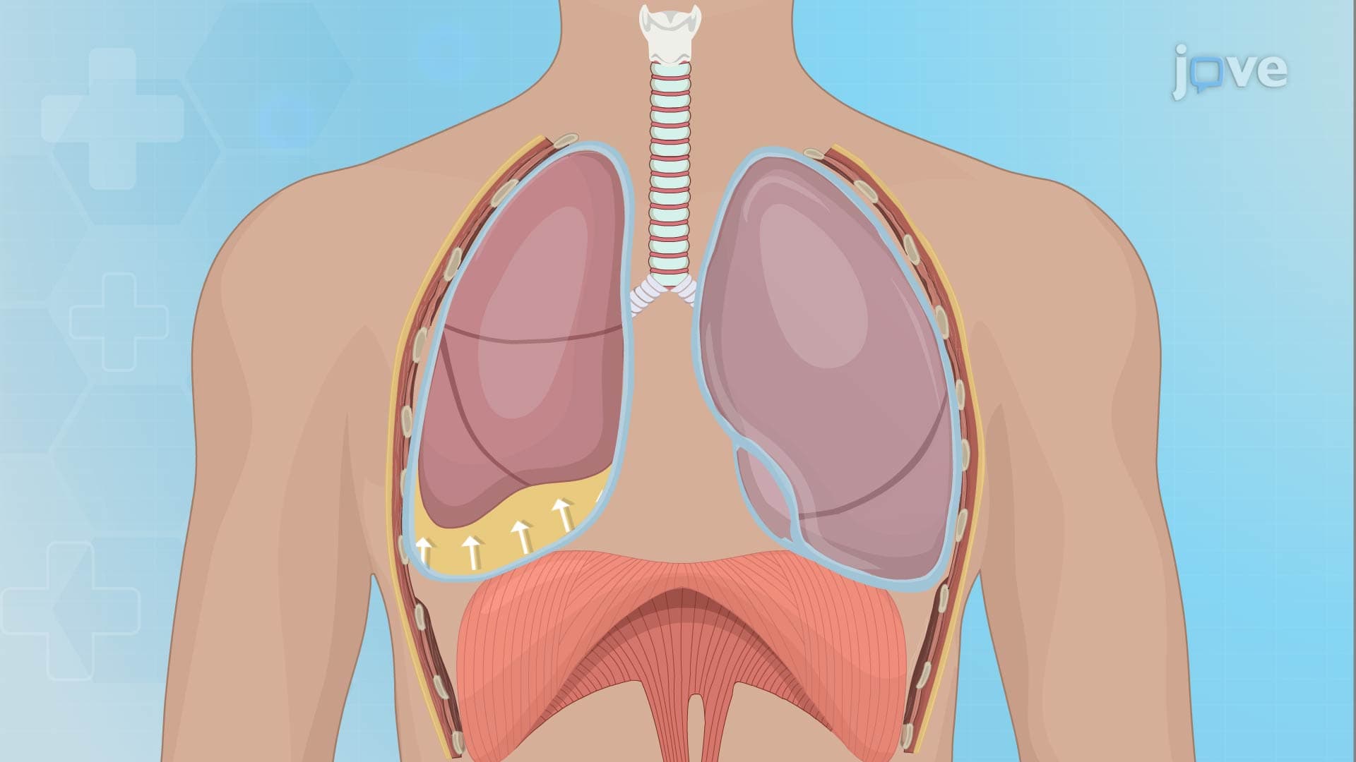

Video Summary: What Is Pleural Effusion I Introduction
Did you know that over 1.5 million Americans develop pleural effusion annually, with heart failure being the leading cause? Pleural effusion i introduction covers the fundamental concept of fluid accumulation between the lungs and chest wall. This condition affects thousands of patients in US hospitals daily, from congestive heart failure cases at Johns Hopkins to pneumonia complications at UCLA Medical Center. Understanding what is pleural effusion i introduction helps explain why patients experience shortness of breath and chest pain. Watch the full video on JoVE Coach to master this concept with expert-led visuals and step-by-step explanations.
Pleural effusion i introduction begins with understanding the pleural space anatomy. The pleural cavity normally contains only 10-20 mL of lubricating fluid between the visceral pleura (covering the lungs) and parietal pleura (lining the chest wall). When this delicate balance is disrupted, excess fluid accumulates, creating a pleural effusion that can compress lung tissue and impair breathing.
The protein content distinction is crucial for diagnosis and treatment. Transudative effusions contain low protein levels (less than 3 g/dL) and result from systemic factors affecting fluid movement. Light's criteria, developed at Brown University, helps clinicians differentiate these types using pleural fluid-to-serum protein and LDH ratios. This classification system is essential for MCAT preparation and appears frequently on USMLE Step 1 examinations.
Heart failure accounts for approximately 40% of all pleural effusions in US emergency departments. When the left ventricle fails to pump effectively, increased venous pressure forces fluid into the pleural space. At Mayo Clinic, cardiologists regularly see patients with bilateral pleural effusions secondary to congestive heart failure. Liver cirrhosis creates a different mechanism-reduced albumin synthesis decreases oncotic pressure, allowing fluid to shift from blood vessels into body cavities. Similarly, nephrotic syndrome causes massive proteinuria, dramatically lowering serum oncotic pressure and promoting fluid accumulation.
Exudative effusions contain high protein content (greater than 3 g/dL) due to increased capillary permeability. Tuberculosis remains a significant cause in urban US populations, particularly in cities like New York and Los Angeles. The mycobacterium triggers intense pleural inflammation, increasing vascular permeability and allowing proteins and inflammatory cells to enter the pleural space. Pneumonia-related effusions, common in patients admitted to hospitals like Cleveland Clinic, result from direct pleural involvement or parapneumonic inflammation. Malignant effusions, often seen in lung cancer patients at MD Anderson Cancer Center, occur when tumor cells invade pleural surfaces or obstruct lymphatic drainage.
Understanding these mechanisms helps students excel on AP Biology exams and college-level pathophysiology courses while providing foundation knowledge for healthcare career preparation.
Related Micro-courses