Loading video…

 6 Concepts
 872 views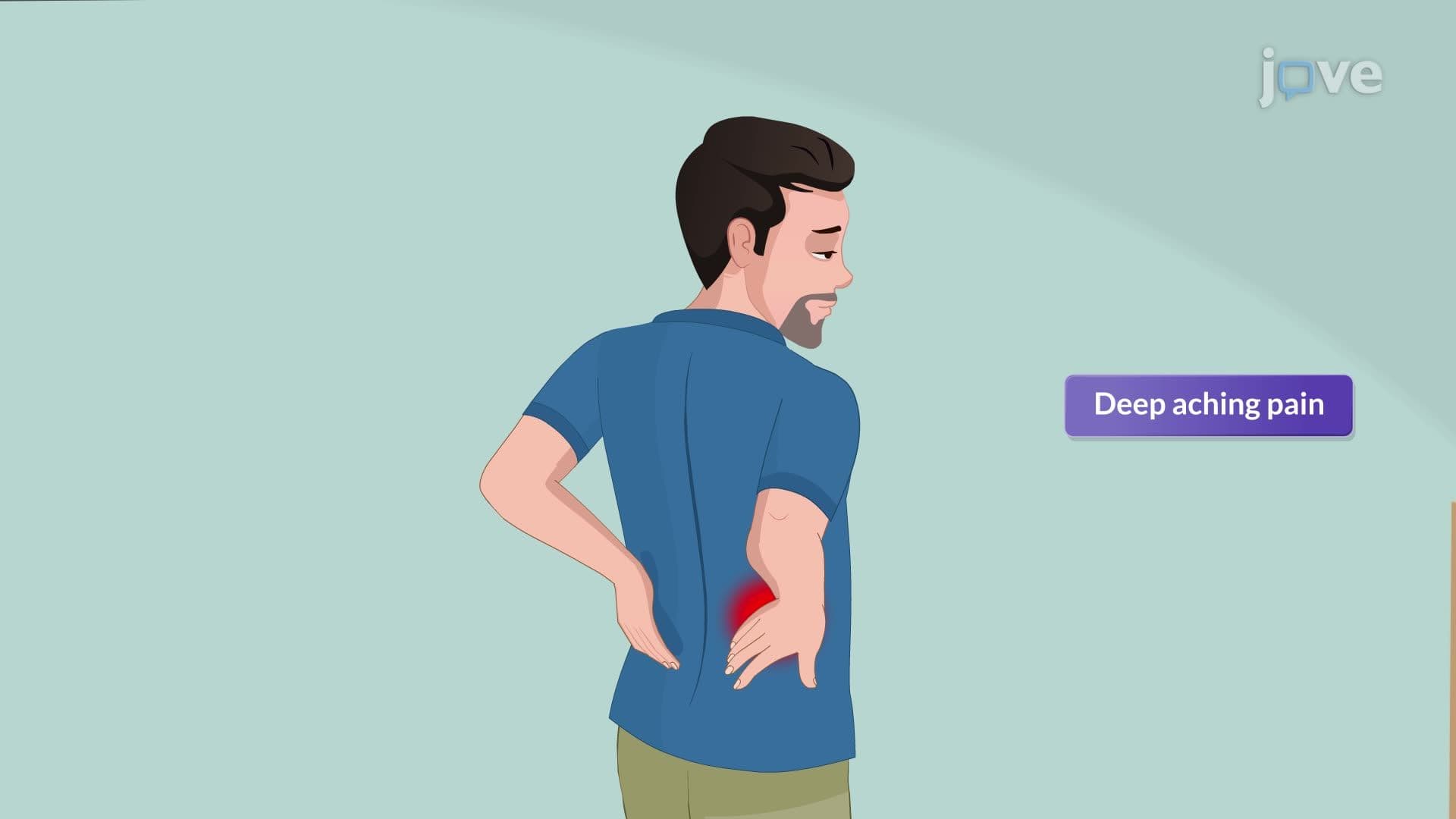

Video Summary: Urinary Tract Calculi Ii Pathophysiology Explained
Did you know that kidney stones affect over 10% of Americans at some point in their lives, with emergency departments seeing thousands of cases annually? Understanding urinary tract calculi ii pathophysiology reveals how mineral concentrations, pH levels, and supersaturation create these painful crystalline formations that can range from microscopic particles to golf ball-sized obstructions. Watch the full video on JoVE Coach to master this concept with expert-led visuals and step-by-step explanations.
The pathophysiology of urinary tract calculi represents a complex interplay of chemical, physical, and anatomical factors that healthcare professionals encounter daily in clinical practice. Students preparing for the MCAT or advanced placement biology exams must grasp these interconnected mechanisms to excel in nephrology-related questions.
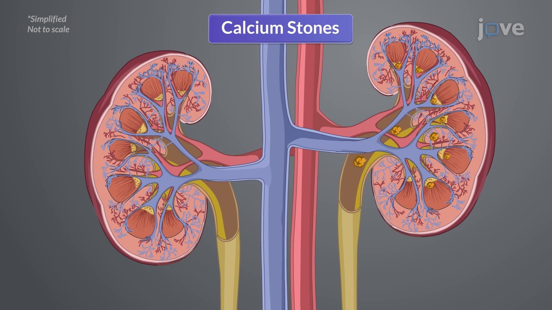
At the cellular level, stone formation begins when urine becomes supersaturated with stone-forming substances. This occurs when the concentration of minerals like calcium oxalate, calcium phosphate, or uric acid exceeds the urine's natural capacity to maintain them in solution. Think of this like adding sugar to iced tea-initially it dissolves, but beyond a certain point, crystals begin forming at the bottom of the glass.
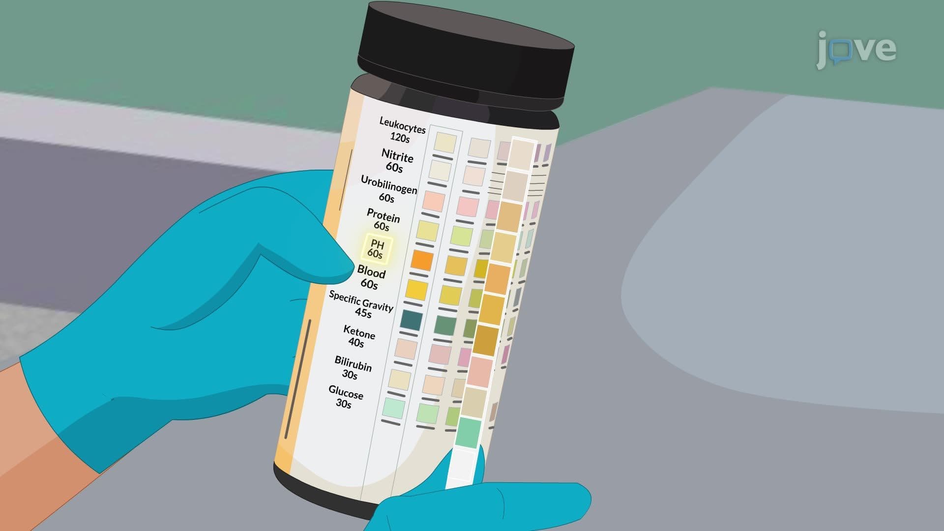
The supersaturation threshold varies significantly based on urine pH. For instance, uric acid stones predominantly form in acidic urine (pH < 5.5), which is why patients at Walter Reed National Military Medical Center with gout often develop these calculi. Conversely, calcium phosphate stones favor alkaline conditions (pH > 7), commonly seen in patients with urinary tract infections involving urease-producing bacteria.
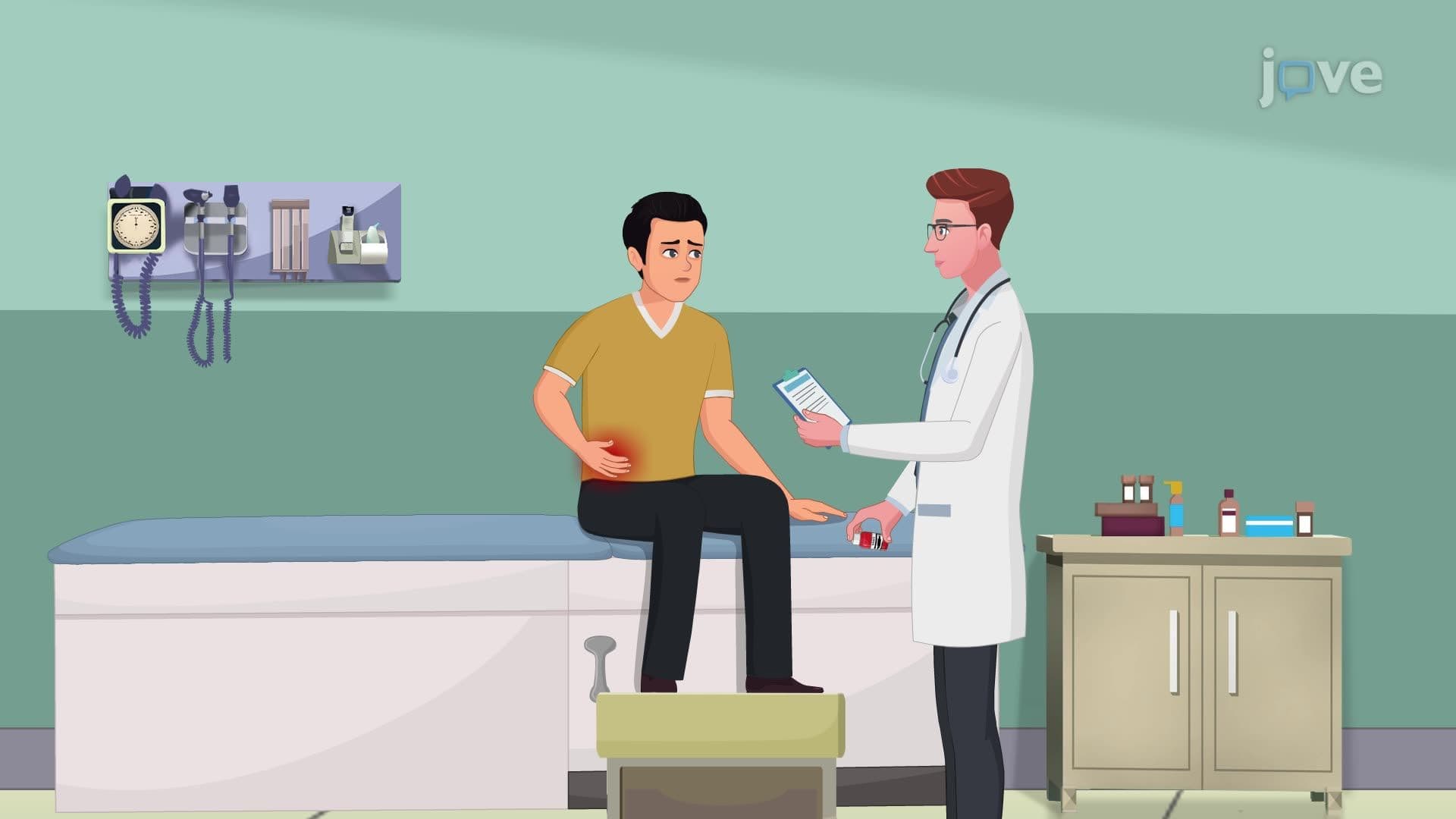
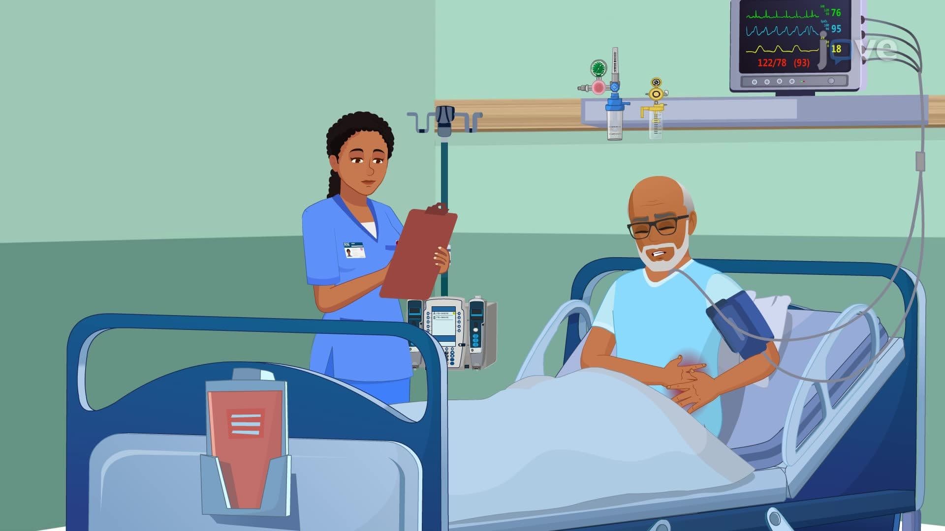
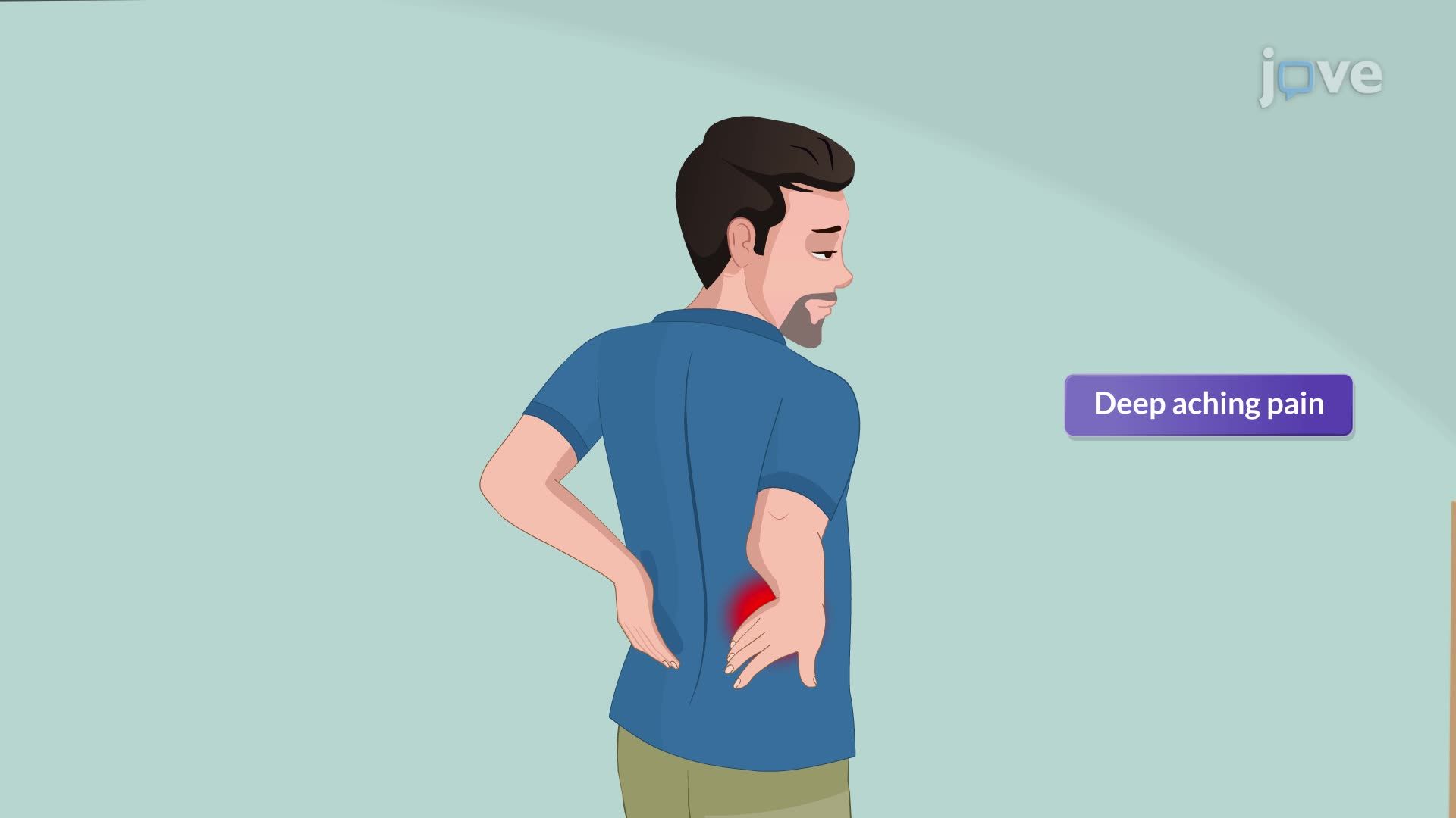
The pathophysiology directly correlates with symptom presentation, a crucial concept for USMLE Step 1 preparation. Renal pelvis stones typically cause deep, aching costovertebral angle pain due to distension of the renal capsule and activation of stretch receptors. This pain pattern helps emergency physicians at institutions like Johns Hopkins distinguish kidney stones from other causes of flank pain.
Ureteral stones present the most dramatic clinical picture due to smooth muscle spasm and obstruction. The severe, colicky pain that radiates from the flank to the groin follows dermatomal patterns, specifically following the path of the genitofemoral nerve. This anatomical correlation explains why male patients often experience testicular pain, while female patients may report labial discomfort.
Understanding these mechanisms guides therapeutic interventions. For example, knowing that calcium oxalate stones form more readily in concentrated urine explains why increased fluid intake remains the cornerstone of prevention. Similarly, the pH-dependent solubility of different stone types informs medical management-potassium citrate therapy alkalinizes urine to prevent uric acid stone formation, while thiazide diuretics reduce calcium excretion for calcium stone prevention.
Related Micro-courses