Loading subject page…

- Anatomy and Physiology
- The Axial Skeleton
- Cranial Bones: Superior and Posterior View
Micro-course: The Axial Skeleton
57,801 views
Learn key concepts and earn your certificate.


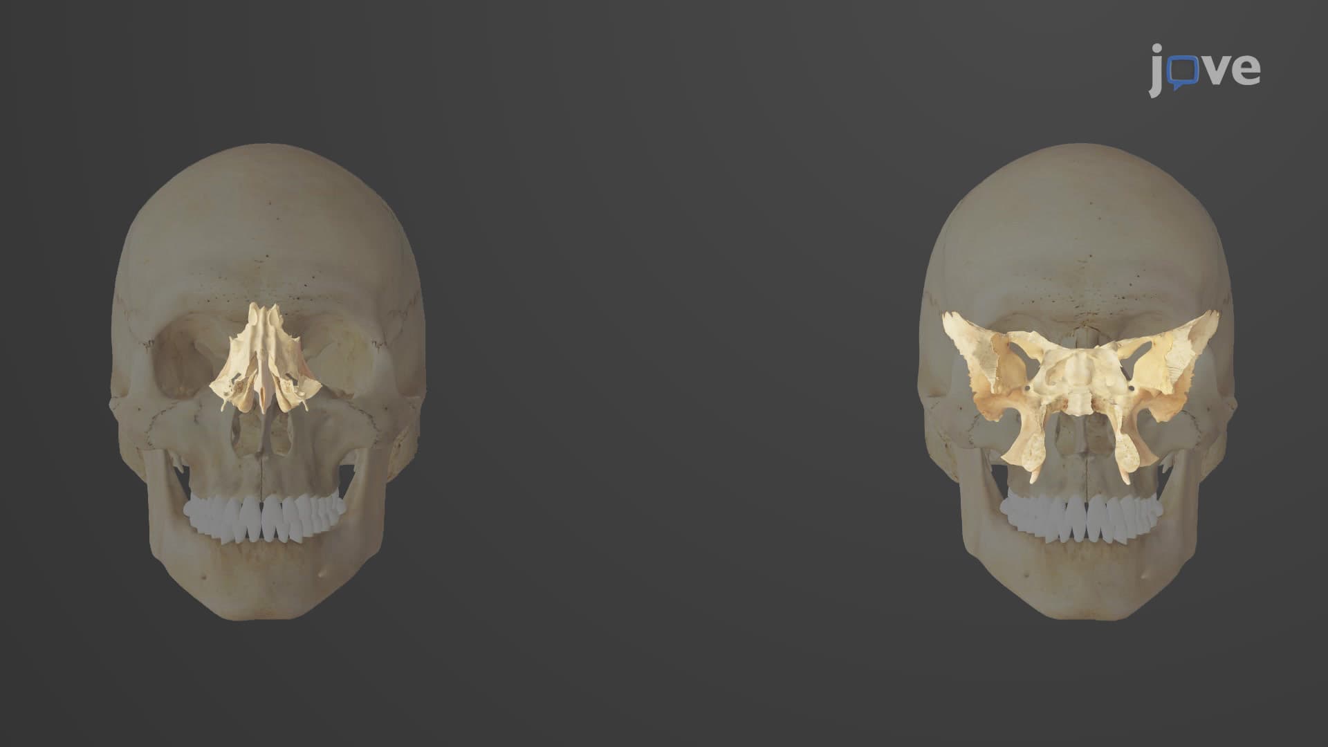
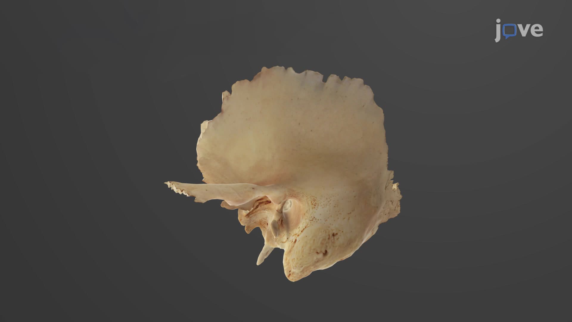


Cranial Bones: Superior and Posterior View

00:00
01:14
Video Summary: Cranial Bones Superior and Posterior View Explained
Ever wondered how forensic scientists at the FBI identify human remains using just skull fragments? Understanding cranial bones superior posterior views is crucial for recognizing the distinctive features that make each skull unique. From the prominent frontal bone forming your forehead to the occipital bone's external protuberance where neck muscles attach, the skull bones top back view reveals intricate anatomical landmarks. Medical students at Johns Hopkins University master these cranial vault features as essential foundations for neuroanatomy. Watch the full video on JoVE Coach to master this concept with expert-led visuals and step-by-step explanations.
- Understand the anatomical features visible in Cranial Bones Superior And Posterior View Explained and their functional significance
- Identify the four primary bones forming the cranial vault: frontal, paired parietals, and occipital
- Analyze the three major sutures connecting cranial bones: coronal, sagittal, and lambdoid
- Recognize key landmarks including the glabella, external occipital protuberance, and nuchal lines
- Compare the structural differences between superior and posterior cranial perspectives
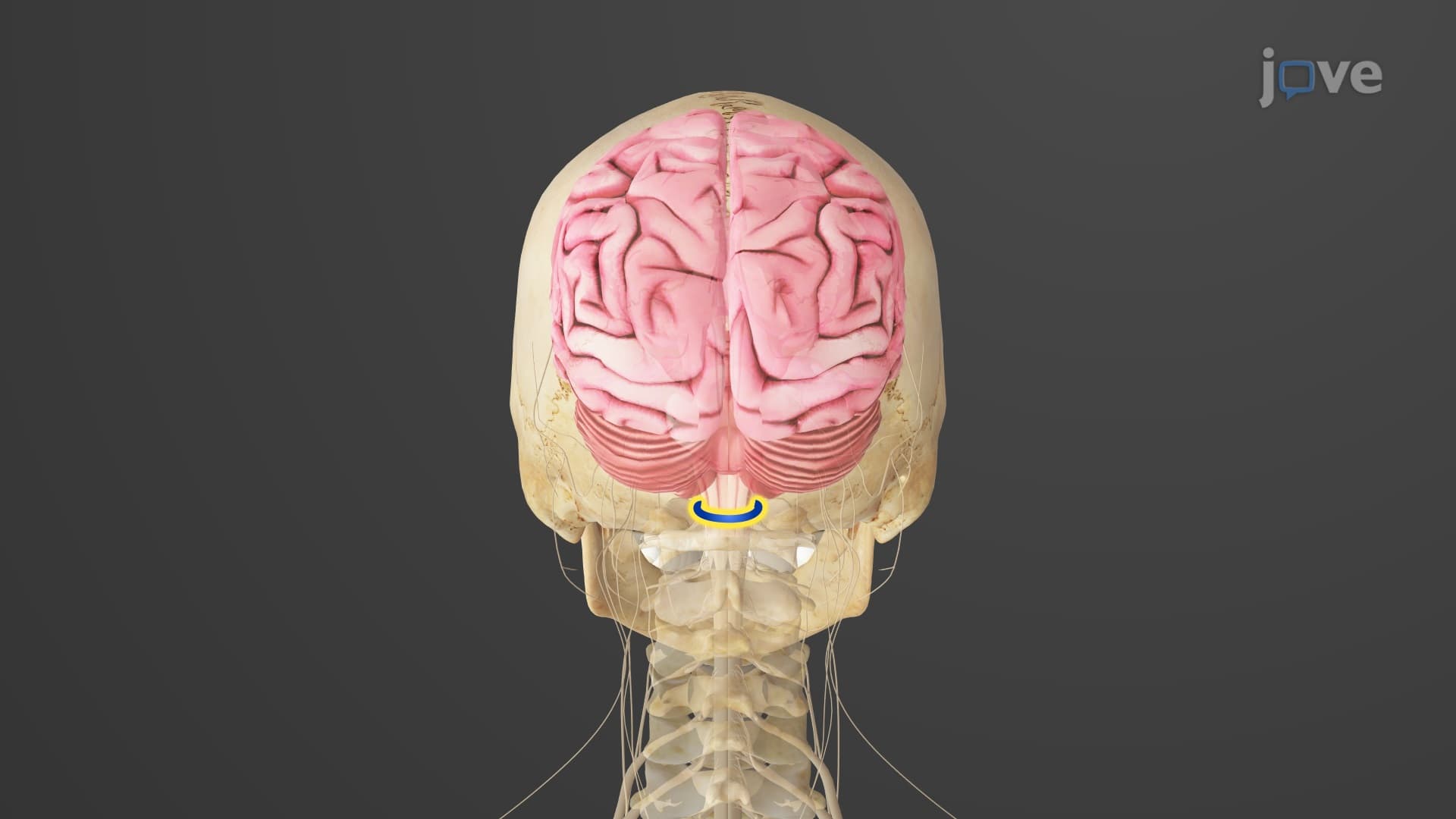
- Apply knowledge of foramen magnum and occipital condyles to understand brain-spinal cord connections
- Evaluate the relationship between bone markings and muscle attachment sites
- Explore how cranial bone identification applies to forensic science and medical diagnosis
Understanding Cranial Bones Superior And Posterior View Explained
The human skull's superior and posterior perspectives reveal the sophisticated architecture of our cranial vault. These viewpoints showcase how multiple bones seamlessly integrate to protect the brain while providing attachment points for muscles and ligaments. For students preparing for the MCAT or Advanced Placement Biology exams, mastering these anatomical relationships forms the foundation for understanding neuroanatomy and head trauma mechanisms.
Superior View: The Cranial Vault's Top Architecture
From above, the skull presents a distinctive pattern dominated by four key bones. The frontal bone creates the forehead's characteristic shape, featuring thick supraorbital margins that protect the eyes from impact. Between these margins lies the glabella, a smooth area that varies significantly between individuals-a feature forensic anthropologists use for identification at crime labs across the United States.
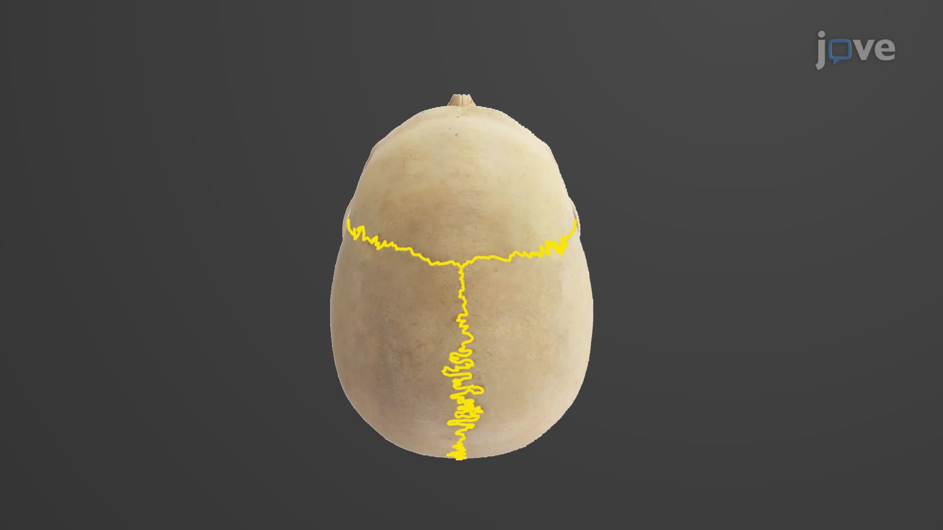
The frontal bone connects posteriorly to the paired parietal bones via the coronal suture, named for its crown-like position. These parietal bones form most of the skull's top and sides, meeting at the midline sagittal suture. This suture pattern is crucial for neurosurgeons at medical centers like Mayo Clinic when planning surgical approaches to brain tumors.
Posterior View: Occipital Bone Dominance and Muscle Attachments
The skull's back view is dominated by the occipital bone, which forms both the posterior cranium and much of the skull base. The lambdoid suture connects the occipital to the parietal bones, creating a distinctive zigzag pattern that helps identify skull fragments in archaeological digs throughout the American Southwest.
The external occipital protuberance serves as the attachment point for the ligamentum nuchae, a powerful elastic ligament supporting head posture. This feature is particularly pronounced in individuals who perform heavy lifting or contact sports, making it relevant for sports medicine physicians treating football players in the NFL.
Clinical and Practical Applications
Understanding these cranial landmarks proves essential in multiple professional contexts. Emergency room physicians at hospitals like Johns Hopkins use these reference points when interpreting CT scans for head trauma patients. The superior and inferior nuchal lines guide plastic surgeons during neck lift procedures, while the foramen magnum's relationship to occipital condyles helps chiropractors understand cervical spine mechanics.
For students taking anatomy courses at universities like UCLA or preparing for nursing entrance exams like the HESI A2, these bone relationships frequently appear in practical examinations where specimens must be identified and labeled under time pressure.
Frequently Asked Questions
These anatomical perspectives show the skull from above (superior) and behind (posterior), revealing how frontal, parietal, and occipital bones connect through sutures. They're essential for understanding brain protection mechanisms and appear frequently on MCAT anatomy sections and college practical exams.
The superior view primarily shows frontal and parietal bones connected by coronal and sagittal sutures. The posterior view is dominated by the occipital bone with its external protuberance and nuchal lines, connected to parietals via the lambdoid suture.
MCAT passages often test suture identification, bone landmarks, and functional relationships between skull features and muscle attachments. Questions may include CT scan interpretation or require matching anatomical structures to their functions in head support and brain protection.
Use physical skull models to trace sutures with your finger while naming each bone aloud. Create flashcards pairing bone landmarks with their functions, and practice drawing superior and posterior views from memory to reinforce spatial relationships.
Emergency physicians use these landmarks to interpret head trauma imaging at trauma centers nationwide. Neurosurgeons rely on suture patterns for surgical planning, while forensic pathologists at FBI labs use bone features for victim identification in criminal investigations.
Not at all-cranial bone anatomy builds logically from basic directional terms you already know. Start with understanding superior means "above" and posterior means "behind," then gradually add bone names and their connections through consistent practice with visual aids.
Progress to lateral skull views to understand temporal bones and zygomatic arches, then explore the skull base (inferior view) to learn about foramina and their contents. This sequence prepares you for advanced neuroanatomy courses and clinical applications.
Related Micro-courses