Loading concepts…


14 Concepts
6 Concepts
21 Concepts
23 Concepts
21 Concepts
15 Concepts
16 Concepts
26 Concepts
18 Concepts
19 Concepts
11 Concepts
6 Concepts
10 Concepts
22 Concepts
20 Concepts
26 Concepts
26 Concepts
14 Concepts
10 Concepts
12 Concepts
23 Concepts
29 Concepts
19 Concepts
38 Concepts
38 Concepts
25 Concepts
40 Concepts
25 Concepts
25 Concepts
20 Concepts
37 Concepts
 22 Concepts
 178.8K Views
 Popular topics


Muscle tissue represents one of the four fundamental tissue types in the human body, comprising specialized cells capable of contraction to enable movement, maintain posture, and support vital physiological functions. This comprehensive course examines the three distinct types of muscle tissue-skeletal, cardiac, and smooth muscle-exploring their unique structural characteristics, molecular mechanisms of contraction, and clinical significance. From the intricate sarcomere structure of skeletal muscle to the autorhythmic properties of cardiac tissue, students will master the complex interactions between myosin, actin, and regulatory proteins that drive muscle function through JoVE Coach's systematic approach.
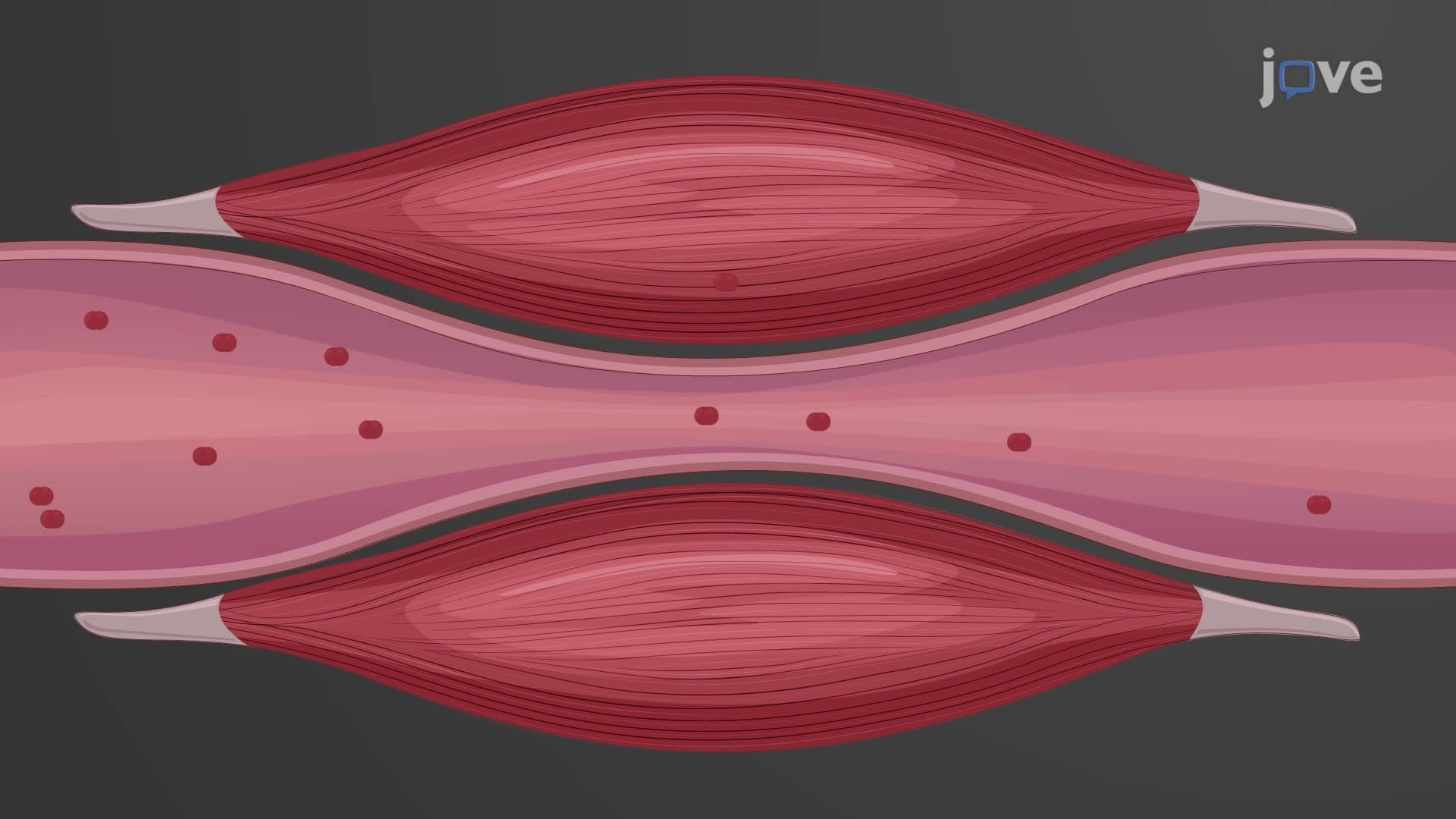
1. Muscle Tissue Classification and General Properties The human body contains three distinct types of muscle tissue, each specialized for specific functions. Skeletal muscle tissue enables voluntary movement and posture maintenance through striated, multinucleated fibers attached to bones via tendons. Cardiac muscle tissue, found exclusively in the heart, displays striations but functions involuntarily with autorhythmic contractions to pump blood throughout the cardiovascular system. Smooth muscle tissue lines hollow organs and blood vessels, exhibiting non-striated appearance and involuntary contractions that control substance passage through body systems. All muscle types contribute to thermoregulation through heat generation during contraction, with skeletal muscle providing the most significant contribution to maintaining normal body temperature of 98.6°F.
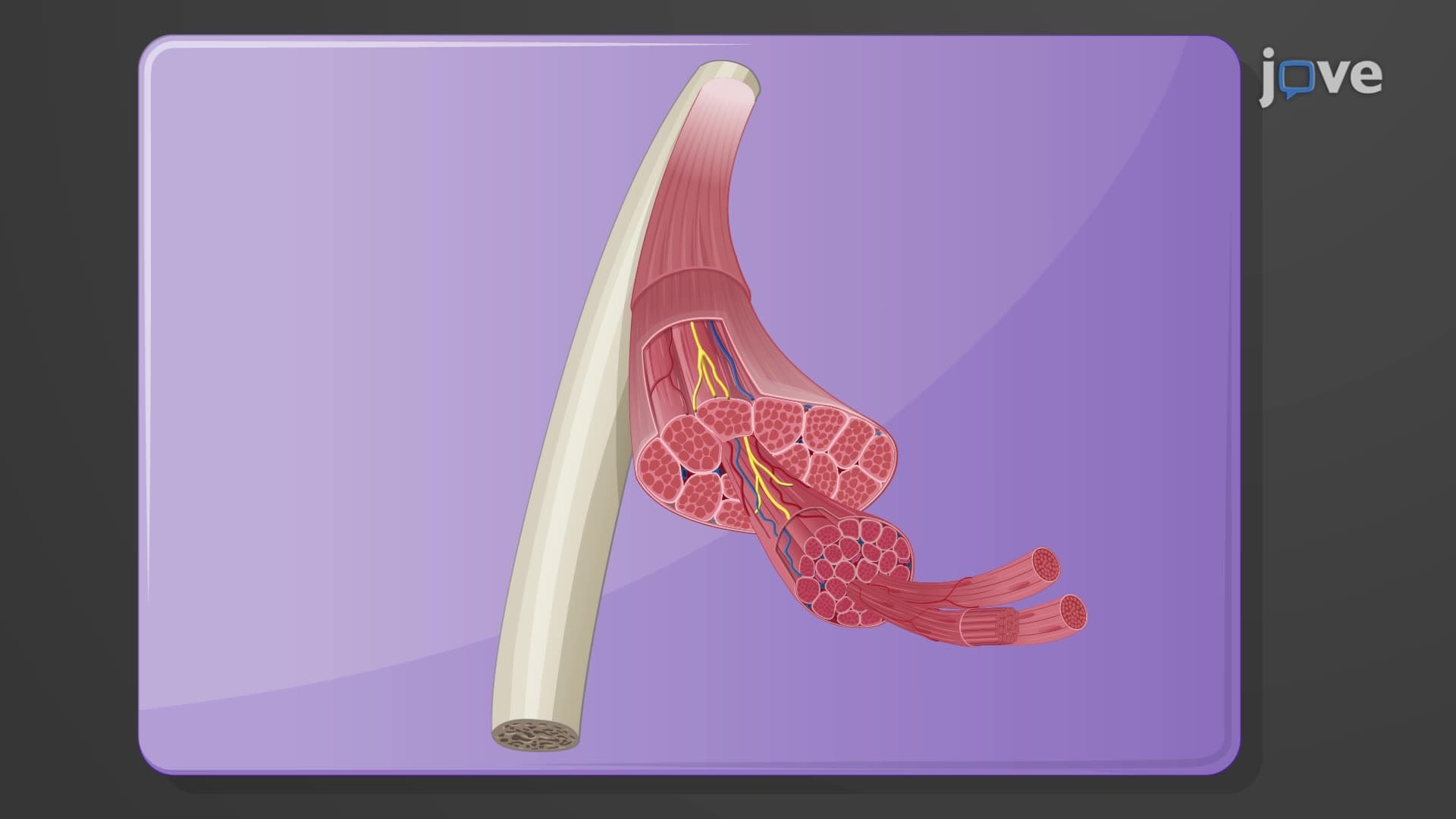
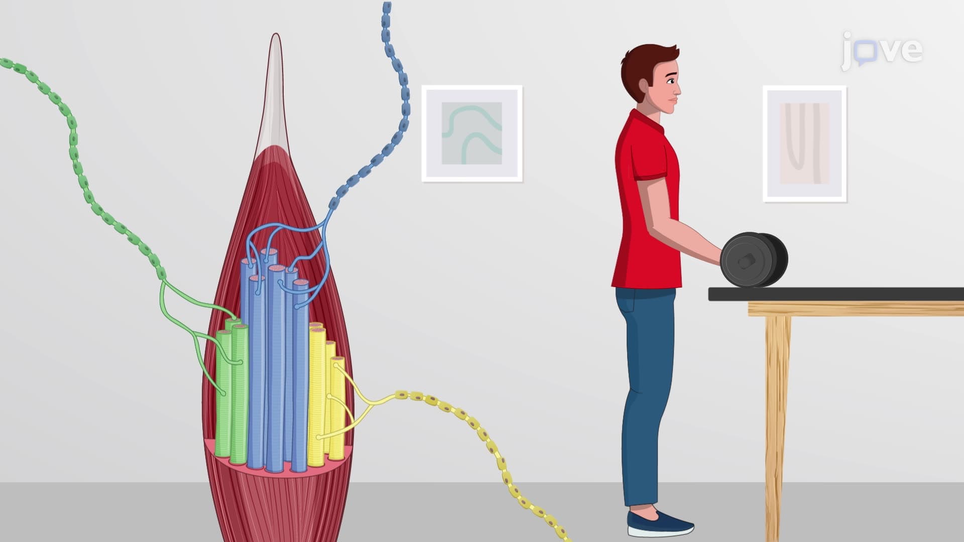
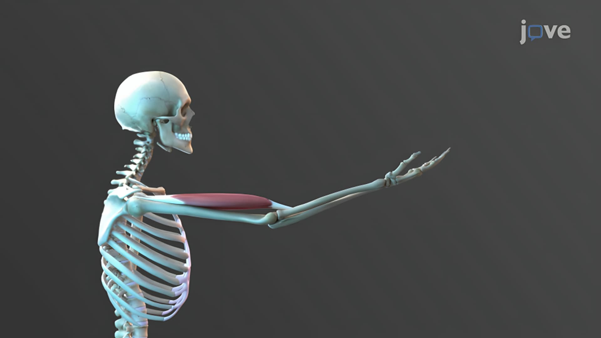
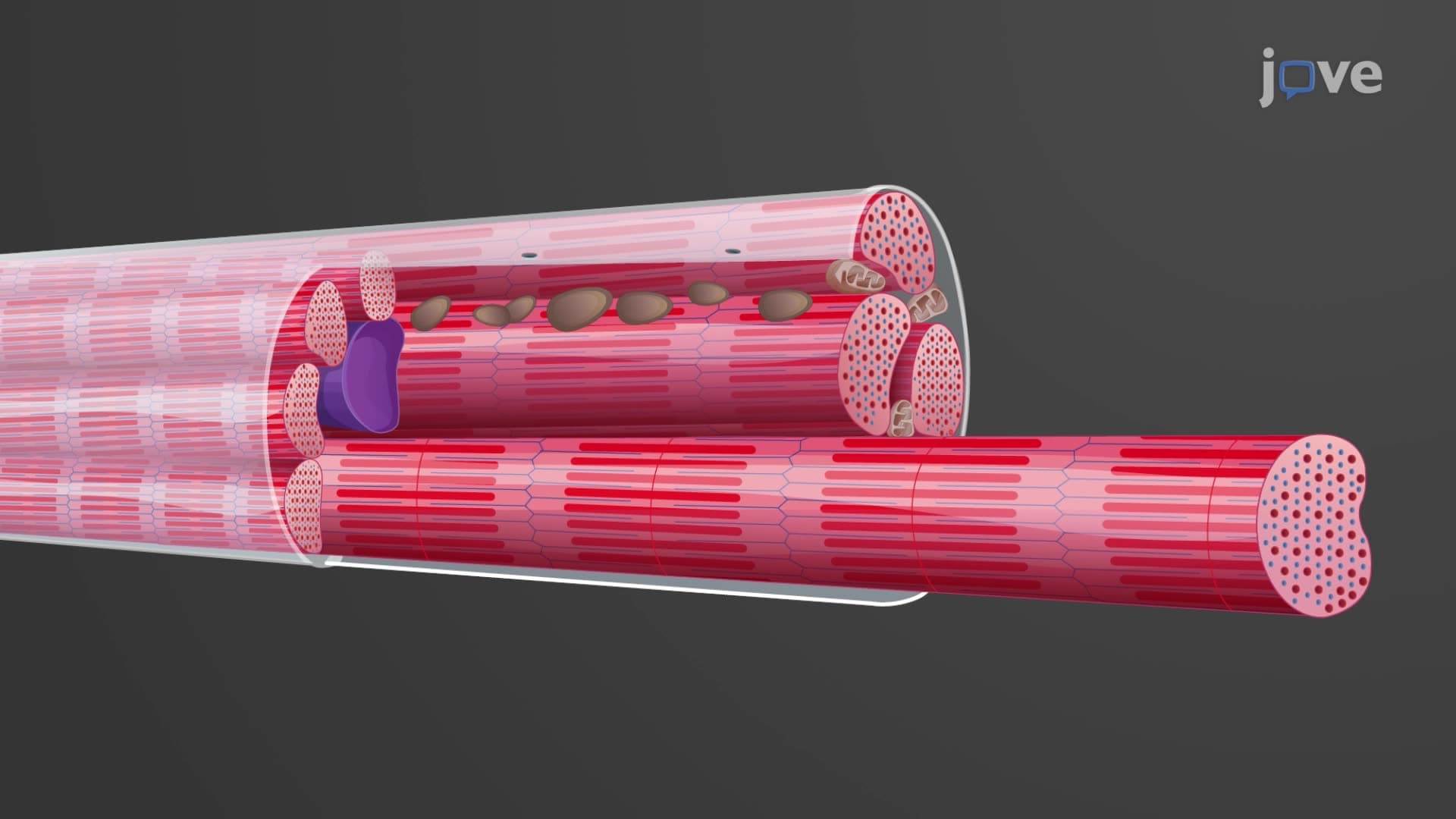
2. Skeletal Muscle Gross Anatomy and Connective Tissue Organization Skeletal muscles exhibit a highly organized structure with three distinct connective tissue layers that provide support, protection, and force transmission. The epimysium surrounds the entire muscle, while the perimysium groups muscle fibers into fascicles, and the endomysium wraps individual muscle fibers. These continuous connective tissue sheaths extend beyond the muscle to form tendons (rope-like structures) or aponeuroses (sheet-like structures) that attach muscles to bones. During contraction, force generated by muscle fibers transmits through these connective tissue layers to move skeletal structures. This organization is clearly demonstrated in muscles like the biceps brachii, where the tendon attachment to the radius bone enables forearm flexion during activities such as lifting weights or performing pull-ups in American fitness centers.
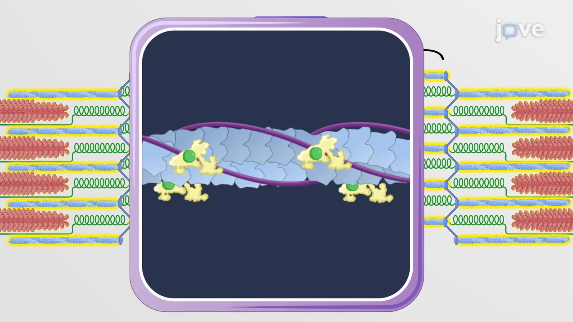
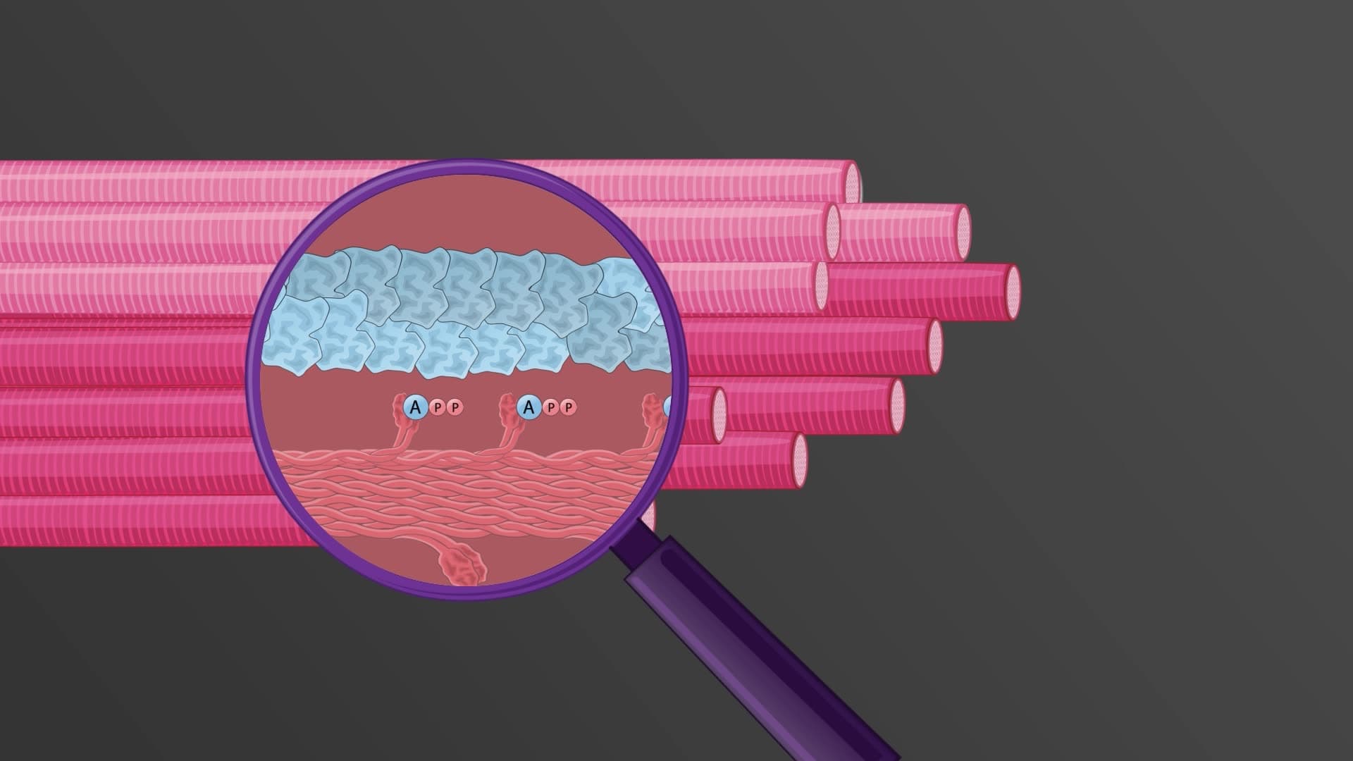
3. Sarcomere Structure and Sliding Filament Mechanism The sarcomere represents the fundamental contractile unit of striated muscle, containing precisely arranged thick (myosin) and thin (actin, troponin, tropomyosin) filaments. The characteristic banding pattern includes the A band (containing myosin), I band (containing only actin), H zone (myosin only), and Z-discs (sarcomere boundaries). Titin proteins provide elasticity and anchor thick filaments, while the M line connects adjacent myosin filaments. During contraction, myosin heads bind to actin filaments in the presence of calcium ions, forming cross-bridges that use ATP hydrolysis to slide filaments past each other. This sliding mechanism shortens sarcomeres without changing individual filament lengths, generating the force necessary for muscle contraction. Understanding this mechanism is crucial for MCAT questions about muscle physiology and ATP utilization in cellular processes.
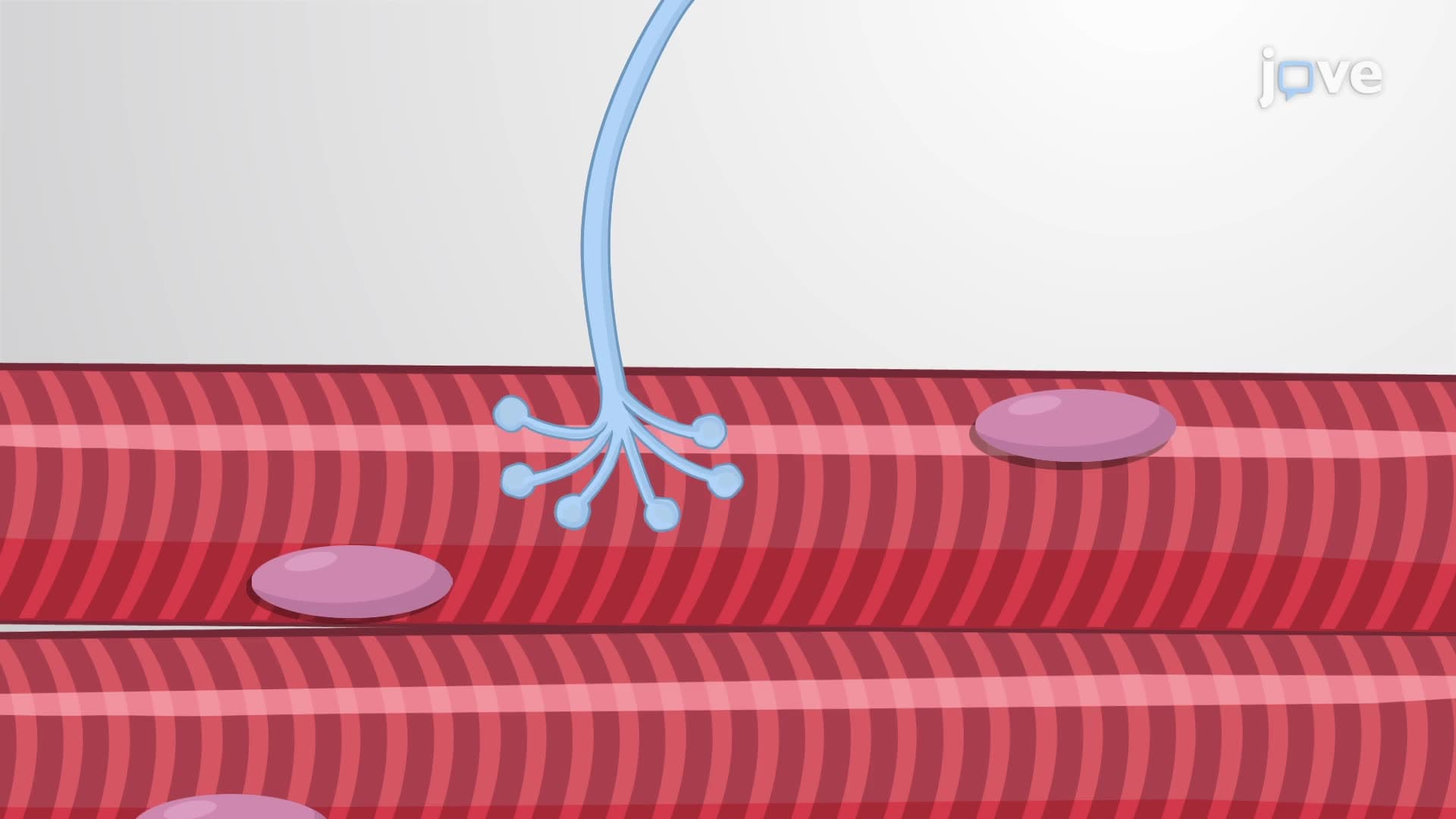
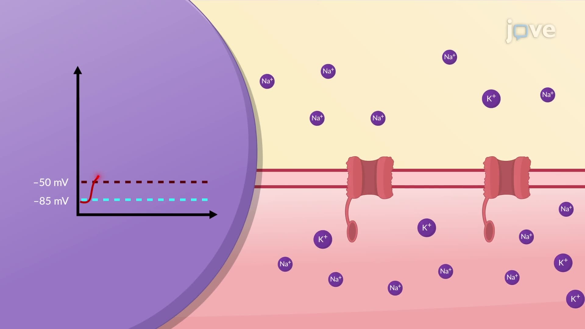
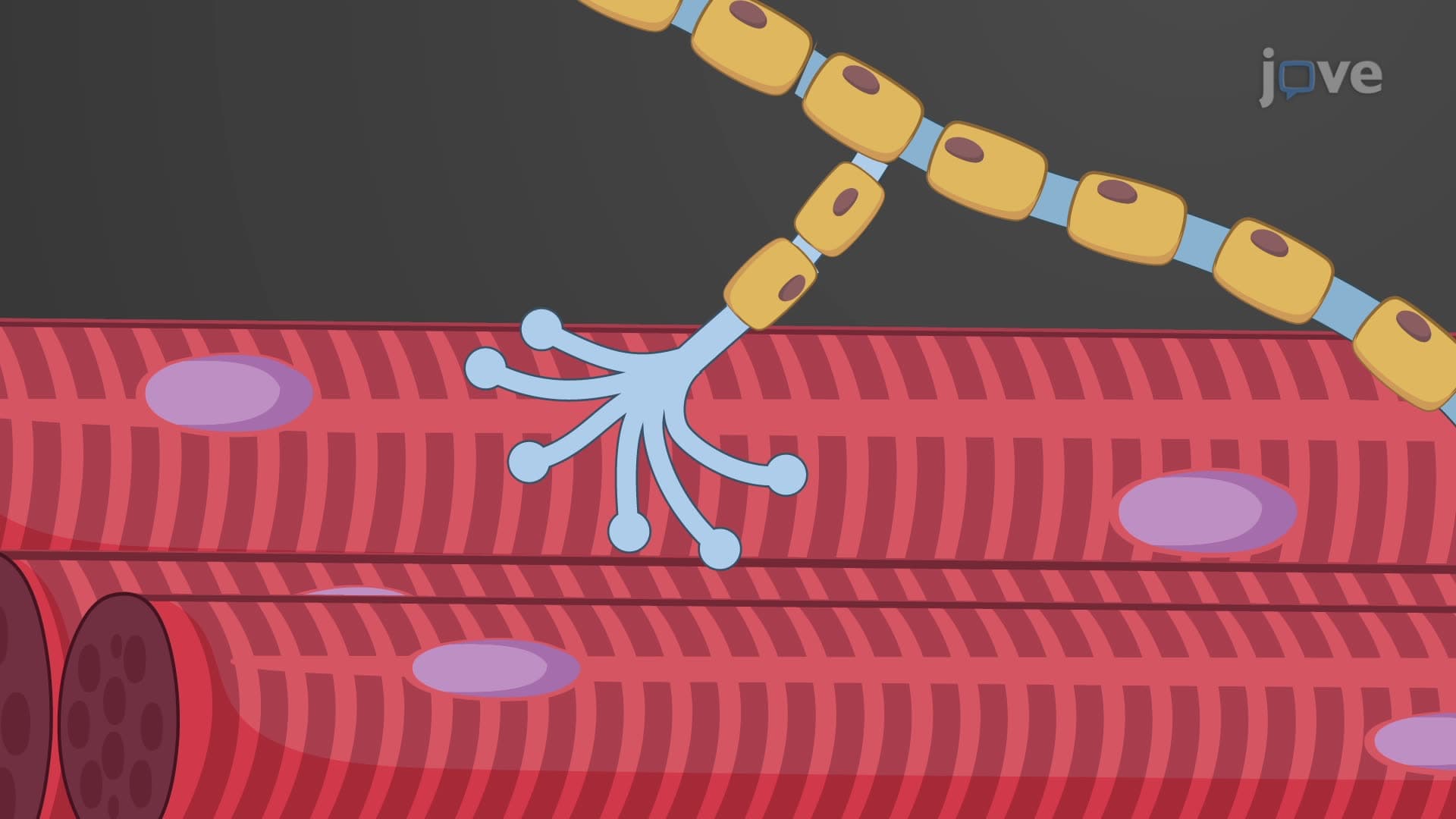
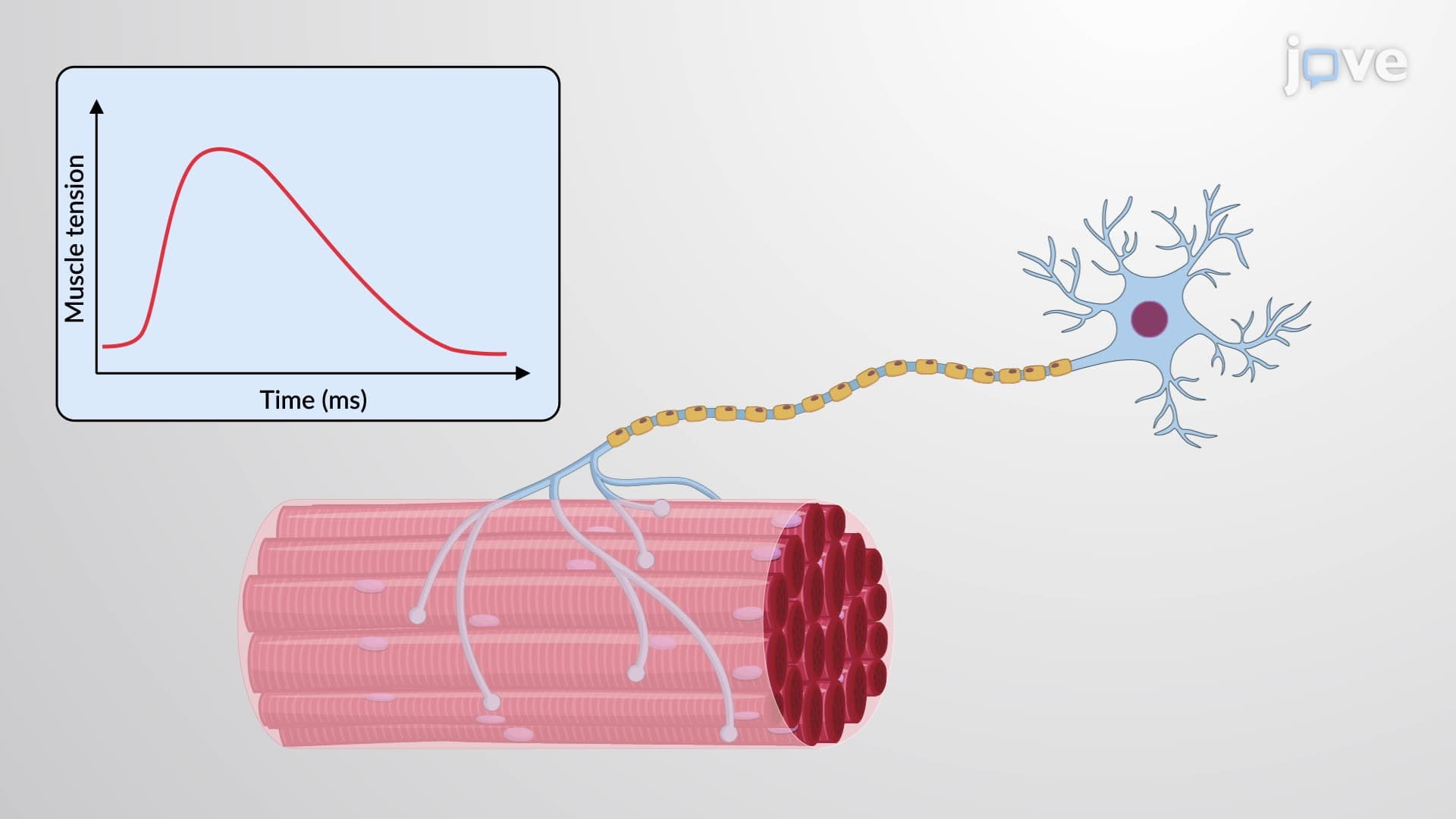
4. Neuromuscular Junction and Action Potential Generation The neuromuscular junction represents a specialized synapse where somatic motor neurons communicate with skeletal muscle fibers through acetylcholine release. Motor neurons originating in the spinal cord extend axons that branch to innervate multiple muscle fibers, with synaptic end bulbs releasing acetylcholine into the synaptic cleft. When acetylcholine binds to nicotinic receptors on the motor end plate, sodium ion influx causes local depolarization. If the threshold potential (-50 to -55 mV) is reached, voltage-gated sodium channels open, generating an action potential that propagates along the entire muscle fiber. This electrical signal initiates the contraction process, demonstrating the integration of nervous and muscular systems. Disruption of this process occurs in conditions like myasthenia gravis, commonly tested on USMLE examinations.
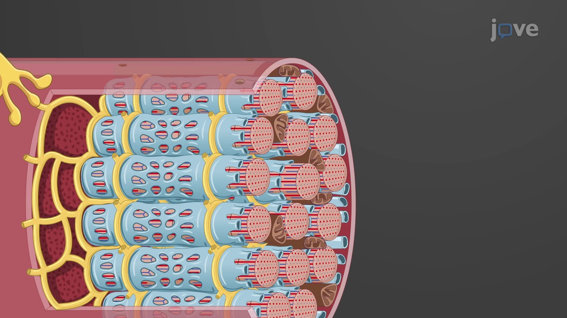
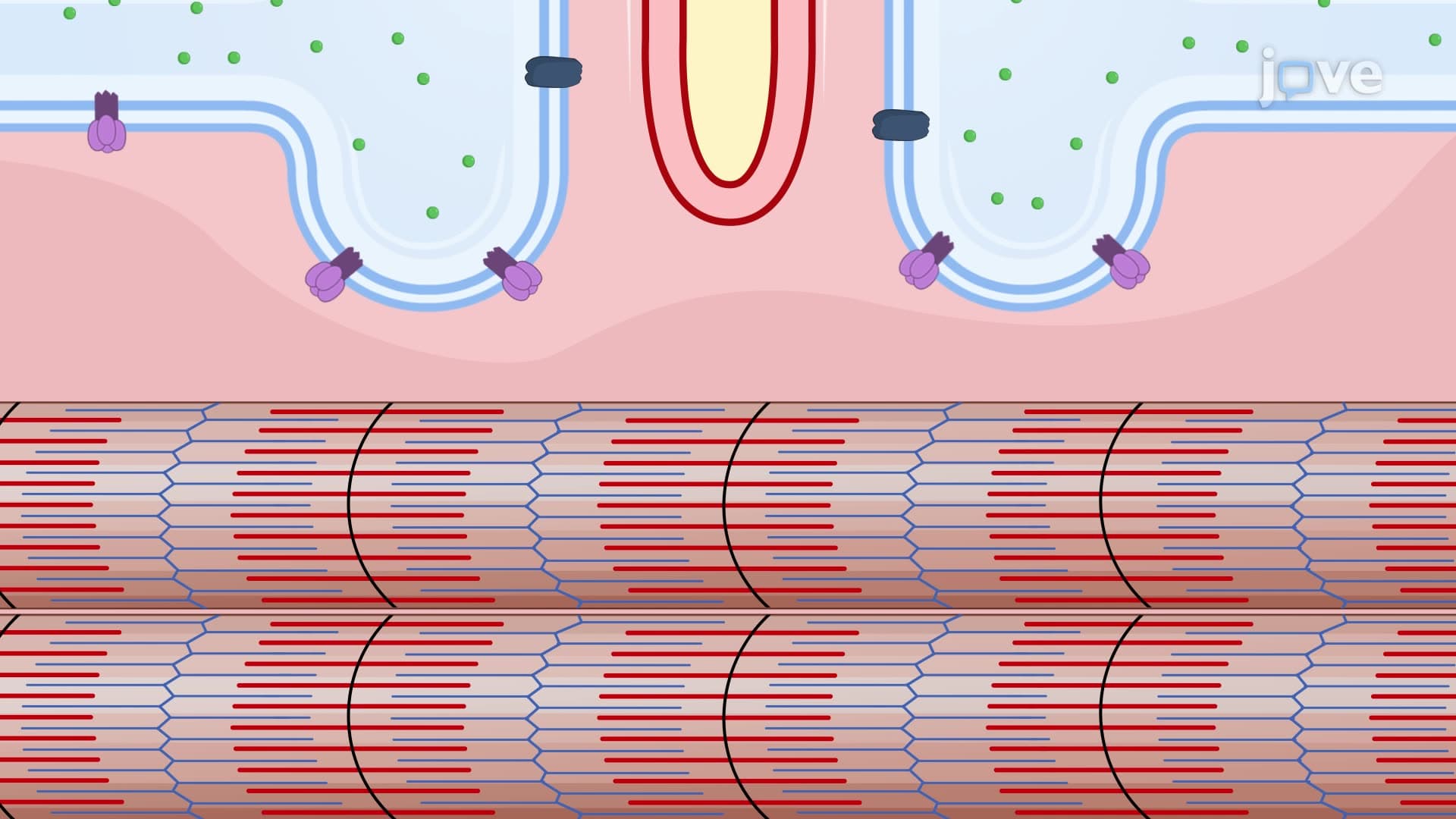
5. Excitation-Contraction Coupling and Calcium Regulation Excitation-contraction coupling links electrical stimulation to mechanical contraction through calcium ion regulation within muscle fibers. Action potentials traveling along T-tubules trigger calcium release from the sarcoplasmic reticulum into the sarcoplasm. Calcium ions bind to troponin, causing tropomyosin to shift and expose myosin-binding sites on actin filaments. This enables cross-bridge formation and subsequent muscle contraction through the sliding filament mechanism. During relaxation, calcium ATPase pumps actively transport calcium back into the sarcoplasmic reticulum, allowing tropomyosin to block binding sites and terminate contraction. This process requires significant ATP expenditure, explaining why muscle fatigue occurs during prolonged exercise sessions common in American high school and college athletics programs.
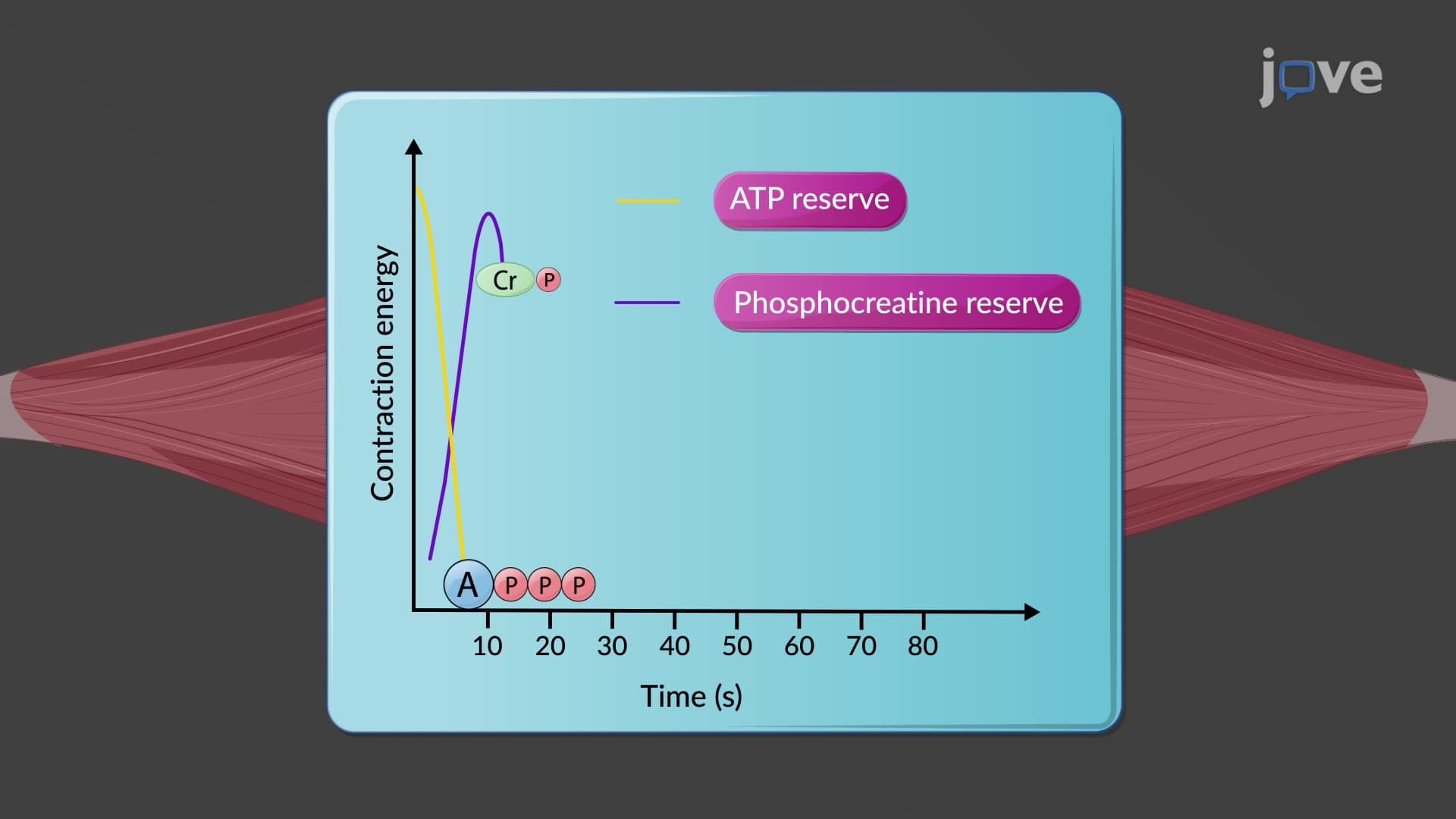
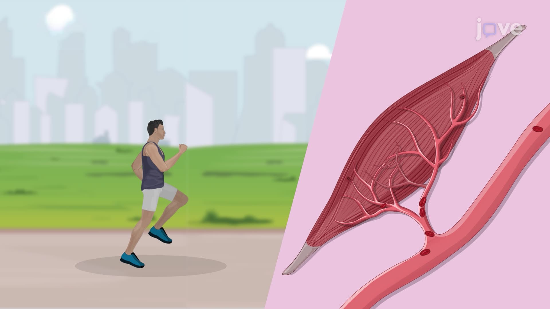
6. Energy Metabolism and Muscle Fatigue Muscle contraction demands enormous ATP quantities, supplied through three distinct pathways: phosphocreatine system, aerobic respiration, and anaerobic glycolysis. The phosphocreatine system provides immediate energy for initial seconds of contraction, utilizing creatine kinase to transfer phosphate groups from phosphocreatine to ADP. Aerobic respiration in mitochondria produces abundant ATP when oxygen supply is adequate, breaking down glucose and fatty acids completely. During intense exercise when oxygen becomes limiting, anaerobic glycolysis provides rapid ATP production but generates lactic acid as a byproduct. Lactic acid accumulation causes muscle fatigue and the characteristic "burn" experienced during high-intensity workouts. Recovery involves converting lactate back to pyruvate and replenishing phosphocreatine stores, explaining the rest periods needed between sets in weightlifting routines popular in American gyms.
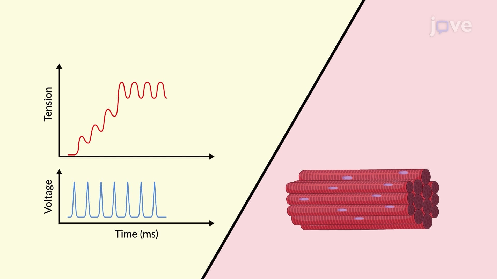
7. Motor Units and Graded Muscle Contractions Motor units consist of a single motor neuron and all muscle fibers it innervates, ranging from few fibers in precision muscles (like those controlling eye movement) to hundreds in powerful muscles (like the quadriceps used in American football). Muscle contraction strength is regulated through motor unit recruitment and stimulation frequency. Low-frequency stimulation produces individual twitches, while increased frequency causes wave summation, incomplete tetanus (20-30 Hz), or complete tetanus (80-100 Hz). This graded response system allows precise force control, from delicate movements required for writing standardized tests like the SAT to powerful contractions needed for activities like shot put throwing in track and field competitions common in American high schools.
8. Skeletal Muscle Fiber Types and Exercise Adaptations Skeletal muscles contain different fiber types optimized for specific activities: slow oxidative (Type I), fast oxidative-glycolytic (Type IIa), and fast glycolytic (Type IIx) fibers. Slow oxidative fibers excel in endurance activities like marathon running, utilizing aerobic respiration and showing high fatigue resistance. Fast oxidative-glycolytic fibers support moderate-intensity activities requiring both strength and endurance, such as middle-distance running or basketball. Fast glycolytic fibers generate maximum force for brief periods, powering explosive movements like sprinting or powerlifting. Exercise training adaptations include increased mitochondrial density and capillarization for endurance training, and fiber hypertrophy with enhanced glycolytic capacity for resistance training. Understanding these adaptations explains why American athletes specialize in specific sports and training regimens.
9. Cardiac Muscle Structure and Autorhythmic Function Cardiac muscle exhibits unique structural features that enable its specialized function in blood circulation. Cardiomyocytes are smaller, typically mononucleated, and interconnected through intercalated discs containing gap junctions and desmosomes. Gap junctions facilitate rapid electrical conduction, while desmosomes prevent cell separation during forceful contractions. Pacemaker cells in the sinoatrial node generate spontaneous action potentials approximately 75 times per minute, creating the intrinsic heart rate independent of nervous stimulation. The cardiac muscle's extended refractory period prevents tetanic contractions, ensuring adequate ventricular filling between beats. This autorhythmic property explains why transplanted hearts function without nervous connections, a concept frequently tested on MCAT cardiology questions and relevant to understanding cardiac pathophysiology in American healthcare settings.
10. Smooth Muscle Organization and Contraction Mechanisms Smooth muscle tissue exists in two organizational patterns: visceral (single-unit) smooth muscle in hollow organs like the intestines and blood vessels, and multi-unit smooth muscle in structures requiring precise control like the iris and large arteries. Smooth muscle cells lack organized sarcomeres, giving them a non-striated appearance, with actin and myosin filaments arranged irregularly throughout the cytoplasm. Contraction occurs through calcium-calmodulin activation of myosin light chain kinase, which phosphorylates myosin heads to enable cross-bridge formation. The slow calcium removal process creates sustained contractions that maintain organ tone, such as the constant tension in arteriolar walls that helps regulate blood pressure. This mechanism is essential for understanding hypertension treatment approaches commonly used in American cardiovascular medicine and frequently appears on NCLEX examinations.