Loading video…


 22 Concepts
 178.8K views

Video Summary: What Is Overview of Muscle Tissues
Why does your heart beat 100,000 times daily without you thinking about it, while you consciously flex your biceps at the gym? This muscle tissue overview biology reveals the fascinating differences between three distinct muscle types that power everything from a marathon runner's stride in the Boston Marathon to automatic digestion. Understanding what is an overview of muscle tissues in biology unlocks how your body coordinates voluntary movements with life-sustaining involuntary functions. Watch the full video on JoVE Coach to master this concept with expert-led visuals and step-by-step explanations.
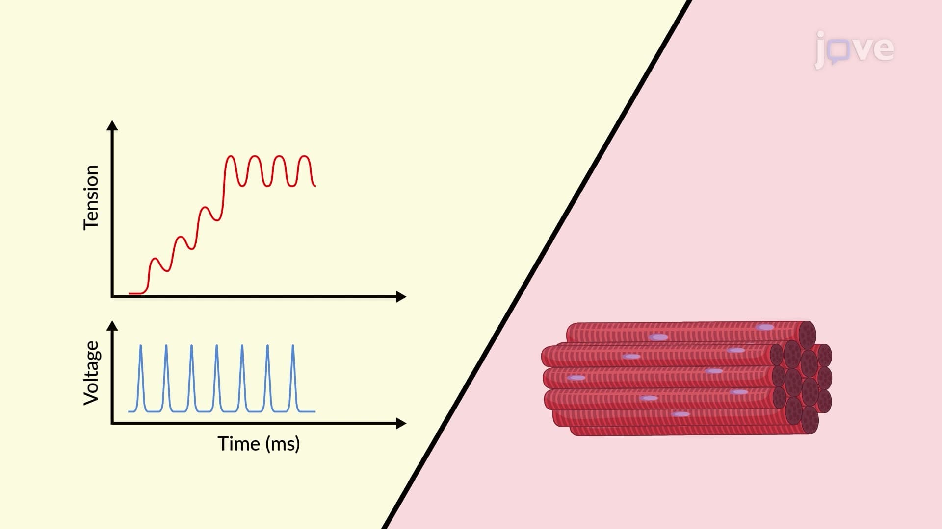
Muscle tissue overview biology forms a cornerstone of human physiology, representing one of four fundamental tissue types that enable complex multicellular life. These specialized tissues share the unique ability to contract and generate force, yet their diverse structures and control mechanisms allow for remarkably different functions throughout the body.
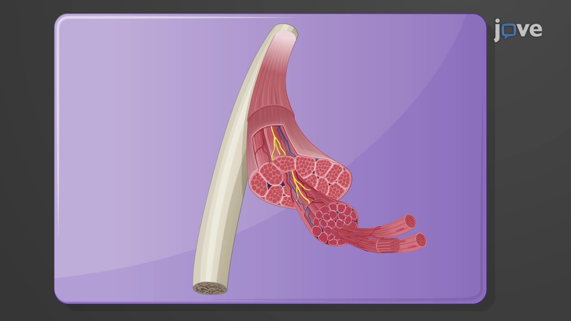
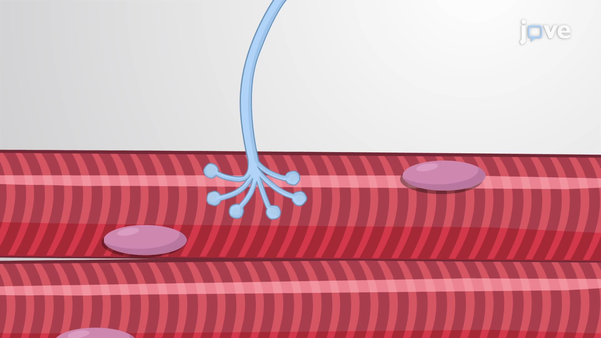
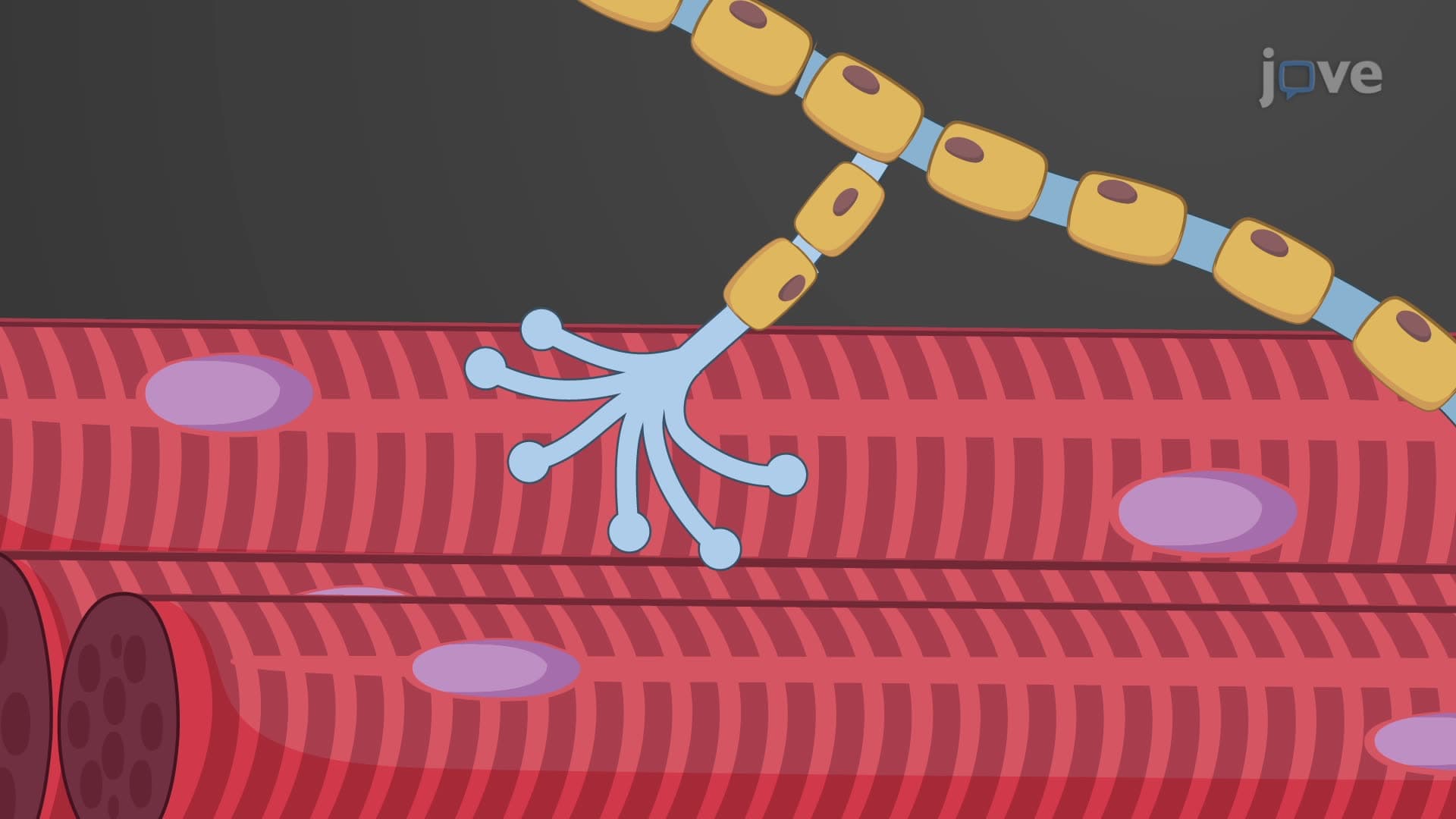
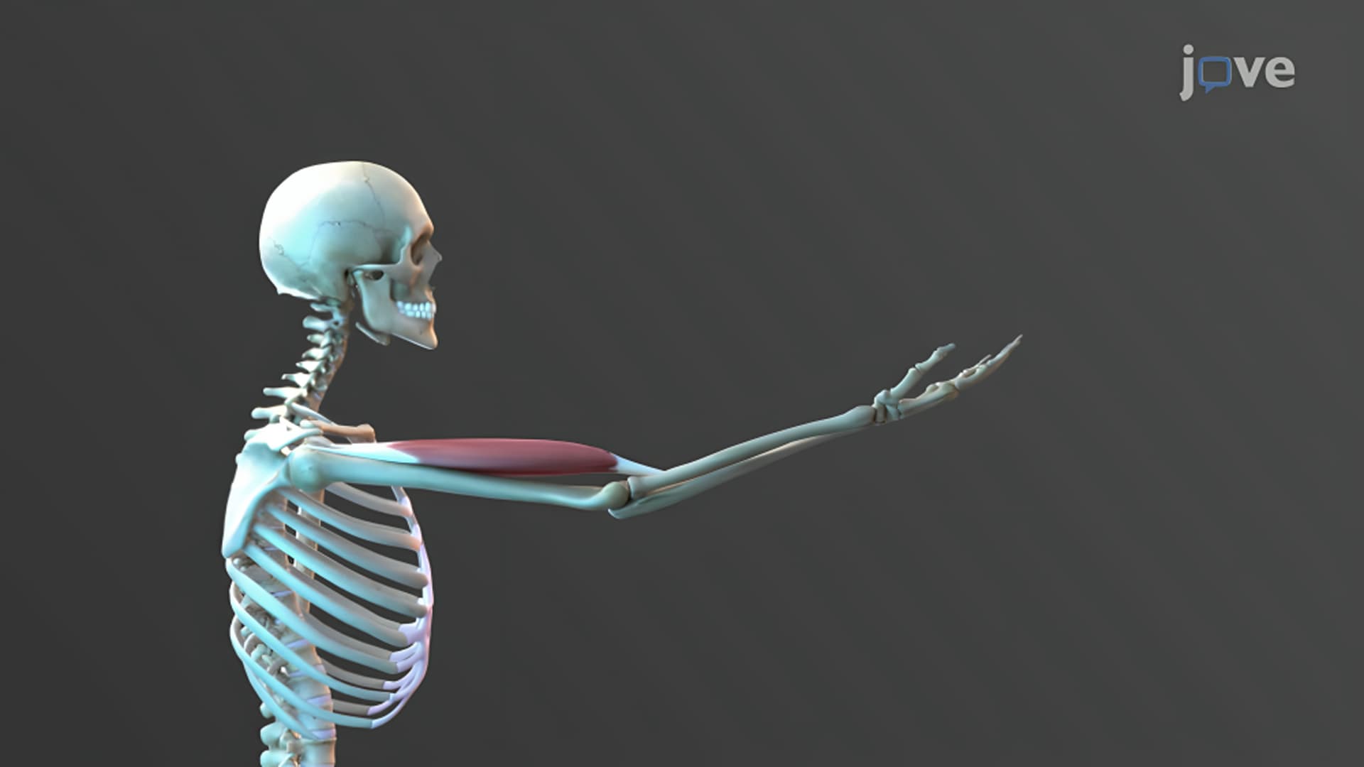
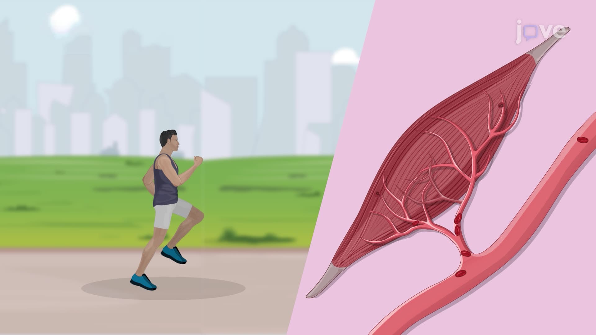
Skeletal muscles represent the most recognizable muscle tissue types overview, comprising approximately 40% of total body weight in healthy adults. These striated muscles attach to bones via tendons, creating the leverage system that enables everything from a tennis player's serve at the US Open to a surgeon's precise hand movements during procedures at Johns Hopkins Hospital.
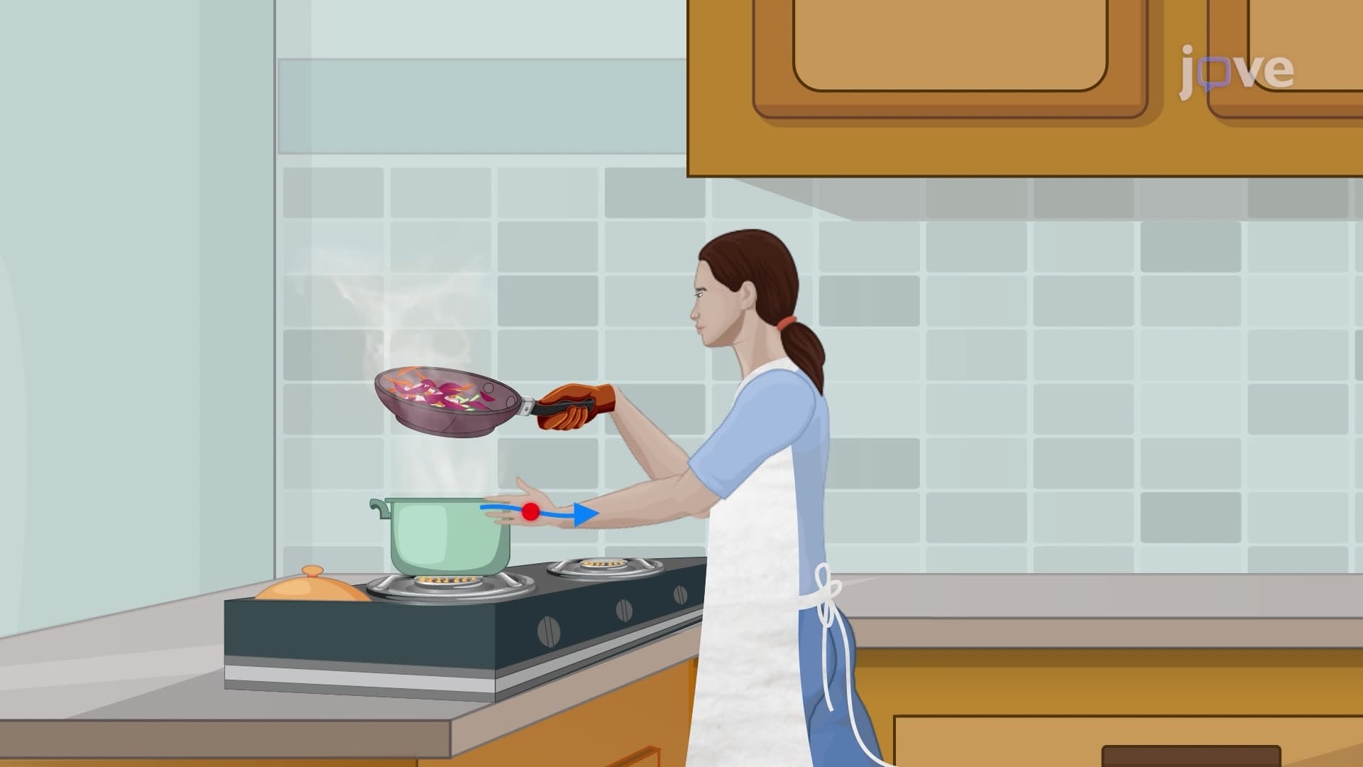
The voluntary involuntary muscle tissue distinction becomes crucial here. Skeletal muscles respond to conscious neural commands from the motor cortex, allowing athletes like Olympic swimmers to fine-tune their stroke technique. However, these muscles also maintain constant low-level contractions (muscle tone) that occur involuntarily, keeping us upright against gravity even while standing still.
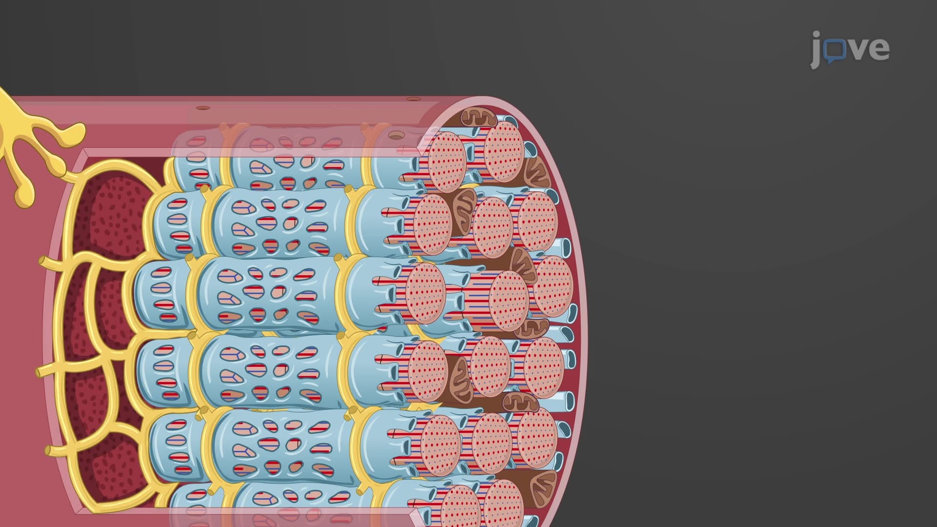
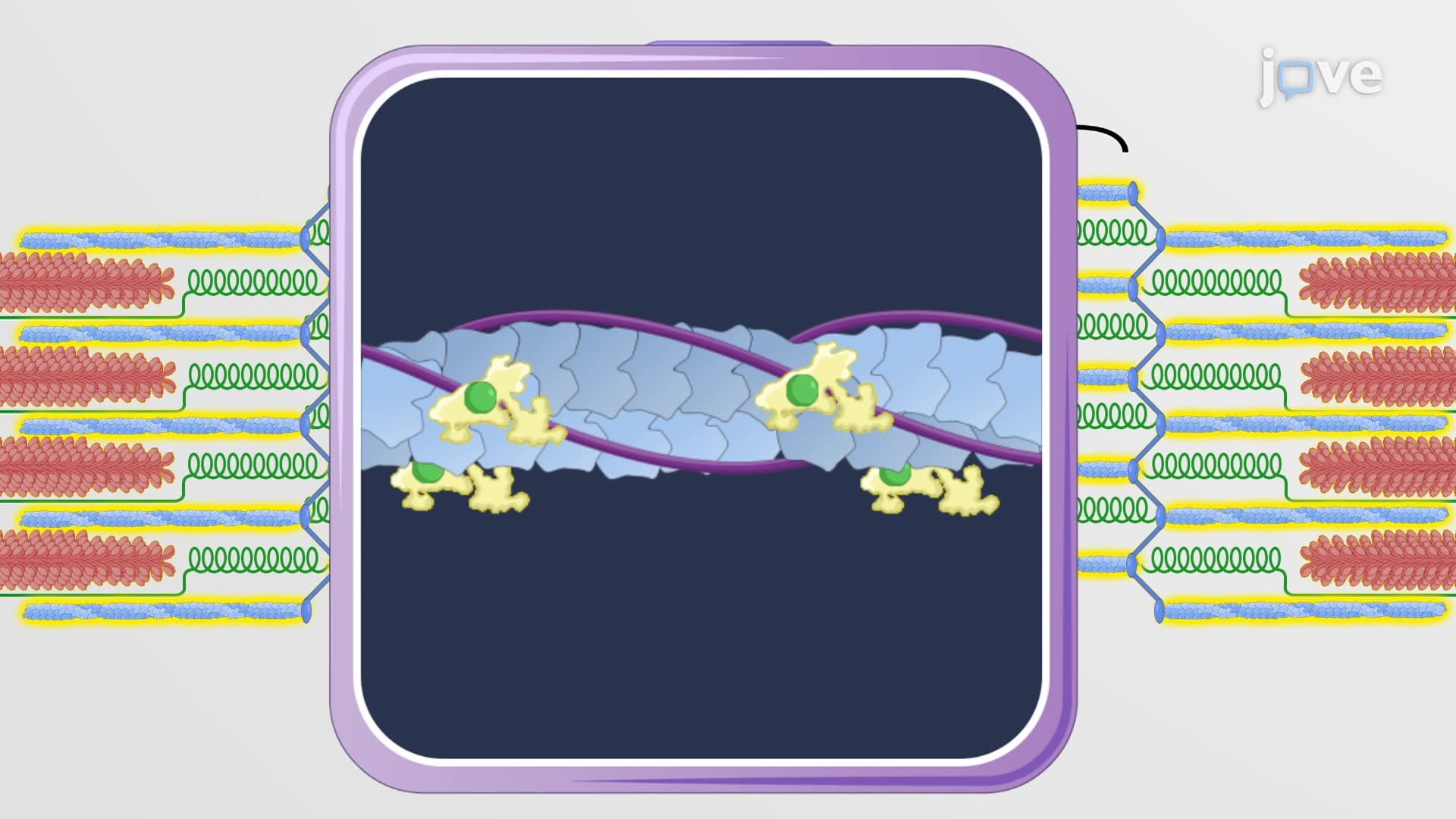
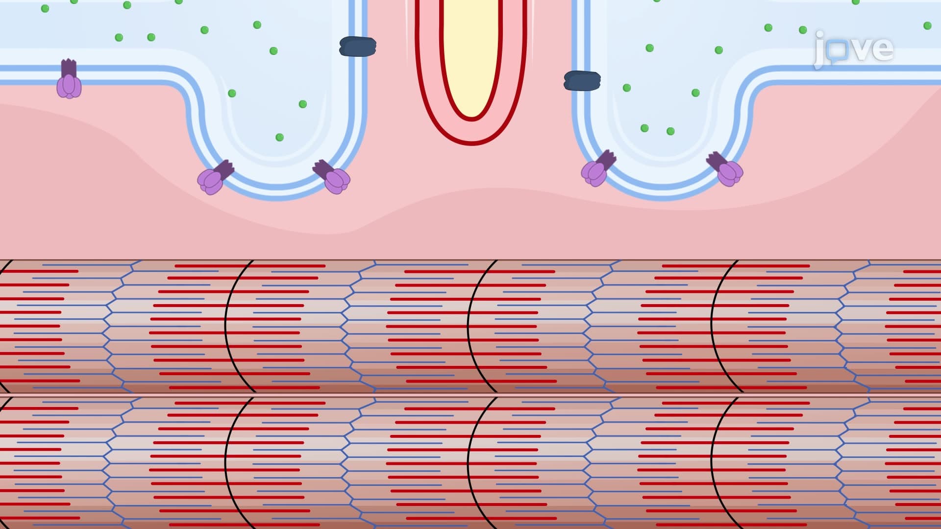
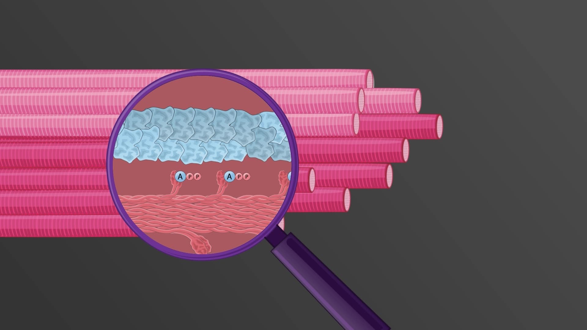
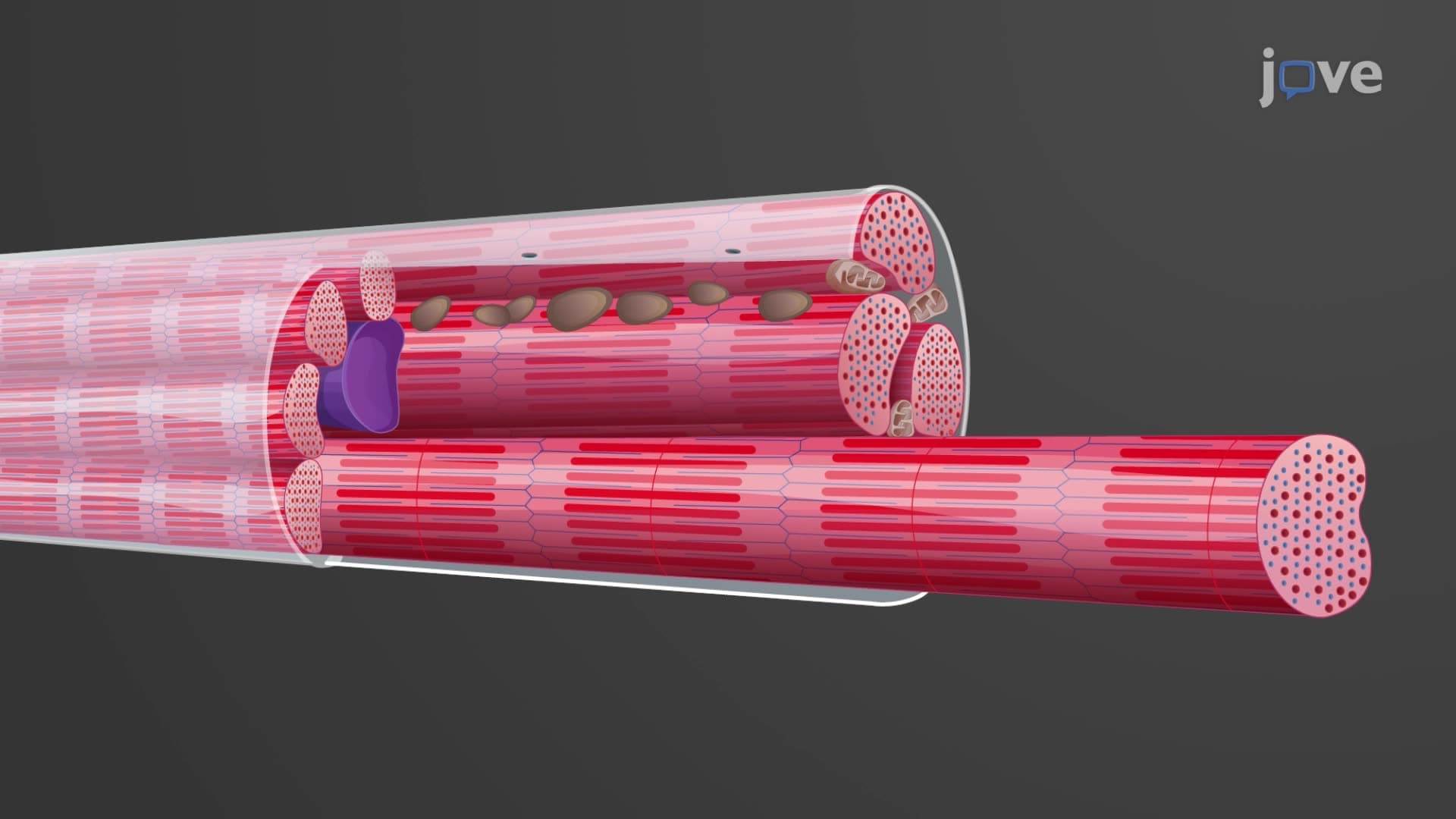
For students preparing for the MCAT or AP Biology exams, understanding skeletal muscle's striated muscle tissue appearance under microscopy is essential. The alternating light and dark bands result from the organized arrangement of actin and thin myosin filaments, creating the contractile apparatus that generates force.
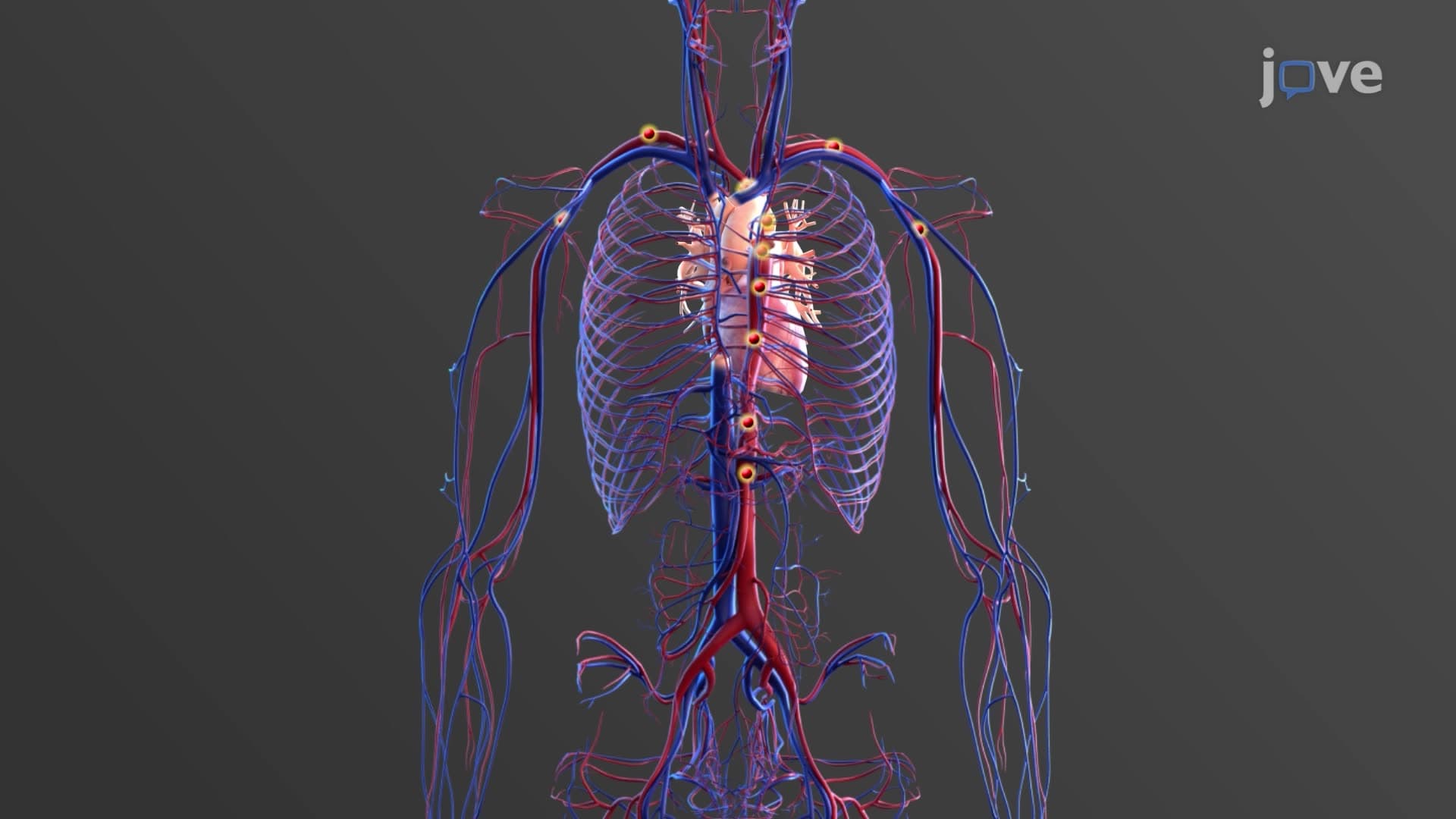
Cardiac muscle tissue exclusively forms the heart's muscular walls (myocardium), pumping approximately 2,000 gallons of blood daily through the cardiovascular system. Unlike skeletal muscle, cardiac tissue demonstrates autorhythmicity-the ability to generate its own electrical impulses without external neural stimulation.
This skeletal smooth cardiac muscle comparison highlights a fascinating evolutionary adaptation. While cardiac muscle appears striated like skeletal muscle, it functions involuntarily like smooth muscle. The sinoatrial (SA) node in the right atrium acts as the heart's natural pacemaker, establishing the rhythm that keeps blood flowing to vital organs in patients across American hospitals from Mayo Clinic to Cleveland Clinic.
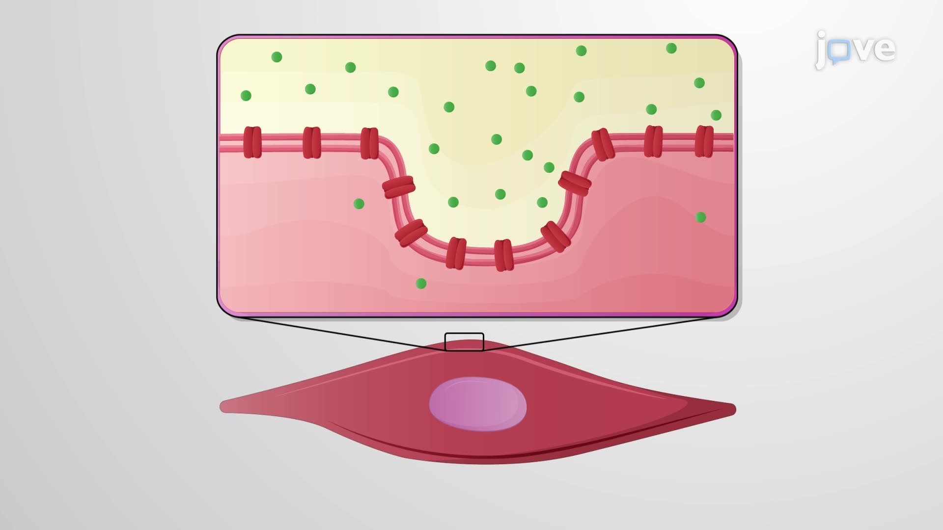
Smooth muscle tissue lines hollow organs throughout the body, including blood vessels, the digestive tract, and respiratory airways. This muscle tissue function overview reveals how smooth muscles regulate internal processes without conscious awareness-from the peristaltic waves moving food through your esophagus during lunch to the bronchodilation occurring during exercise.
For college anatomy students, understanding smooth muscle's non-striated appearance and involuntary control proves crucial for courses like Human Physiology. Unlike the rapid contractions of skeletal muscle, smooth muscle generates sustained contractions that can maintain tension for extended periods with minimal energy expenditure.
The muscle cell type biology concept extends beyond academic study into real clinical applications. Physicians treating patients with muscular dystrophy focus primarily on skeletal muscle degeneration, while cardiologists addressing heart failure examine cardiac muscle function. Emergency medicine residents studying for USMLE Step 1 must understand how smooth muscle dysfunction in blood vessels contributes to hypertensive crises.
Related Micro-courses