Loading video…


 22 Concepts
 178.8K views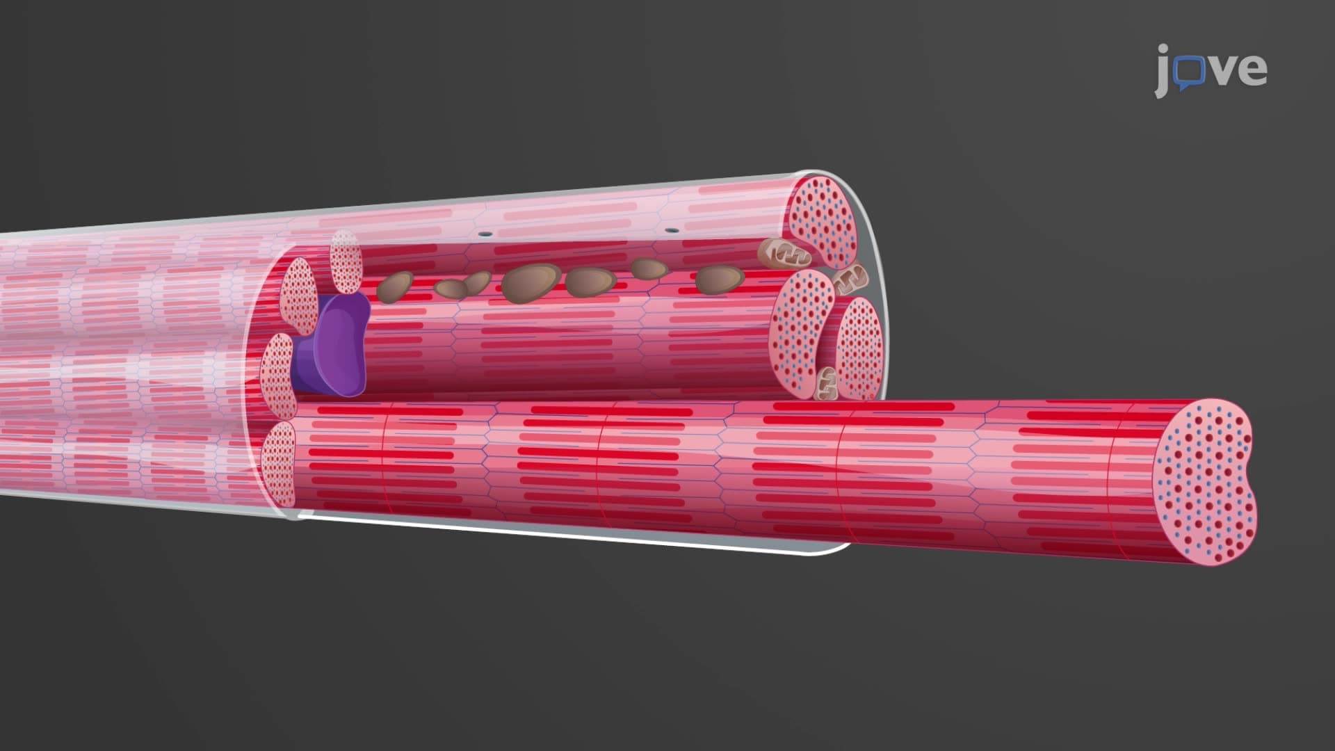

Video Summary: What Is Structure of Cardiac Muscles
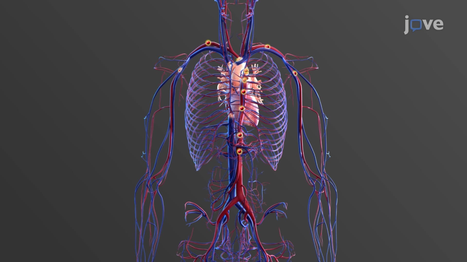
Every minute, your heart beats approximately 70 times, pumping blood through a cardiovascular system that would span 60,000 miles if laid end-to-end. This incredible feat relies on the unique cardiac muscle structure anatomy found exclusively in heart tissue. Unlike the skeletal muscles that move your arms and legs, cardiac muscle cells feature specialized branching patterns and intercalated discs that enable the synchronized contractions keeping Americans alive from coast to coast. Watch the full video on JoVE Coach to master this concept with expert-led visuals and step-by-step explanations.
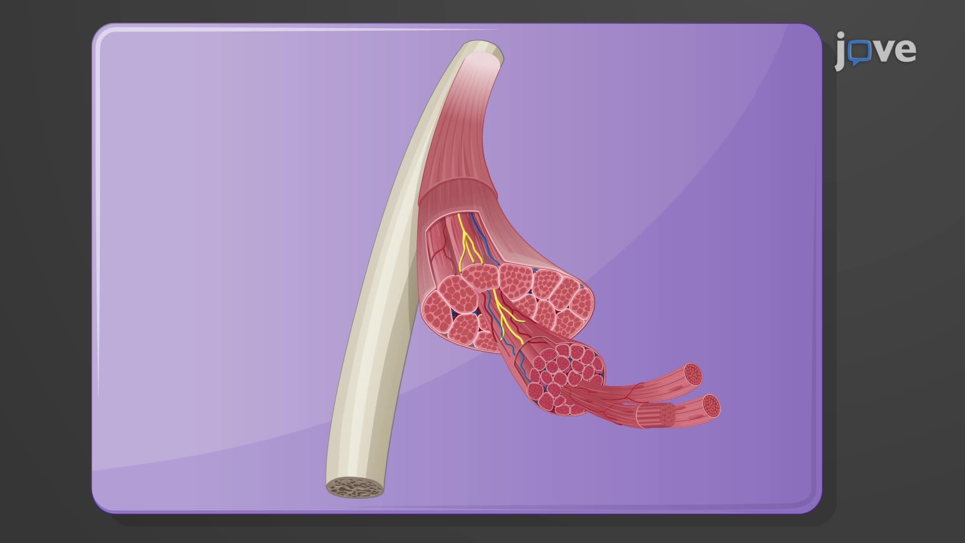
The structure of cardiac muscles represents one of nature's most sophisticated biological engineering solutions. Found exclusively in the myocardium (heart wall), these specialized cells must contract continuously for an entire lifetime-approximately 2.5 billion beats for the average American. This demanding function requires unique structural adaptations that distinguish cardiac muscle from both skeletal and smooth muscle tissues.
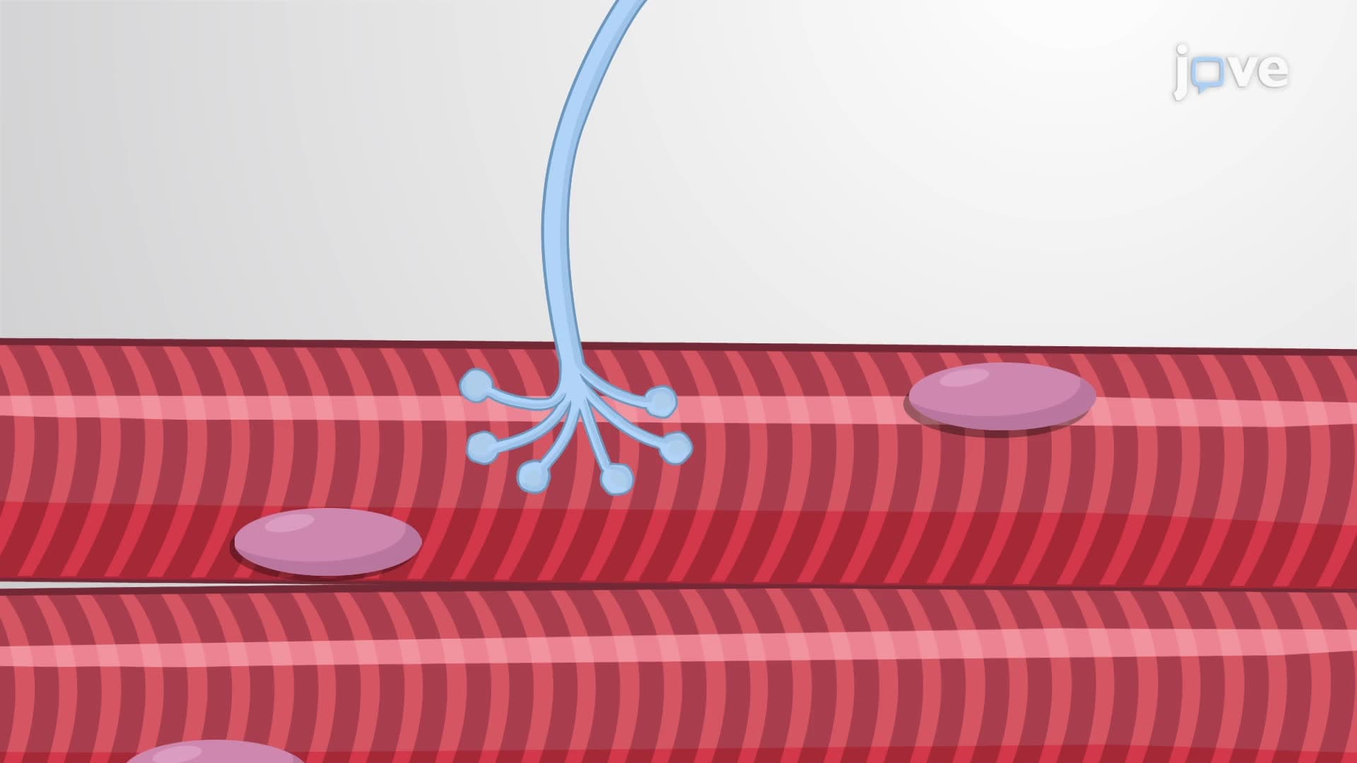
Heart muscle tissue structure begins at the cellular level with cardiomyocytes, which are notably smaller than skeletal muscle fibers. These cells typically measure 50-100 micrometers in length and 10-20 micrometers in width. Most importantly, cardiac muscle cells are mononucleated (single nucleus), though some may contain two nuclei. This single nucleus cardiac cell arrangement differs markedly from the multinucleated skeletal muscle fibers.
The cardiac muscle branching pattern creates a three-dimensional network that ensures mechanical and electrical coupling throughout the heart wall. This branched architecture allows force transmission in multiple directions, essential for the heart's pumping efficiency. Students preparing for the MCAT or AP Biology exams should note that this branching pattern is a key distinguishing feature tested on standardized assessments.
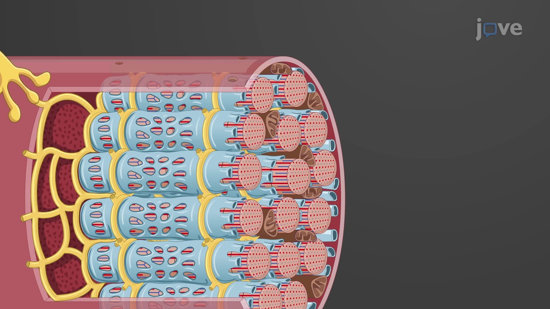
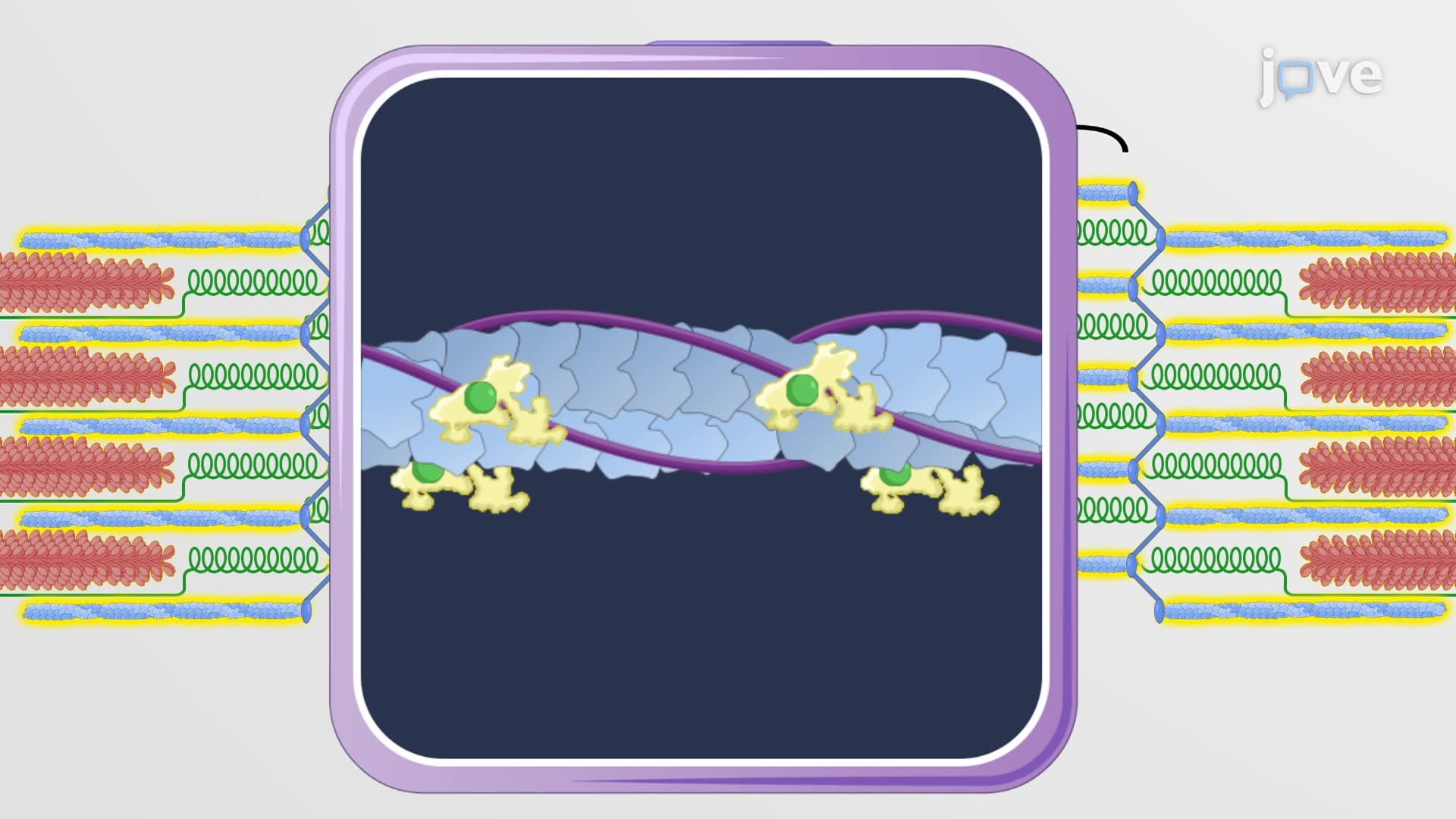
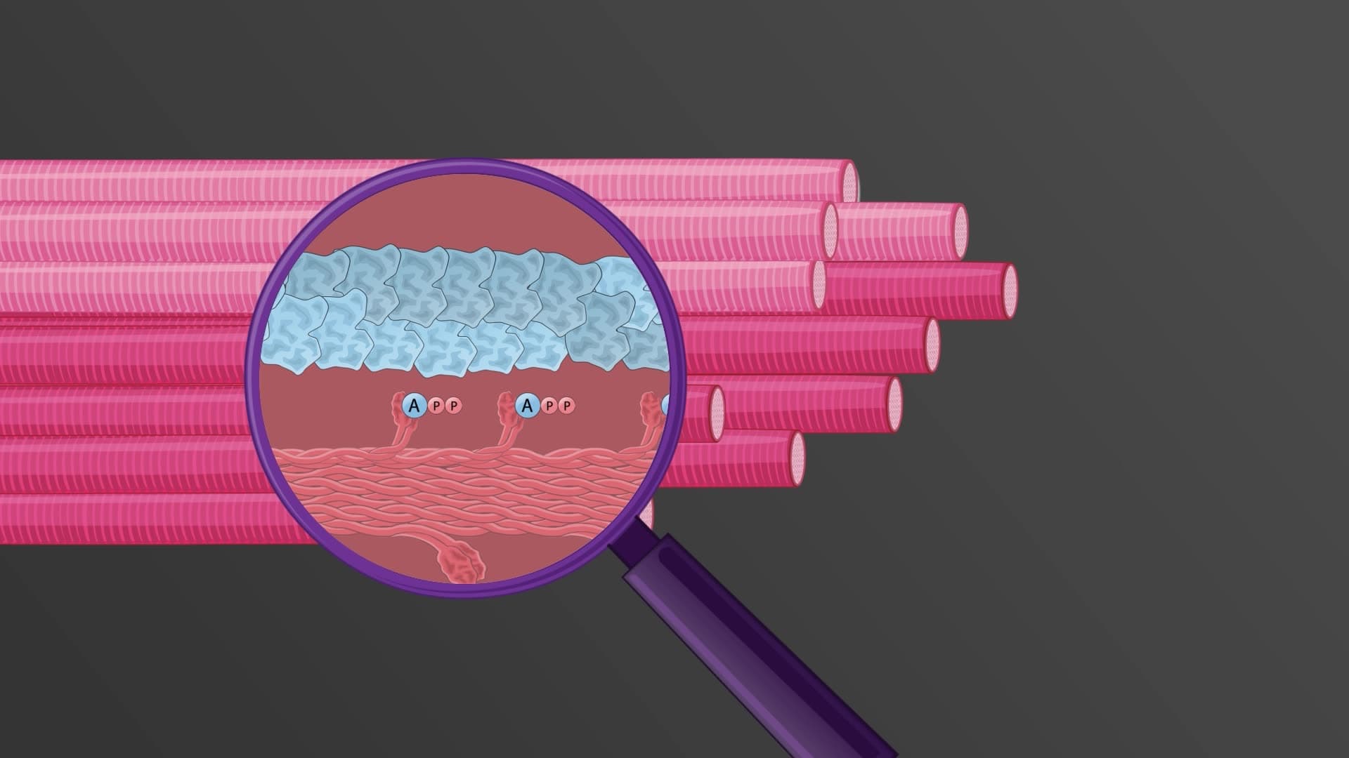
Like skeletal muscle, cardiac muscle exhibits a cardiomyocyte striated structure due to the regular arrangement of sarcomeres within myofibrils. However, cardiac sarcomeres are typically shorter (1.8-2.2 micrometers) compared to skeletal muscle. The striated appearance results from alternating dark (A) bands containing thick myosin filaments and light (I) bands containing thin actin filaments.
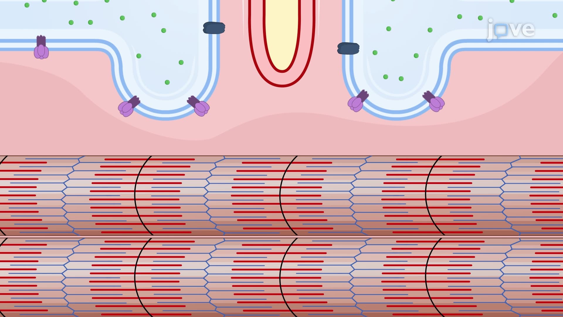
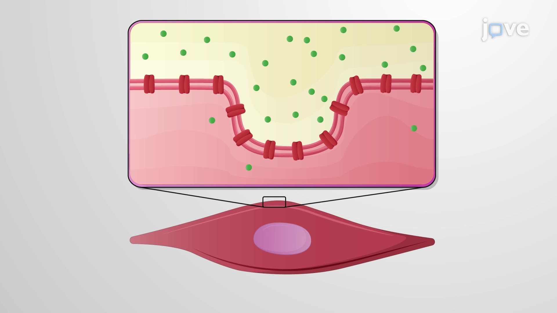
T-tubules in cardiac muscle are broader but less numerous than in skeletal muscle, arising at the Z-lines rather than at A-I junctions. This arrangement facilitates calcium release from the sarcoplasmic reticulum during excitation-contraction coupling, a process crucial for understanding cardiac physiology in college-level courses.
Perhaps the most distinctive feature of heart muscle cell structure is the presence of intercalated discs. These specialized junctions occur where adjacent cardiomyocytes meet end-to-end. Each intercalated disc cardiac structure contains three types of connections: gap junctions (for electrical coupling), adherens junctions (for mechanical attachment), and desmosomes (for structural integrity).
Gap junctions contain connexin proteins that form channels allowing ions to pass freely between cells, enabling the heart to function as an electrical syncytium. This arrangement ensures that electrical signals spread rapidly throughout the heart, coordinating contraction-a concept frequently tested on the USMLE Step 1 examination.
Cardiac muscle cells contain approximately 25-35% mitochondria by volume, far exceeding the mitochondrial content of skeletal muscle. This adaptation reflects the heart's enormous energy demands-cardiac muscle consumes more oxygen per gram than any other tissue. Large glycogen stores and lipid droplets provide additional energy reserves, supporting the heart's preferential use of fatty acids for ATP production under normal conditions.
Related Micro-courses