Loading video…


 22 Concepts
 178.8K views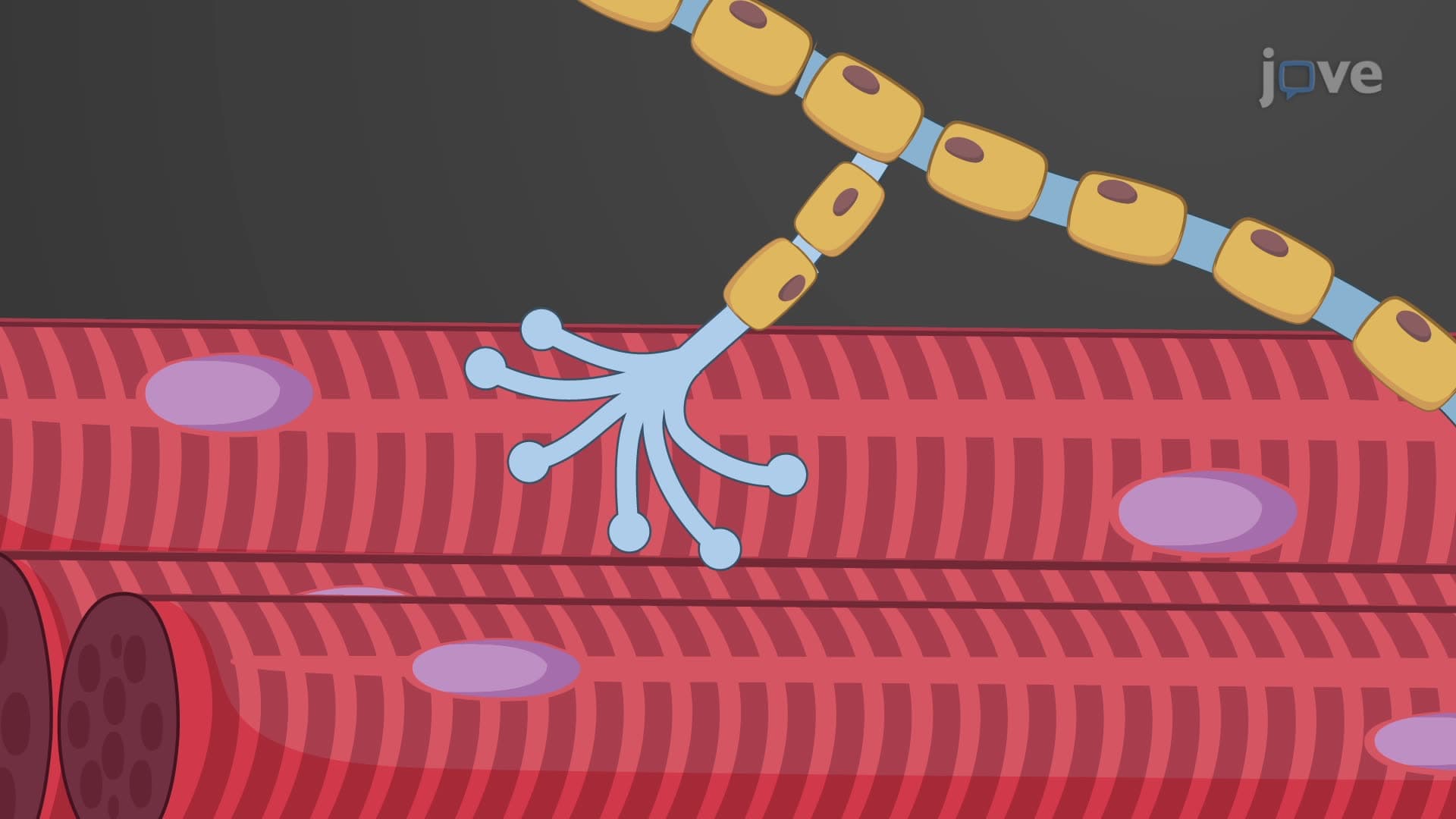

Video Summary: What Is Excitation Contraction Coupling
Ever wonder how your heart beats over 100,000 times daily without you thinking about it? Excitation contraction coupling muscle mechanisms make this possible by converting electrical signals into mechanical force. When a UCLA medical student studies cardiac physiology, they learn that what is excitation contraction coupling represents the critical bridge between nerve stimulation and actual muscle movement in every heartbeat and skeletal muscle contraction. Watch the full video on JoVE Coach to master this concept with expert-led visuals and step-by-step explanations.
Excitation contraction coupling muscle physiology represents one of the most elegant biological processes, seamlessly converting electrical nerve impulses into the mechanical work that powers every movement from a Harvard athlete's sprint to a surgeon's precise hand movements. This process occurs millions of times daily in your body, yet most people never consider the intricate molecular machinery making it possible.
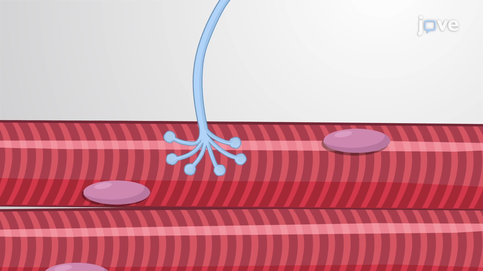
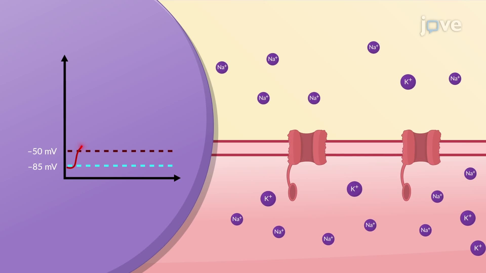
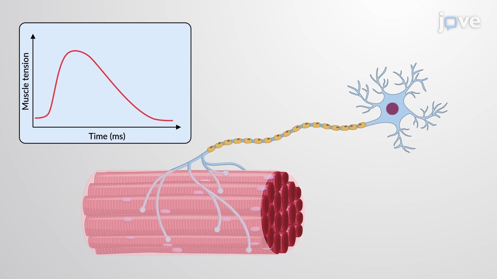
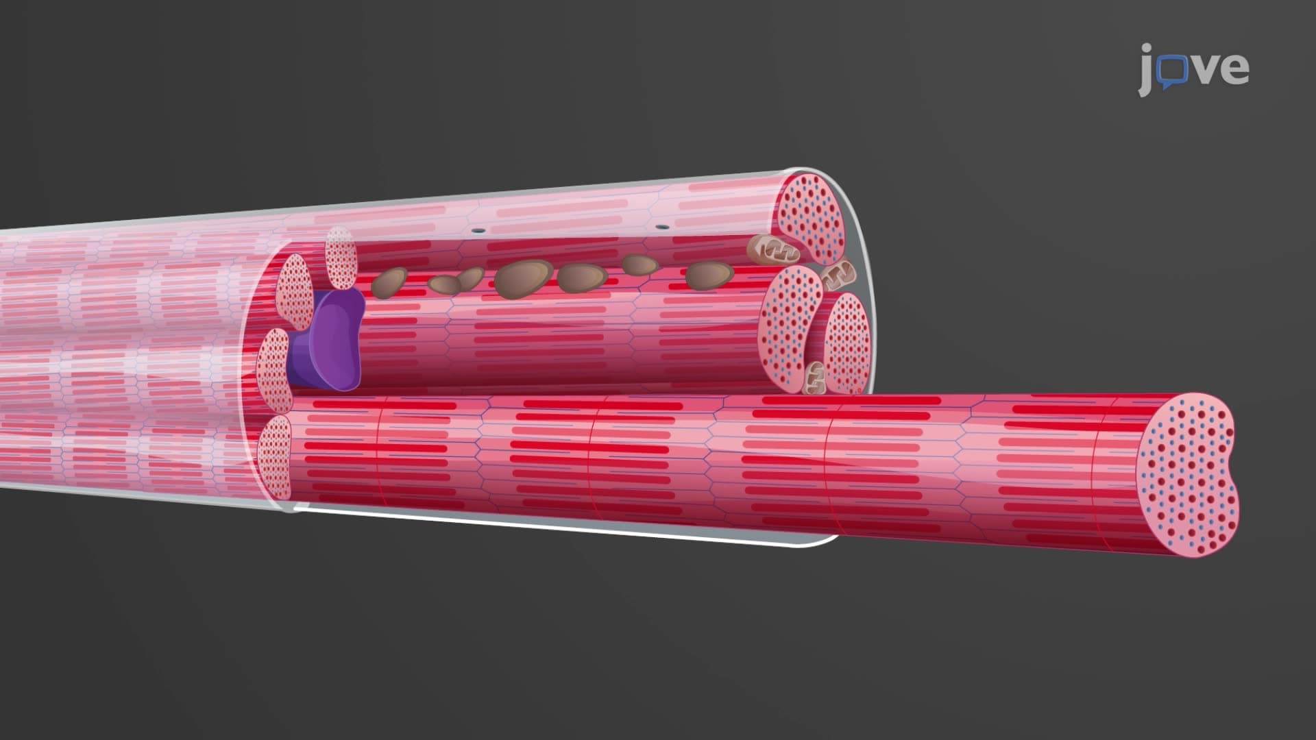
The process begins when motor neurons from your spinal cord release acetylcholine at the neuromuscular junction, creating an action potential that spreads across the muscle fiber's surface membrane (sarcolemma). This electrical signal must somehow reach the interior of large muscle fibers, which can be up to 100 micrometers in diameter-a significant distance at the cellular level.
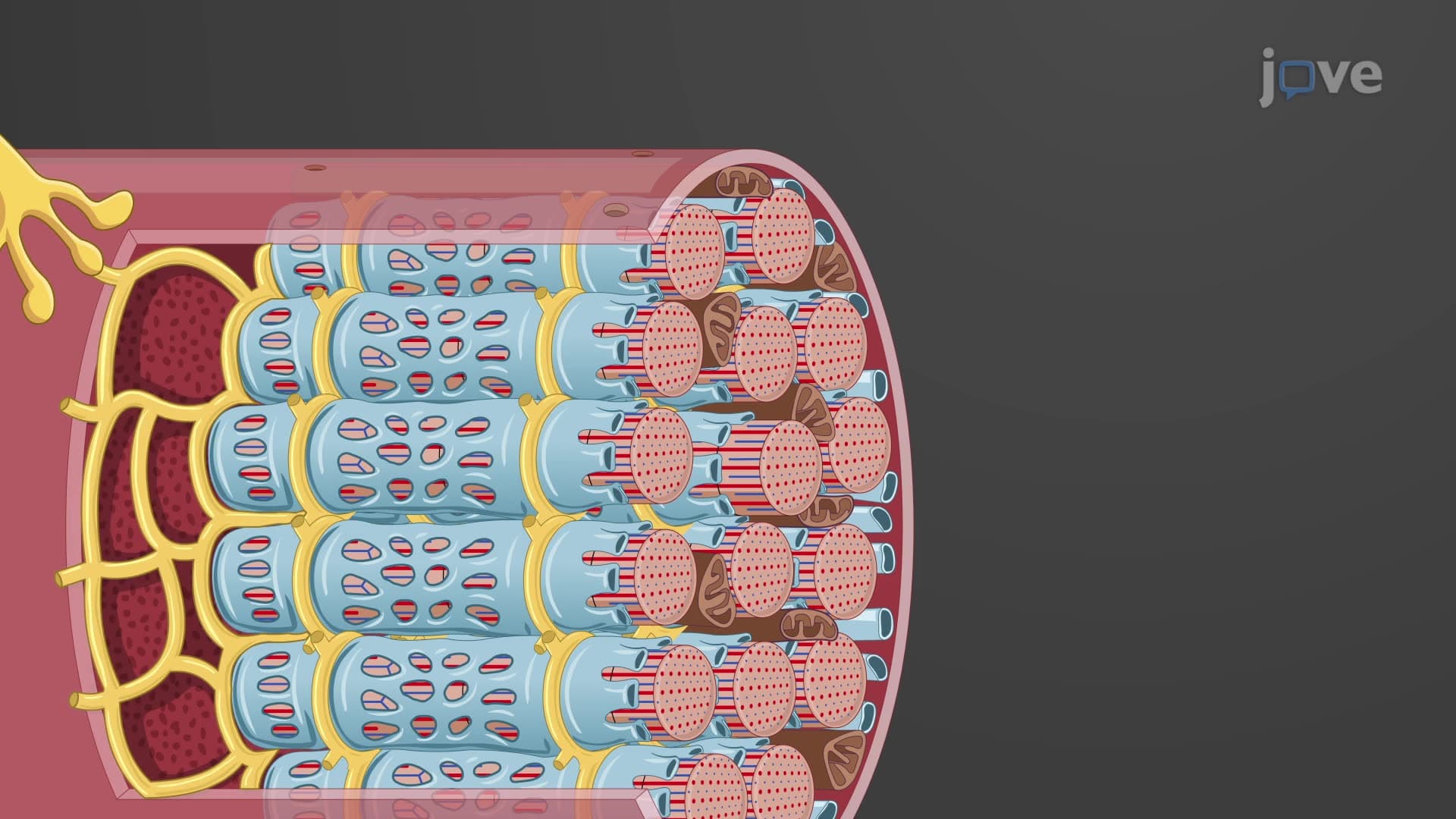
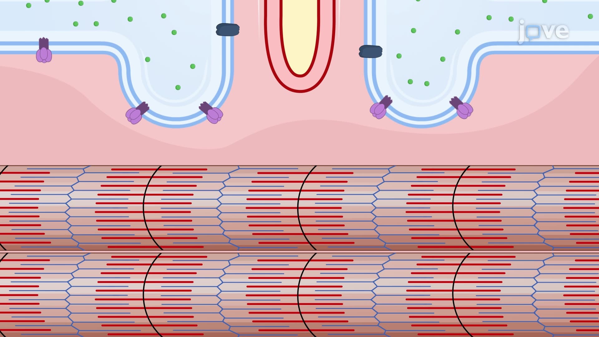
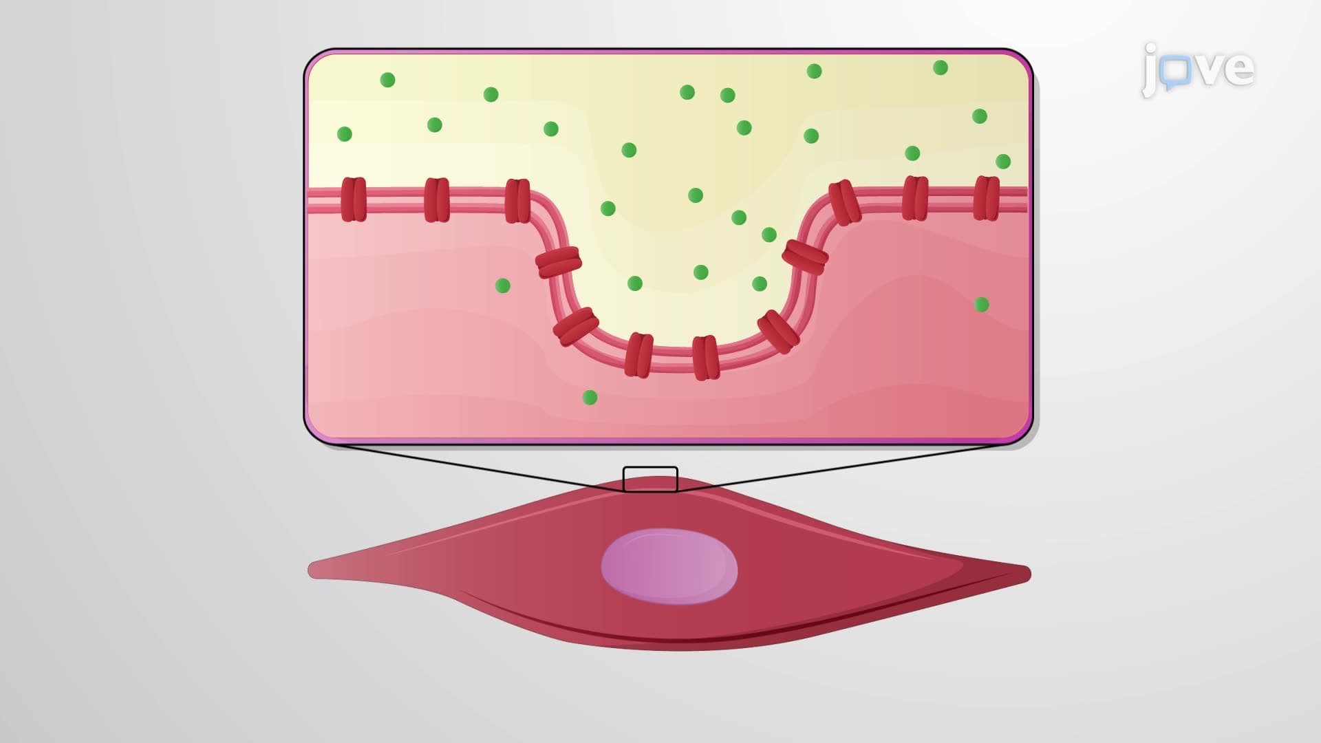
T-tubule calcium release ECC systems solve this transmission problem through specialized membrane invaginations called transverse tubules or T-tubules. These structures act like electrical highways, carrying action potentials deep into muscle fibers where they can trigger calcium release. In the Johns Hopkins physiology lab, students often struggle to visualize how these microscopic tubes enable rapid, synchronized muscle contraction across the entire fiber diameter.
The T-tubules are strategically positioned adjacent to the sarcoplasmic reticulum's terminal cisternae, forming triads-functional units where electrical signals get converted to chemical signals. When action potentials travel down T-tubules, they cause voltage-gated dihydropyridine receptors to change shape, mechanically opening ryanodine receptor channels in the sarcoplasmic reticulum membrane.
Sarcoplasmic reticulum calcium stores are massive-containing millimolar concentrations that dwarf the nanomolar levels found in resting muscle cytoplasm. This 10,000-fold concentration gradient drives rapid calcium release when channels open, flooding the myofibril environment within milliseconds.
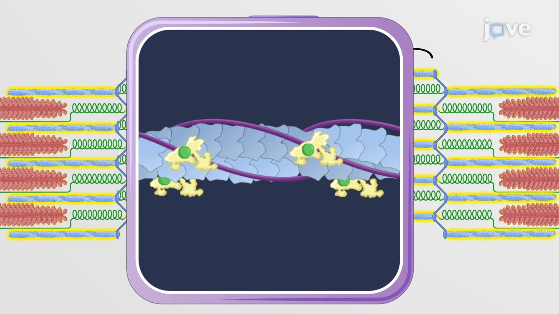
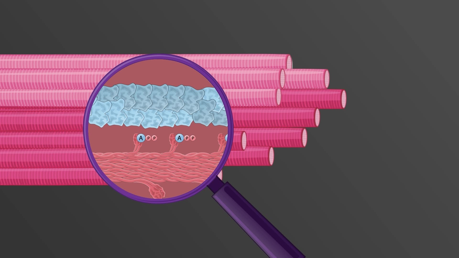
The released calcium binds to troponin tropomyosin ECC regulatory proteins on thin filaments. Troponin C, the calcium-binding subunit, undergoes conformational changes that shift tropomyosin molecules, exposing myosin-binding sites on actin. This molecular choreography enables calcium cross bridge coupling as myosin heads form cross-bridges with actin, hydrolyze ATP, and generate the power stroke that shortens sarcomeres.
Understanding ECC physiology explained becomes crucial for pre-med students preparing for MCAT questions about muscle disorders. Malignant hyperthermia, a potentially fatal condition triggered by certain anesthetics, results from defective ryanodine receptors that cause uncontrolled calcium release. Similarly, muscular dystrophies often involve disrupted excitation contraction coupling due to membrane instability or calcium handling defects.
AP Biology students frequently encounter this topic when studying muscle physiology, particularly how calcium availability determines muscle performance. College anatomy and physiology courses emphasize how this process differs between skeletal, cardiac, and smooth muscle, with cardiac muscle showing unique calcium-induced calcium release mechanisms that amplify the initial signal.
Related Micro-courses