14 Concepts
6 Concepts
21 Concepts
23 Concepts
21 Concepts
15 Concepts
16 Concepts
26 Concepts
18 Concepts
19 Concepts
11 Concepts
6 Concepts
10 Concepts
22 Concepts
20 Concepts
26 Concepts
26 Concepts
14 Concepts
10 Concepts
12 Concepts
23 Concepts
29 Concepts
19 Concepts
38 Concepts
38 Concepts
25 Concepts
40 Concepts
25 Concepts
25 Concepts
20 Concepts
37 Concepts
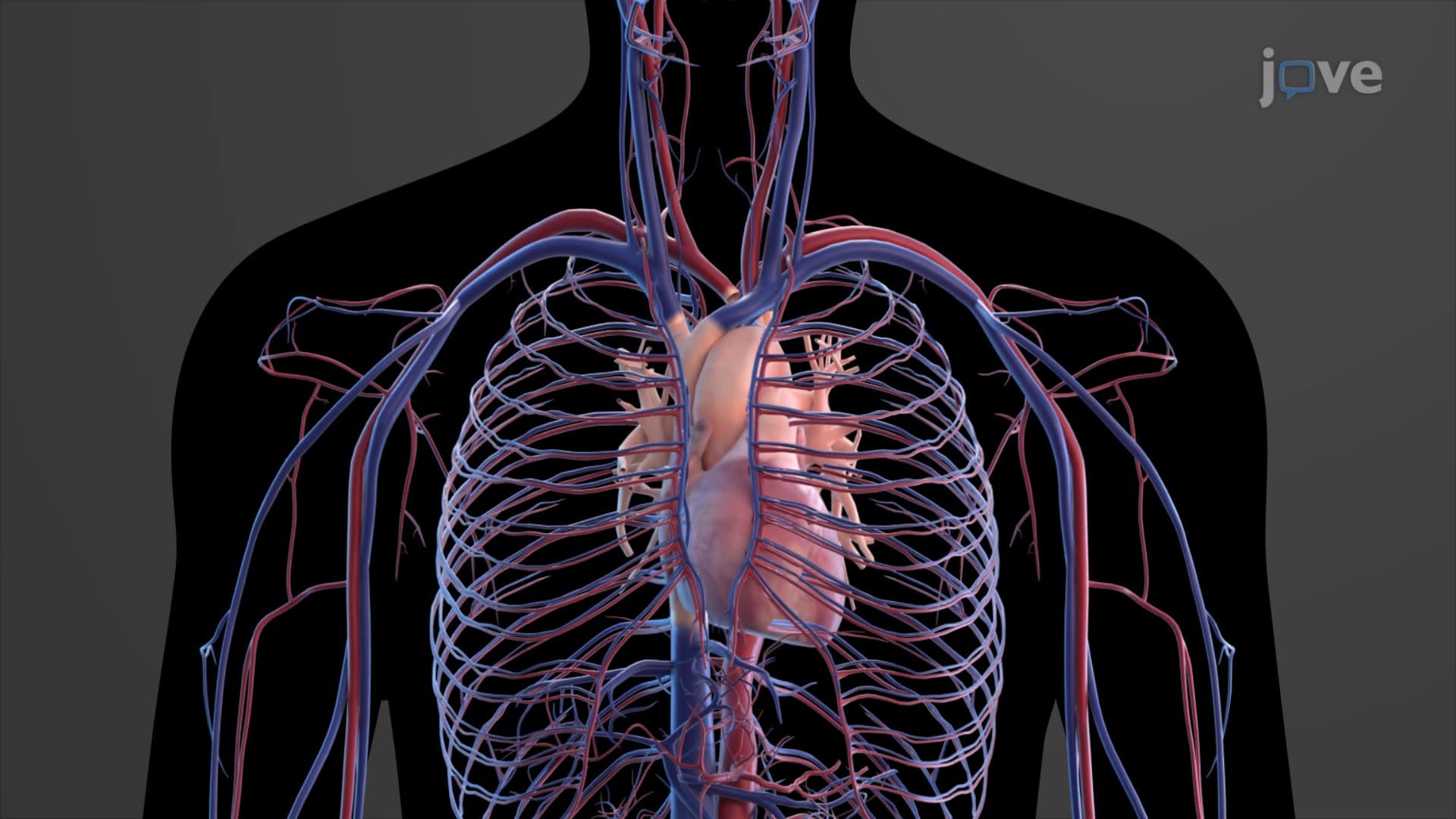
The heart serves as the body's central pump, driving circulation through its four-chambered structure and complex electrical conduction system. This JoVE Coach micro-course explores cardiac anatomy and physiology, from the structure and function of the human heart to clinical applications relevant to US medical education. Students examine heart chambers valves, the cardiac cycle, conduction system, myocardium, and essential heart physiology concepts critical for understanding cardiovascular health and disease.
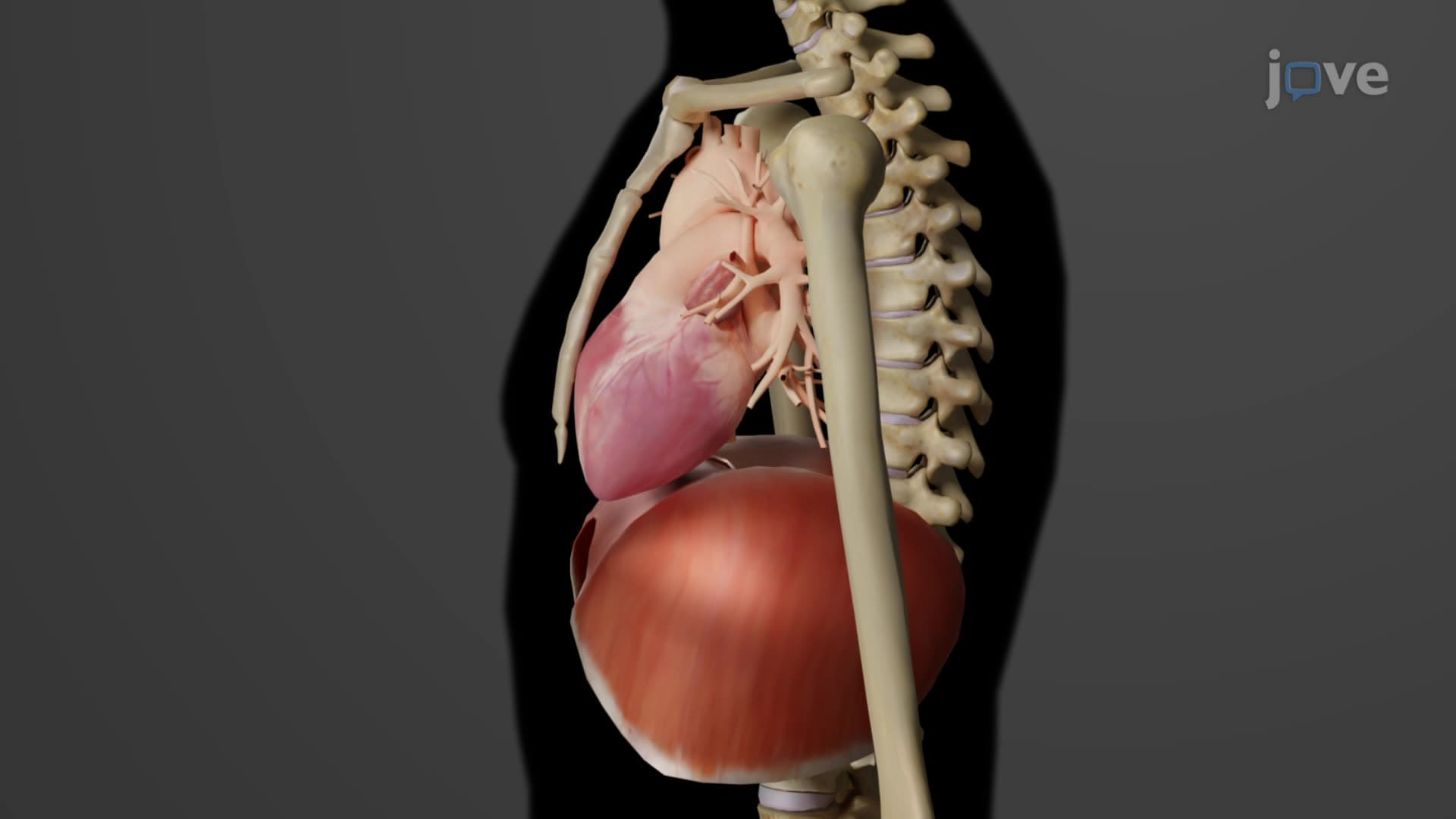
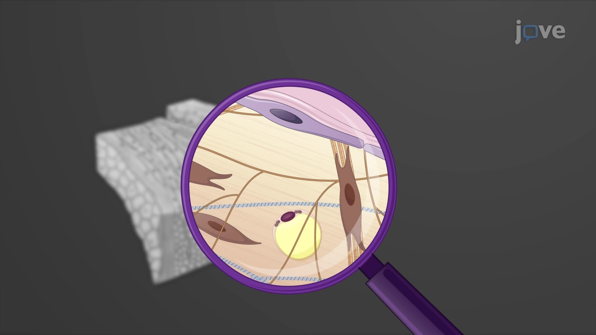
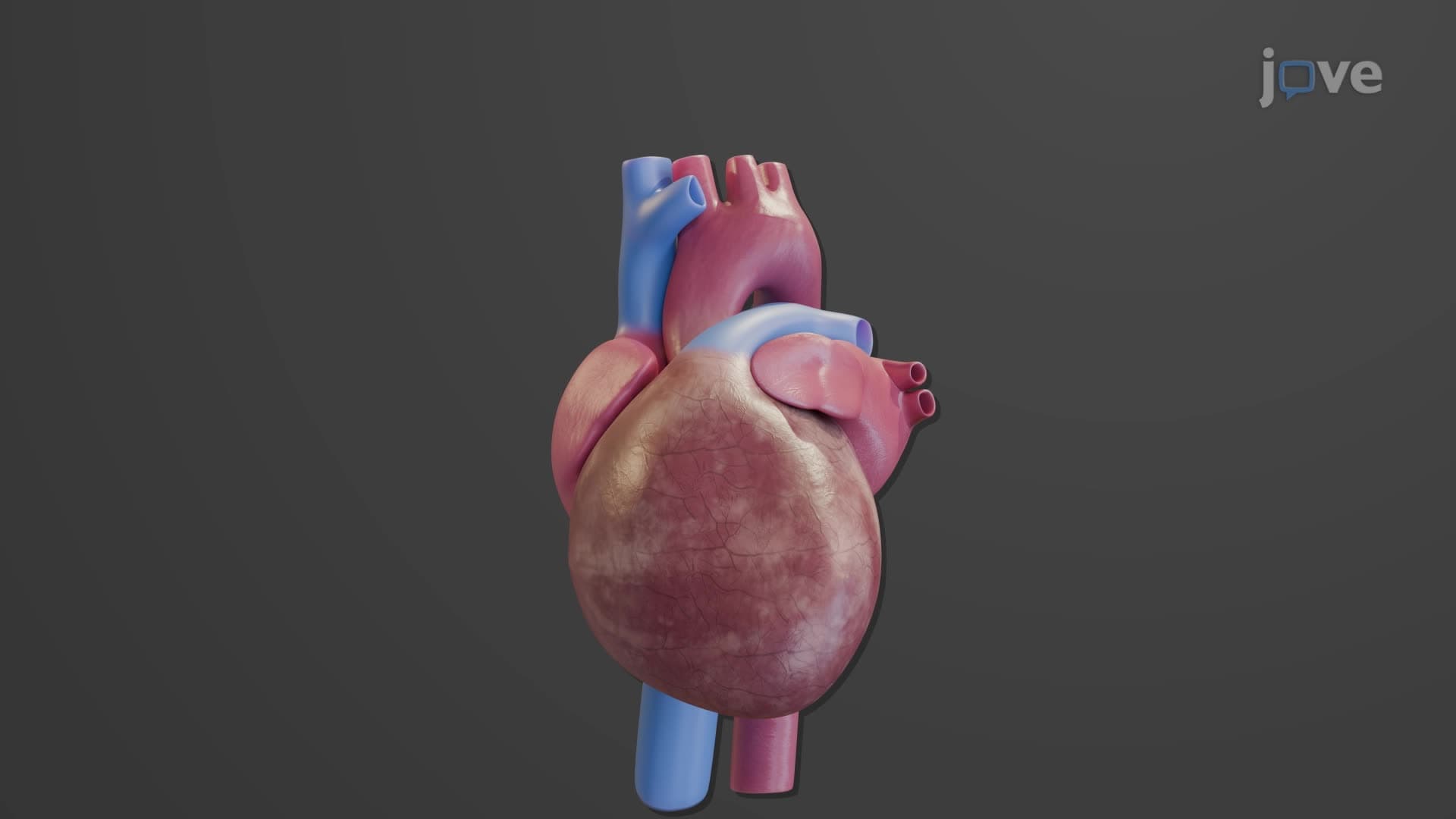
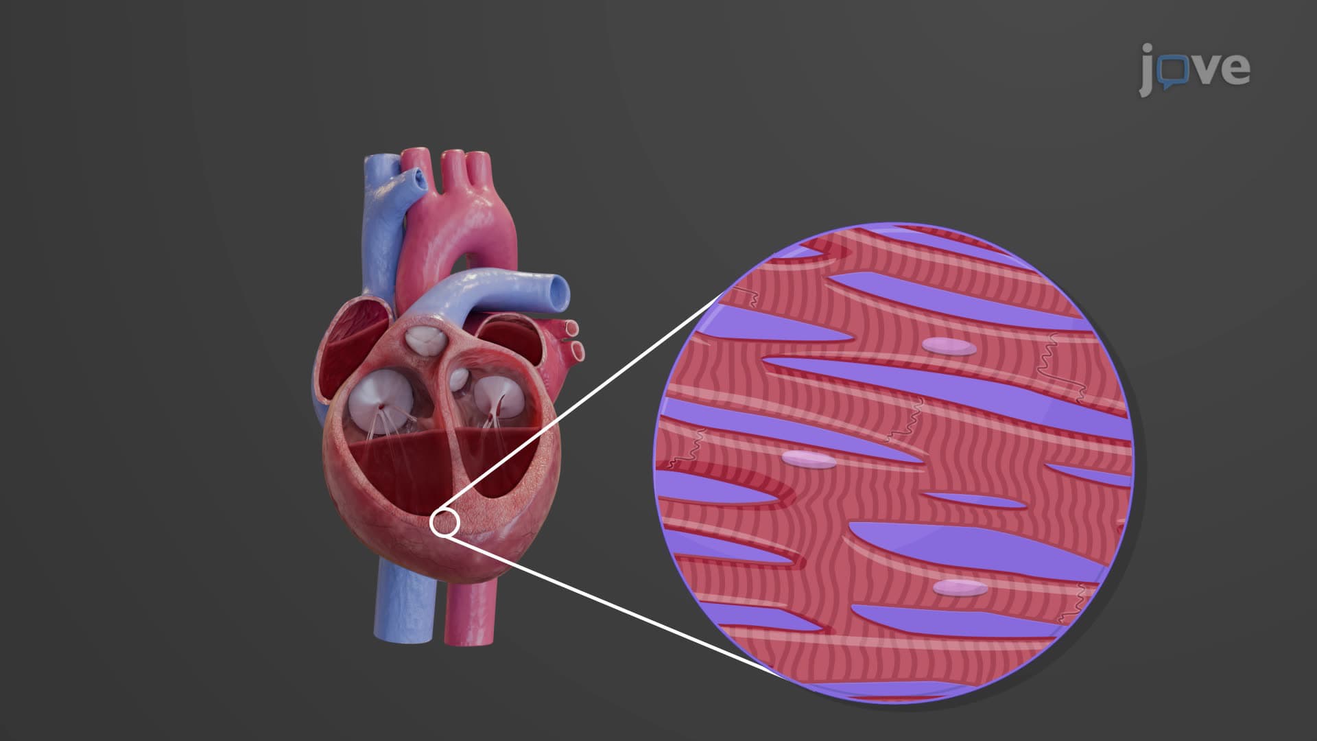
1. Heart Structure and Location: The heart is a cone-shaped, hollow muscular organ weighing 250-300 grams, positioned in the mediastinum between the lungs. Its three-layered wall consists of the epicardium (outer protective layer), myocardium (thick muscular middle layer responsible for contractions), and endocardium (smooth inner lining). The pericardium, a double-walled protective sac, surrounds the heart and contains pericardial fluid to reduce friction during contractions. Understanding this anatomy is essential for interpreting chest X-rays and performing cardiac procedures in US healthcare settings.
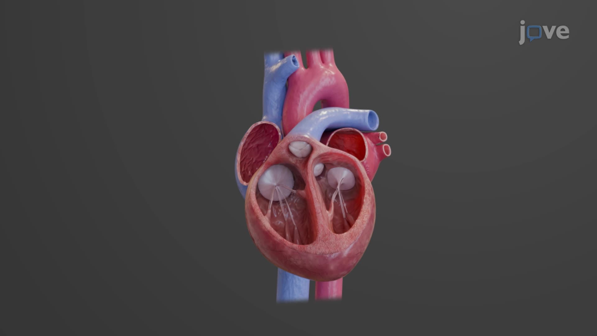
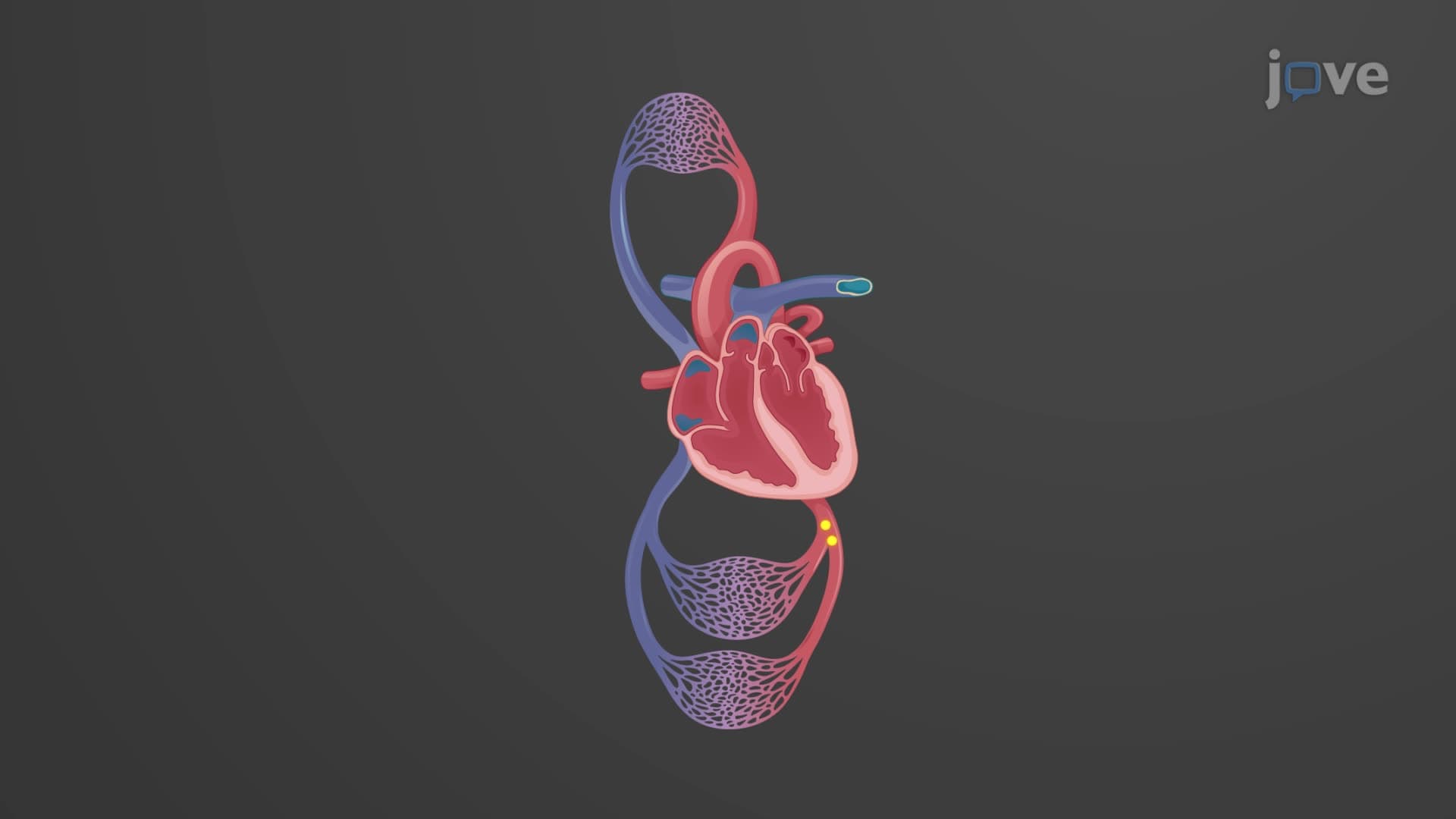
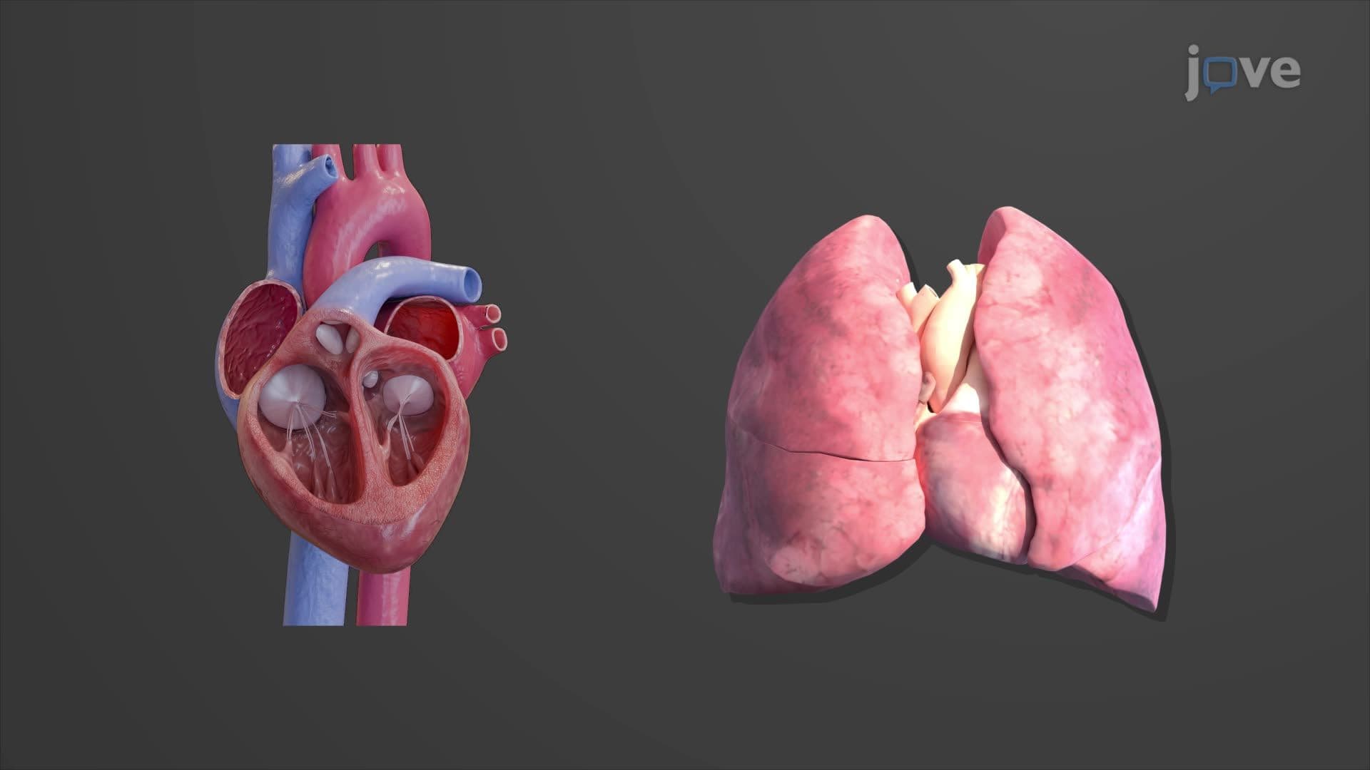
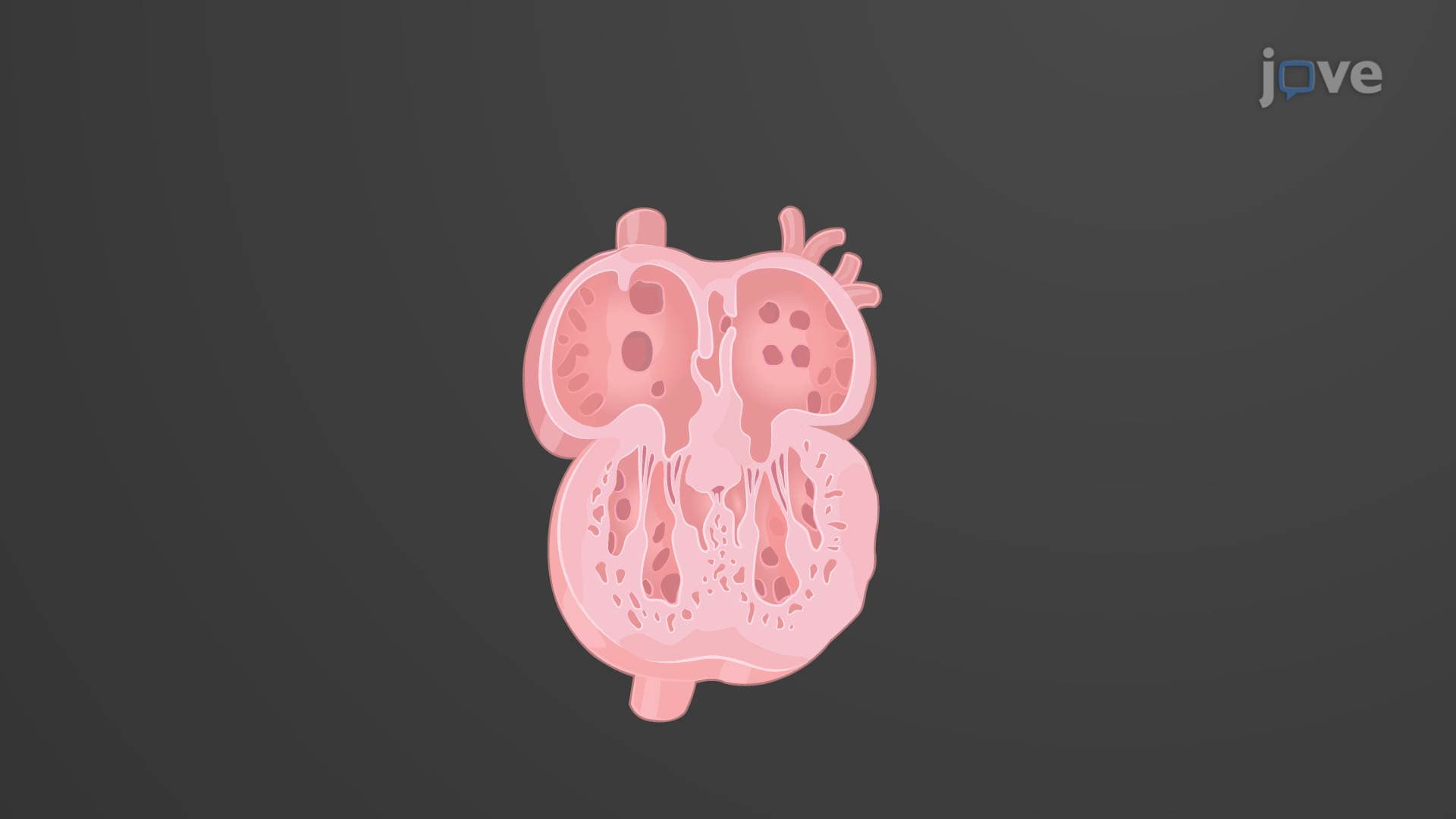
2. Four-Chamber Heart Design and Blood Flow Pathways: The heart contains two atria (receiving chambers) and two ventricles (pumping chambers), separated by the interatrial and interventricular septa. The right side handles deoxygenated blood from systemic circulation, pumping it to the lungs via pulmonary circulation. The left side receives oxygenated blood from the lungs and distributes it throughout the body via systemic circulation. This separation ensures efficient oxygenation and is crucial for understanding congenital heart defects commonly seen in US pediatric cardiology.
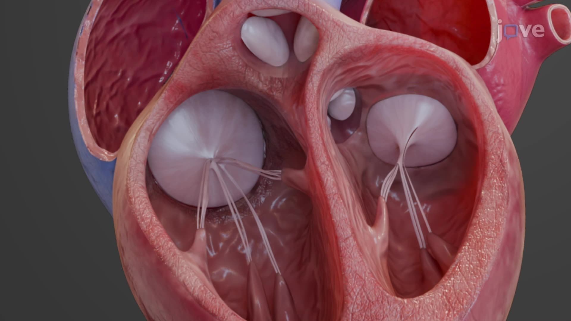
3. Heart Valve Function and Regulation: Four valves maintain unidirectional blood flow: tricuspid and bicuspid (mitral) atrioventricular valves, plus aortic and pulmonary semilunar valves. These valves open and close based on pressure gradients created by chamber contractions. The papillary muscles and chordae tendineae prevent valve prolapse during ventricular systole. Valve dysfunction leads to conditions like mitral valve prolapse or aortic stenosis, commonly encountered in US emergency departments and requiring surgical intervention.
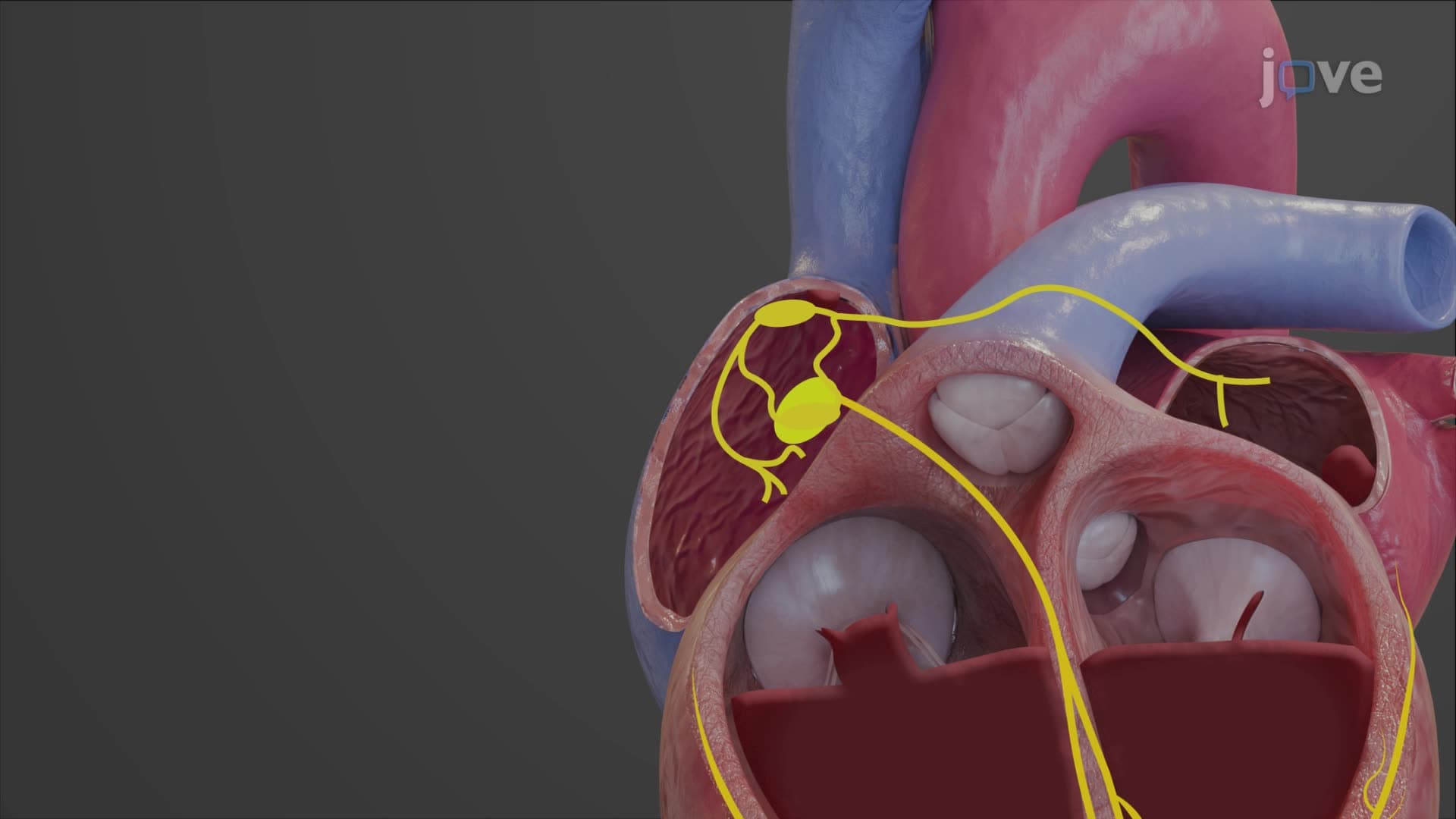
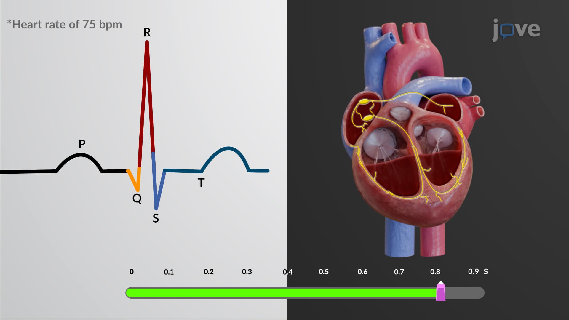
4. Cardiac Conduction System and Electrical Activity: The heart's intrinsic conduction system begins with the sinoatrial (SA) node, the primary pacemaker, followed by the atrioventricular (AV) node, bundle of His, and Purkinje fibers. This system ensures coordinated contractions through electrical impulses. The SA node fires at 60-100 beats per minute in healthy adults, while backup pacemakers have slower rates. Understanding this system is vital for interpreting arrhythmias and implementing treatments like pacemakers in US cardiac care facilities.
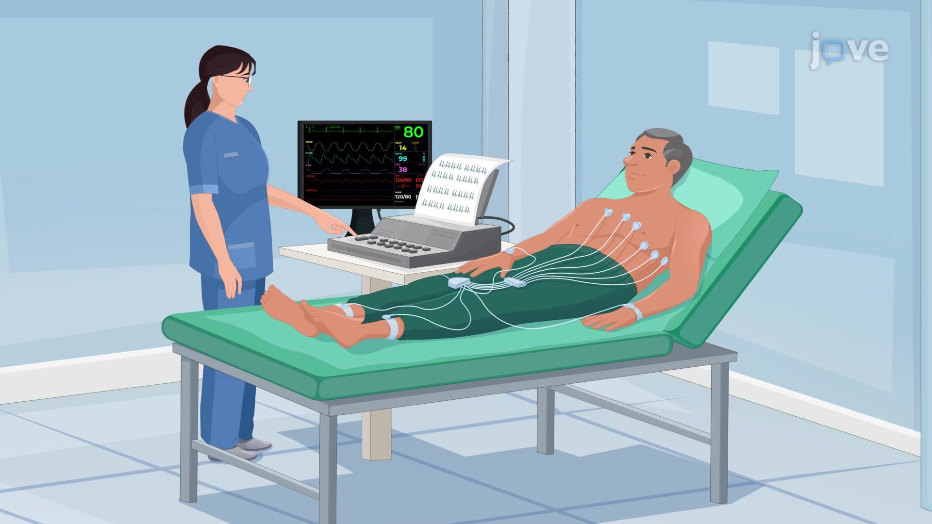
5. Electrocardiogram Interpretation and Clinical Significance: The ECG records the heart's electrical activity through P waves (atrial depolarization), QRS complexes (ventricular depolarization), and T waves (ventricular repolarization). Key intervals like P-R and Q-T provide diagnostic information about conduction delays and repolarization abnormalities. ECG changes can indicate myocardial infarction, arrhythmias, or electrolyte imbalances. Proficiency in ECG interpretation is essential for US healthcare providers, from paramedics to cardiologists.
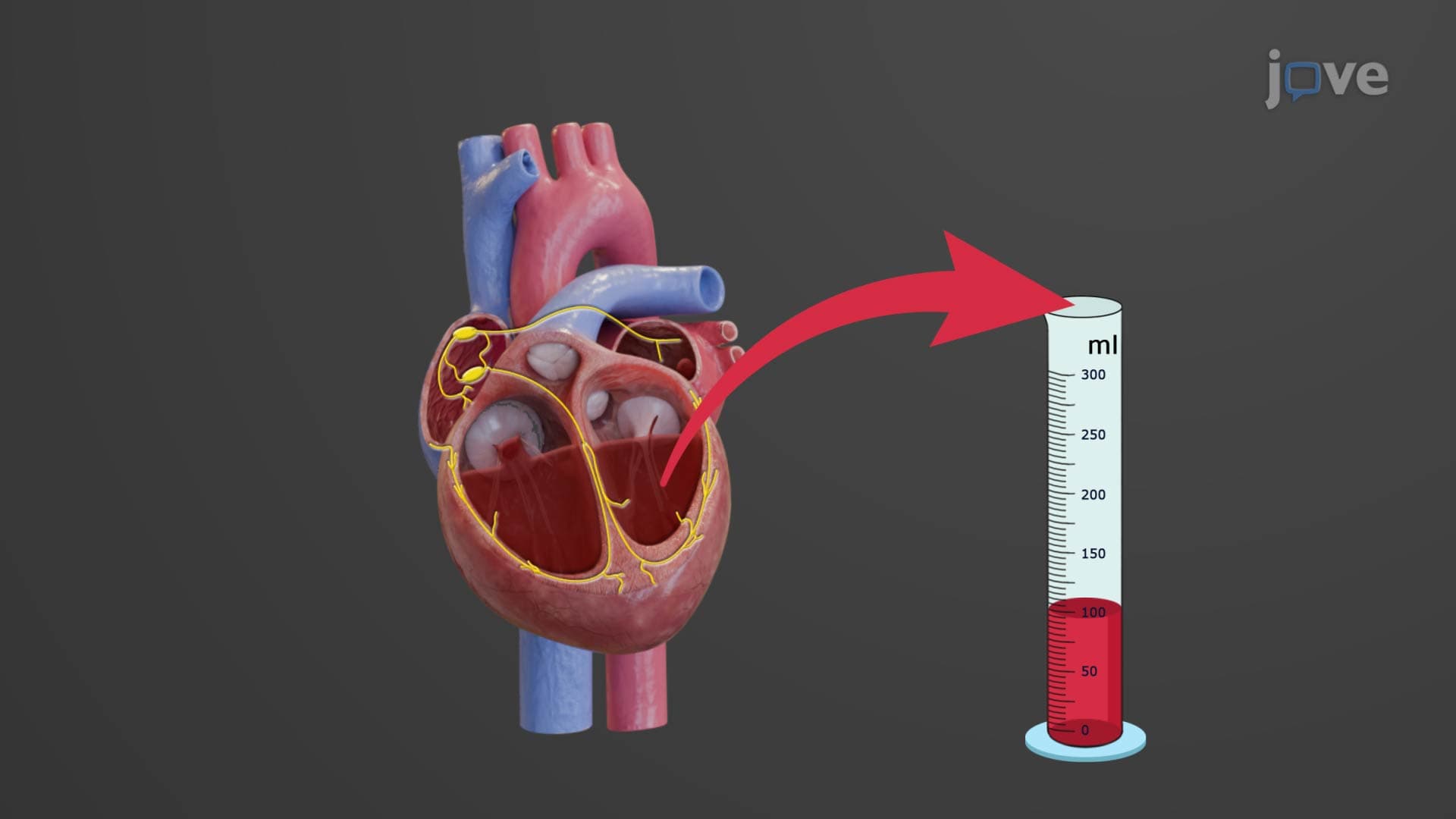
6. Cardiac Cycle Mechanics and Hemodynamics: The cardiac cycle alternates between systole (contraction) and diastole (relaxation), with isovolumetric phases when all valves are closed. End-diastolic volume (approximately 130 mL) and end-systolic volume (approximately 60 mL) determine stroke volume. Ventricular filling occurs during diastole, while ejection happens during systole. Understanding these mechanics helps explain heart failure pathophysiology and guides treatment decisions in US intensive care units.
7. Cardiac Output Regulation and Clinical Applications: Cardiac output equals stroke volume multiplied by heart rate, averaging 5.25 L/min in resting adults. Stroke volume regulation involves preload (ventricular filling), contractility (force of contraction), and afterload (resistance to ejection). The Frank-Starling mechanism ensures that increased venous return leads to stronger contractions. These concepts are fundamental for managing shock, heart failure, and cardiac medications in US hospitals.
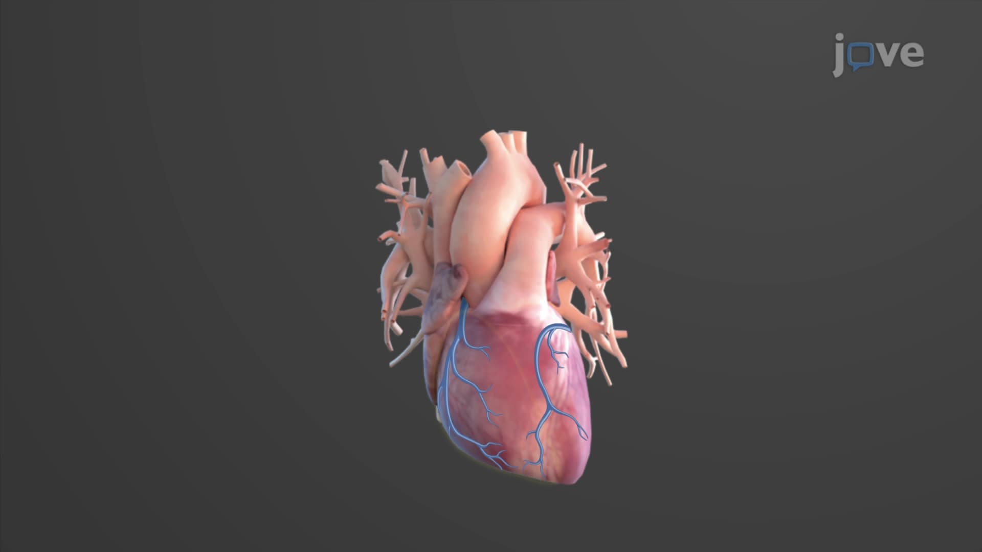
8. Coronary Circulation and Myocardial Blood Supply: The left and right coronary arteries branch from the ascending aorta, supplying the myocardium with oxygenated blood. The left anterior descending (LAD) and circumflex arteries serve the left ventricle, while the right coronary artery supplies the right ventricle and inferior wall. Coronary artery disease, the leading cause of death in the US, results from atherosclerotic narrowing of these vessels, potentially causing myocardial infarction and requiring interventions like angioplasty or bypass surgery.