Loading video…


 19 Concepts
 112.2K views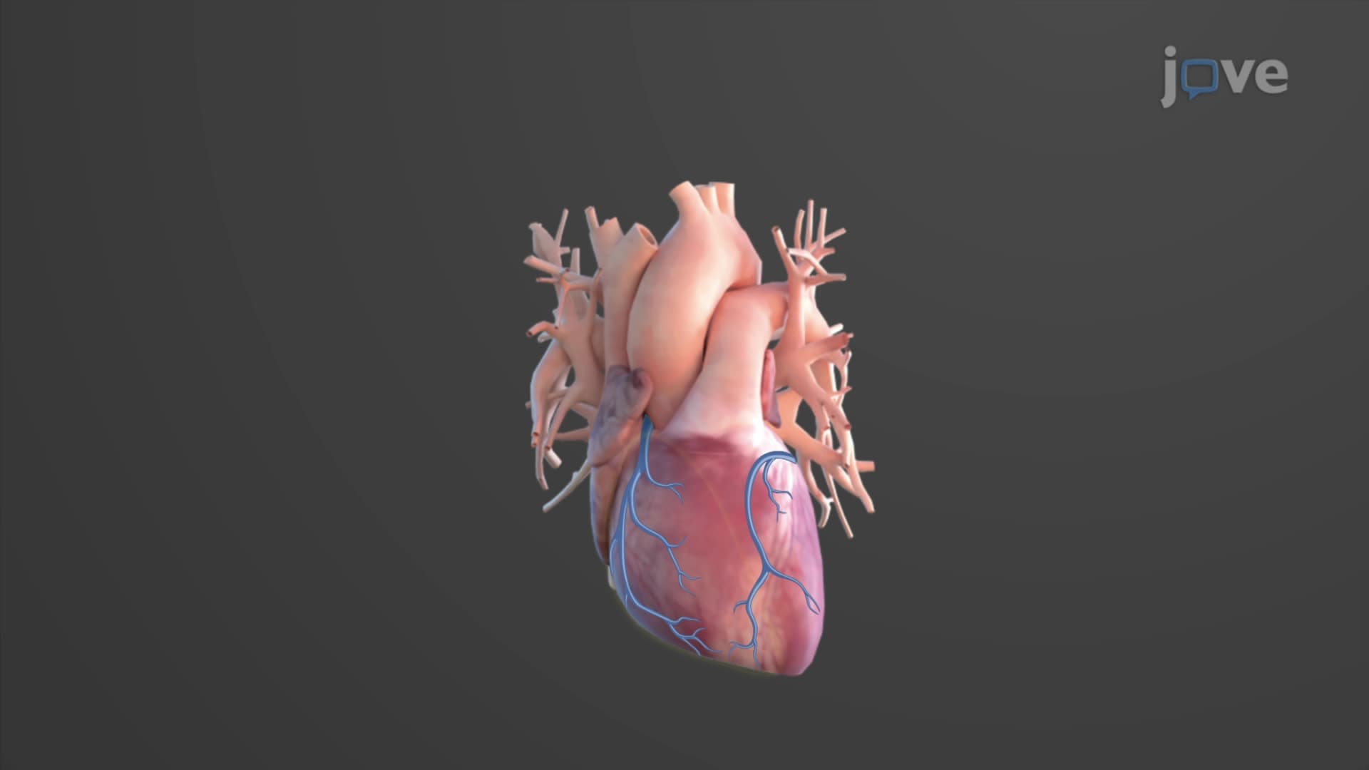

Video Summary: What Is Coronary Circulation
Every 60 seconds, your heart pumps about 5 liters of blood throughout your body, but did you know the heart muscle itself needs its own dedicated blood supply system? Coronary circulation anatomy biology forms a specialized network that keeps your heart muscle alive and functioning. When this system fails, as seen in over 655,000 Americans who die from heart disease annually according to the CDC, the consequences can be fatal. Understanding what is coronary circulation in cardiac anatomy reveals how the heart feeds itself through left and right coronary arteries, their branches, and venous drainage pathways. Watch the full video on JoVE Coach to master this concept with expert-led visuals and step-by-step explanations.
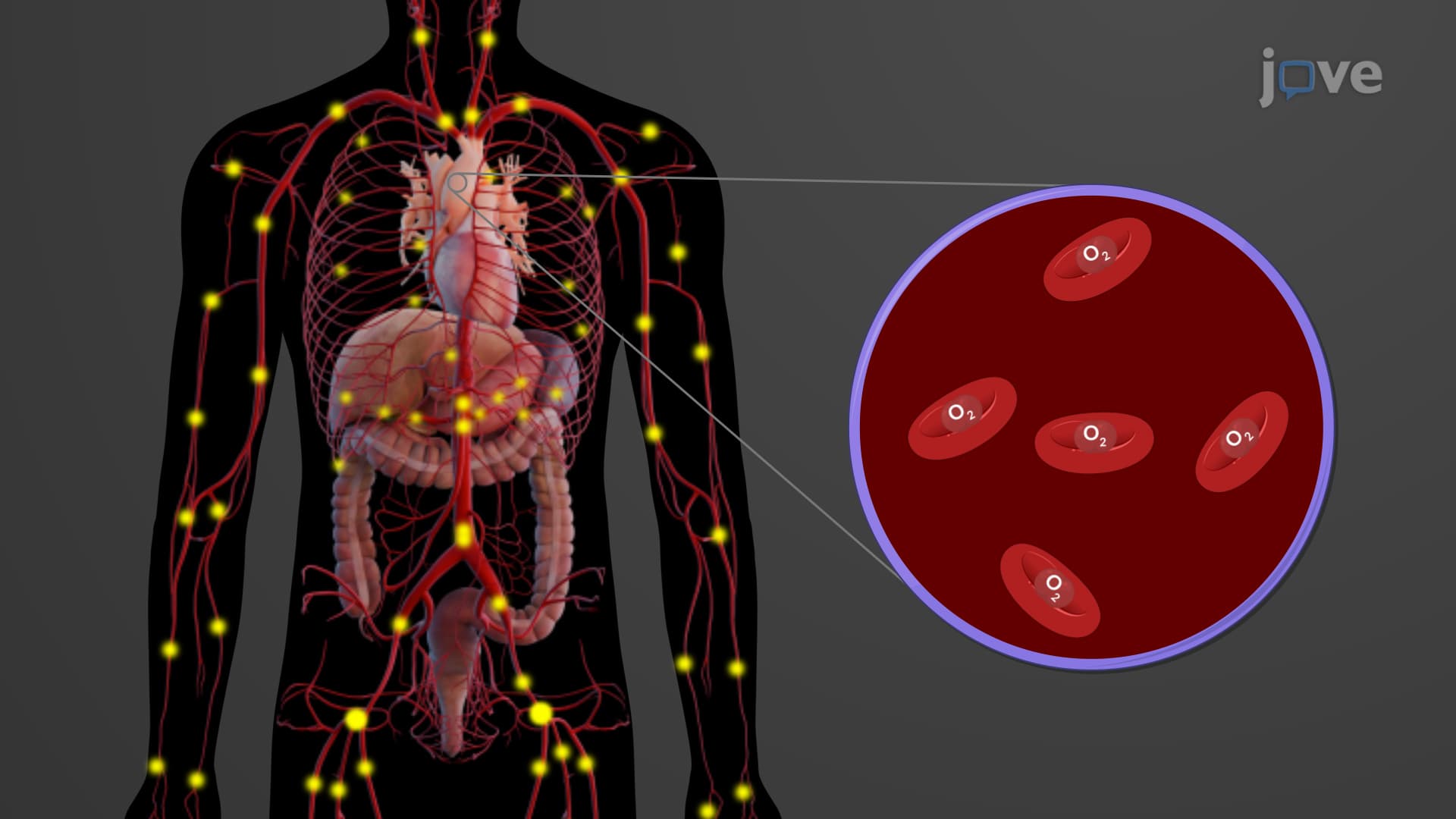
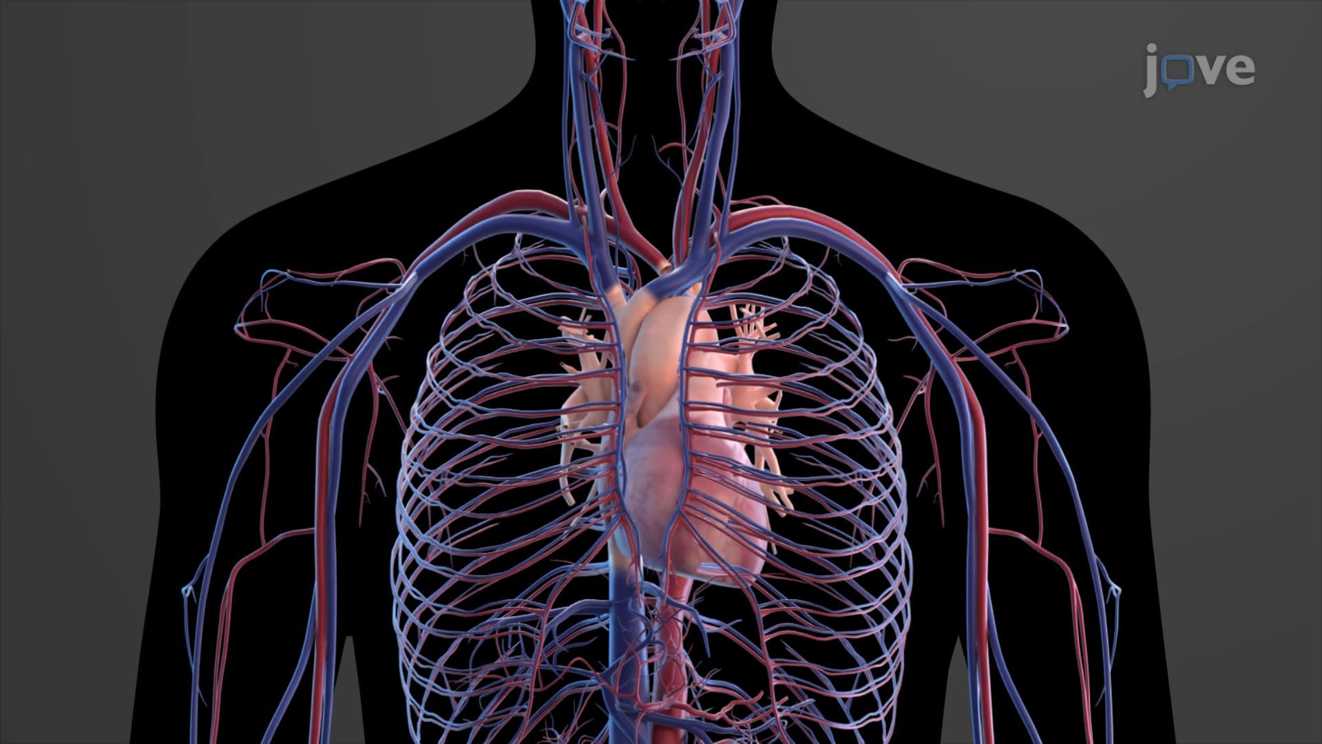
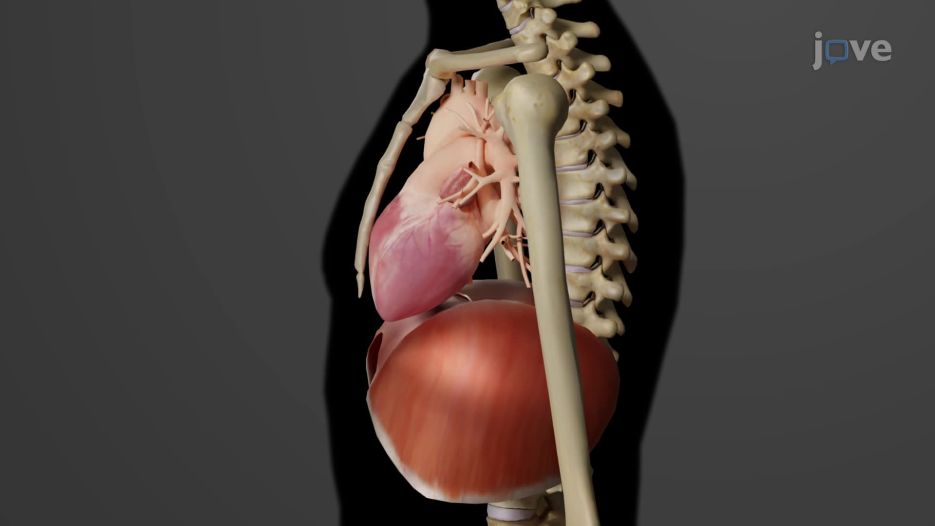
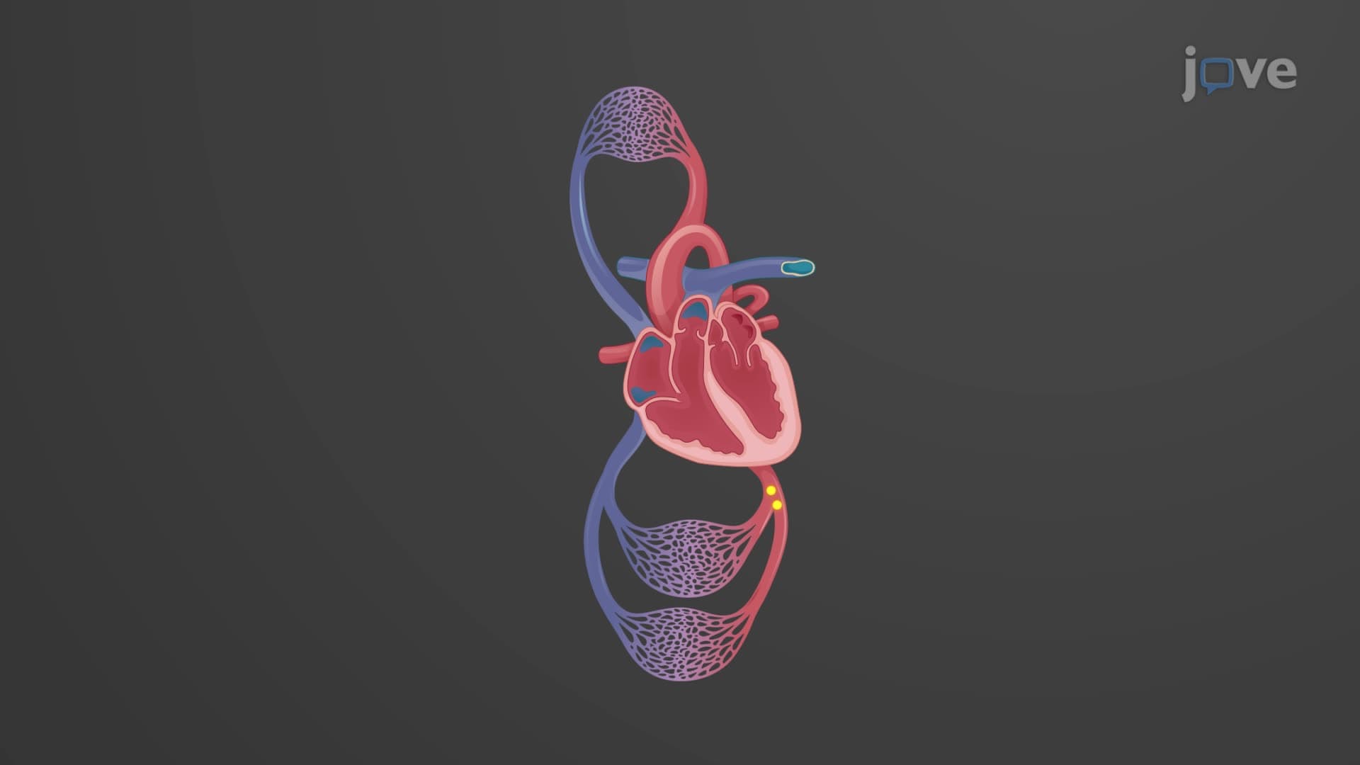
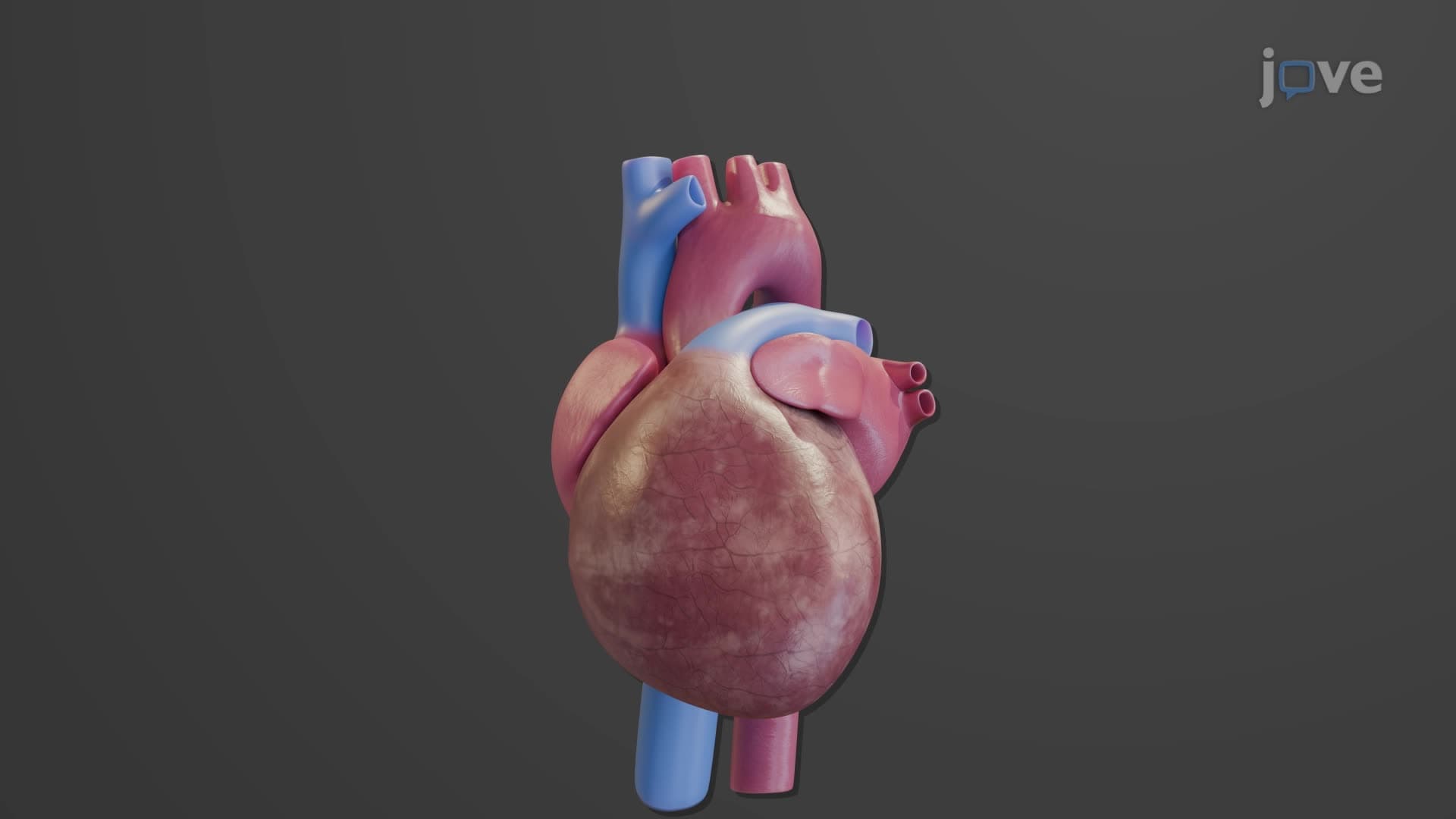
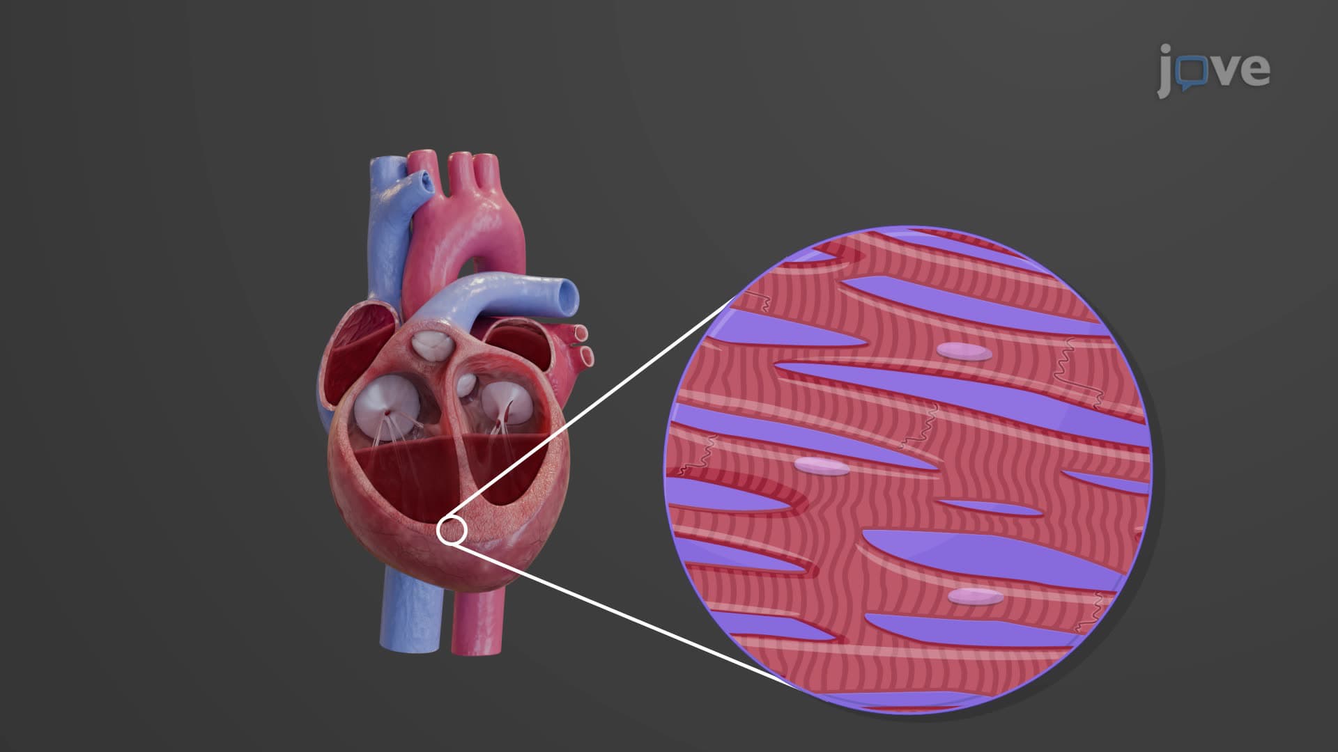
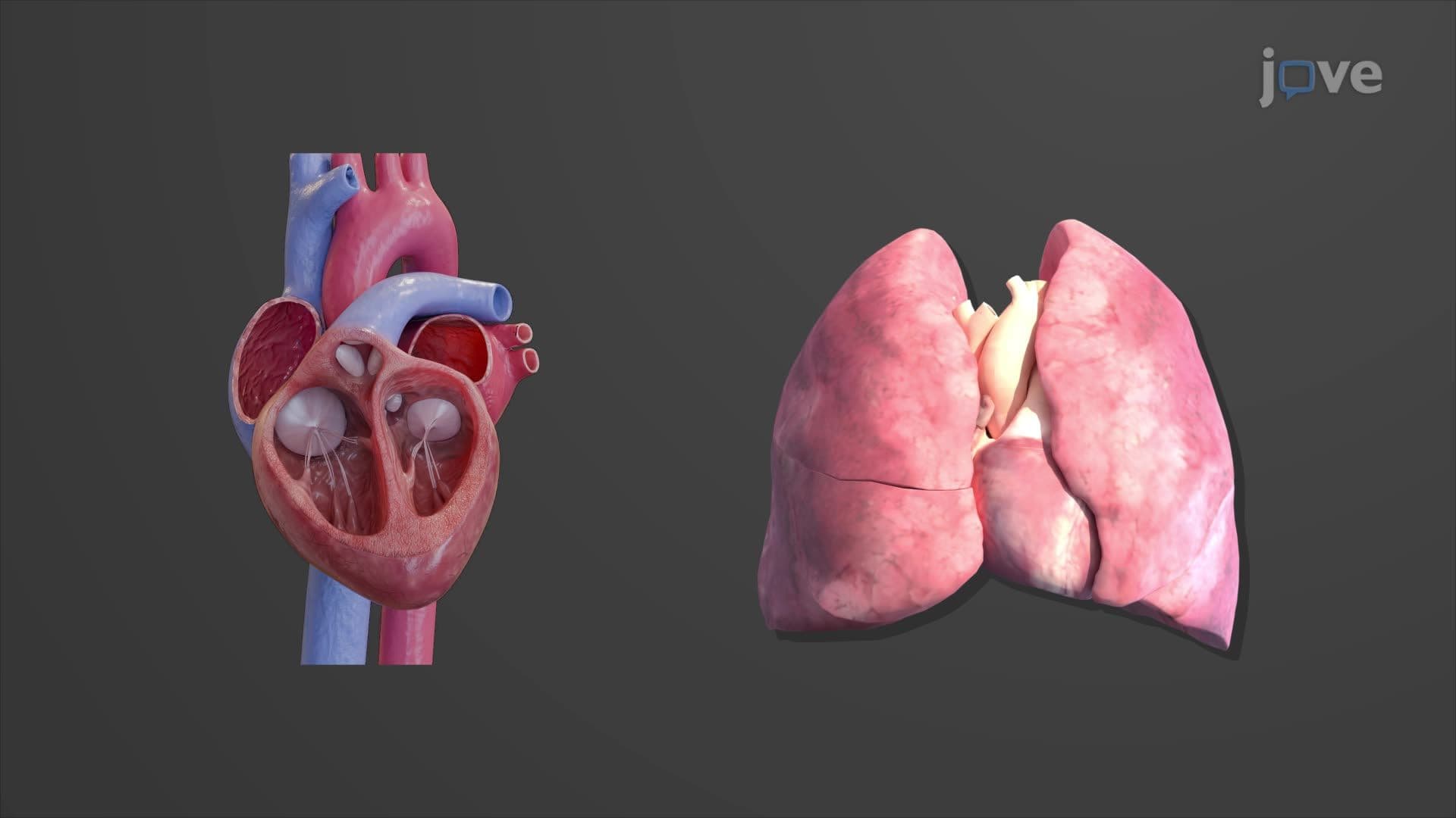
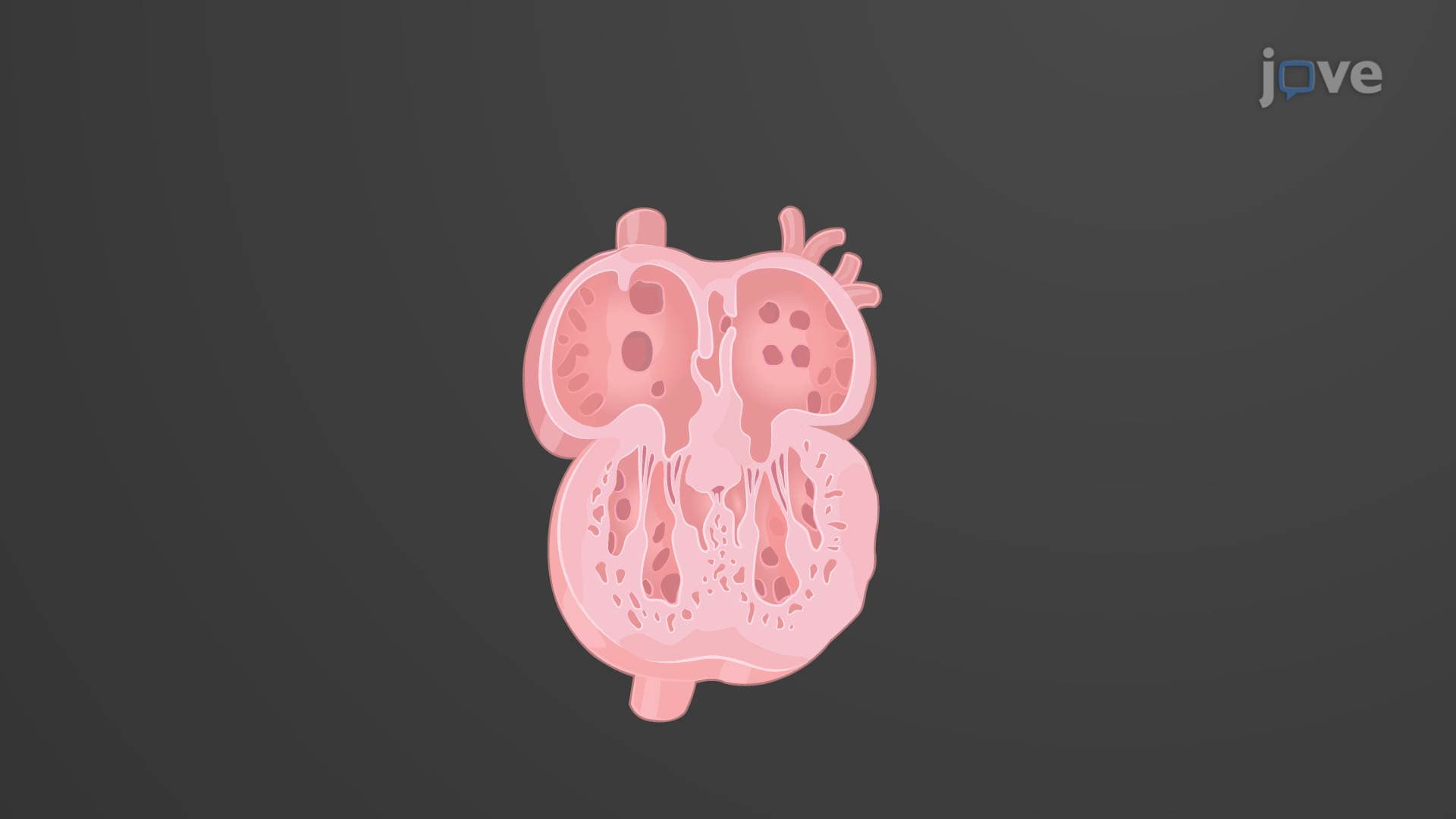
Coronary circulation anatomy biology represents one of the most critical vascular networks in the human body. Unlike other organs that receive blood from distant arteries, the heart muscle (myocardium) requires its own dedicated blood supply system operating independently from systemic and pulmonary circuits. This specialized circulation ensures continuous oxygen and nutrient delivery to cardiac muscle cells that work 24/7 without rest.
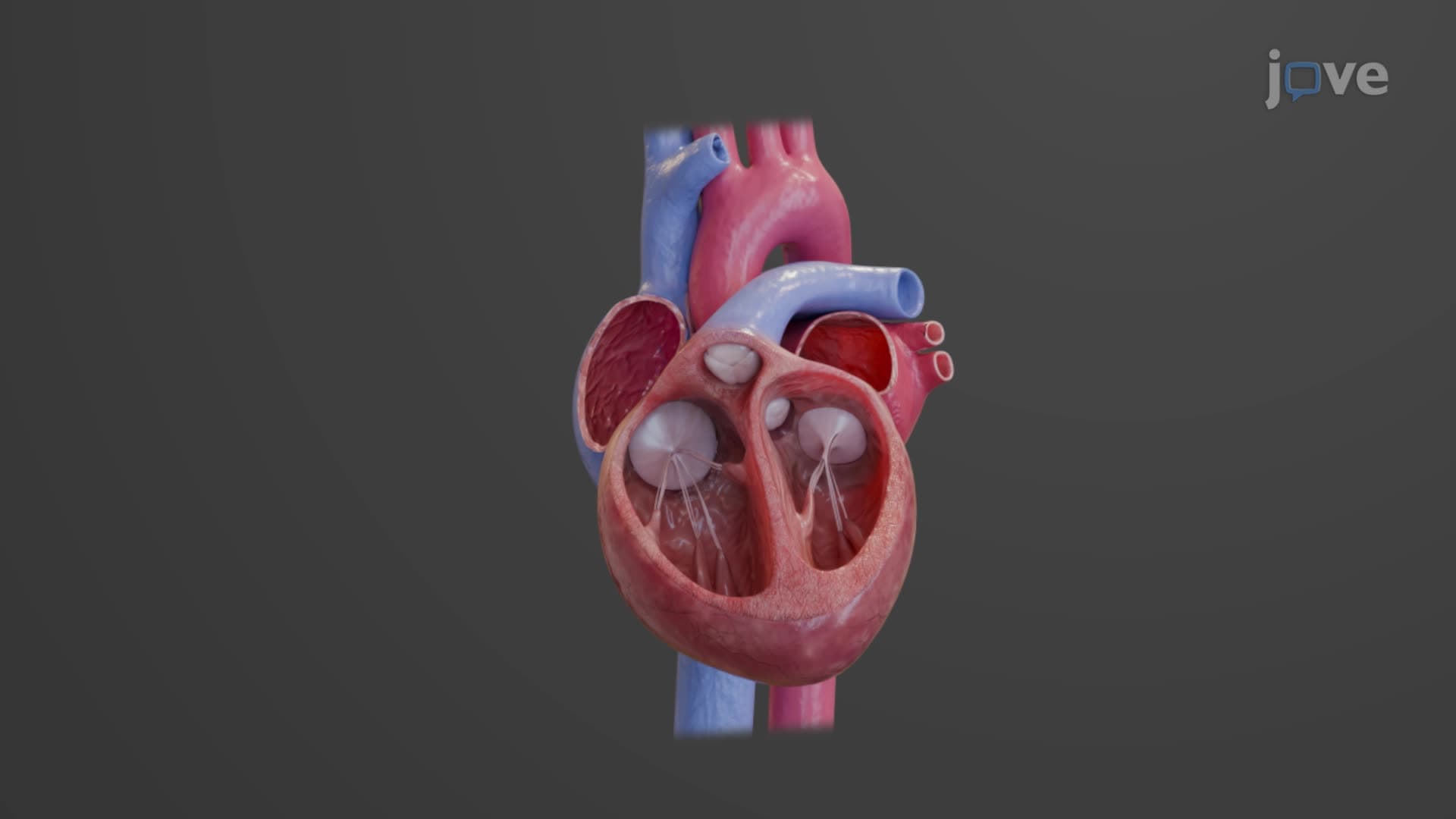
The heart blood supply explained begins with two main coronary arteries originating from the ascending aorta just above the aortic valve. The left main coronary artery quickly bifurcates into the left anterior descending artery (LAD) and circumflex artery. The LAD, often called the "widow maker" due to its critical importance, supplies the anterior wall of both ventricles and the anterior portion of the interventricular septum. Students preparing for the MCAT should note that LAD occlusion causes anterior wall myocardial infarctions, the most common type seen in emergency departments across the United States.
The circumflex artery wraps around the left side of the heart, supplying the lateral and posterior walls of the left ventricle and left atrium. Meanwhile, the right coronary artery (RCA) travels along the right atrioventricular groove, giving rise to the posterior descending artery (PDA) and marginal branches. This LAD RCA coronary system demonstrates functional redundancy through anastomoses-natural bypass connections that can provide alternate blood flow routes when primary vessels become blocked.
Coronary sinus venous return completes the circulation by collecting deoxygenated blood from cardiac muscle. The coronary sinus, located in the posterior wall of the right atrium, receives drainage from the great cardiac vein (LAD territory), middle cardiac vein (RCA territory), and small cardiac vein (right ventricle). Some anterior cardiac veins drain directly into the right atrium, bypassing the coronary sinus entirely.
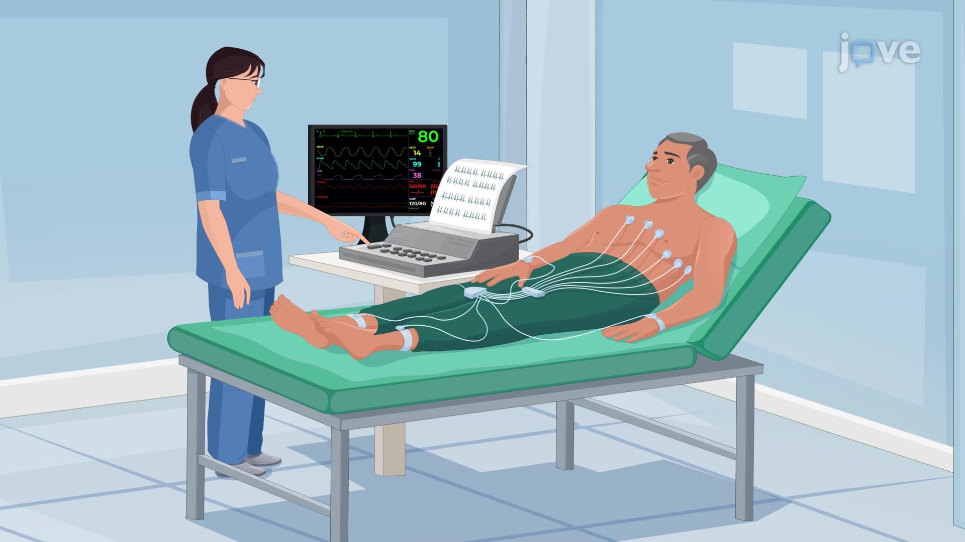
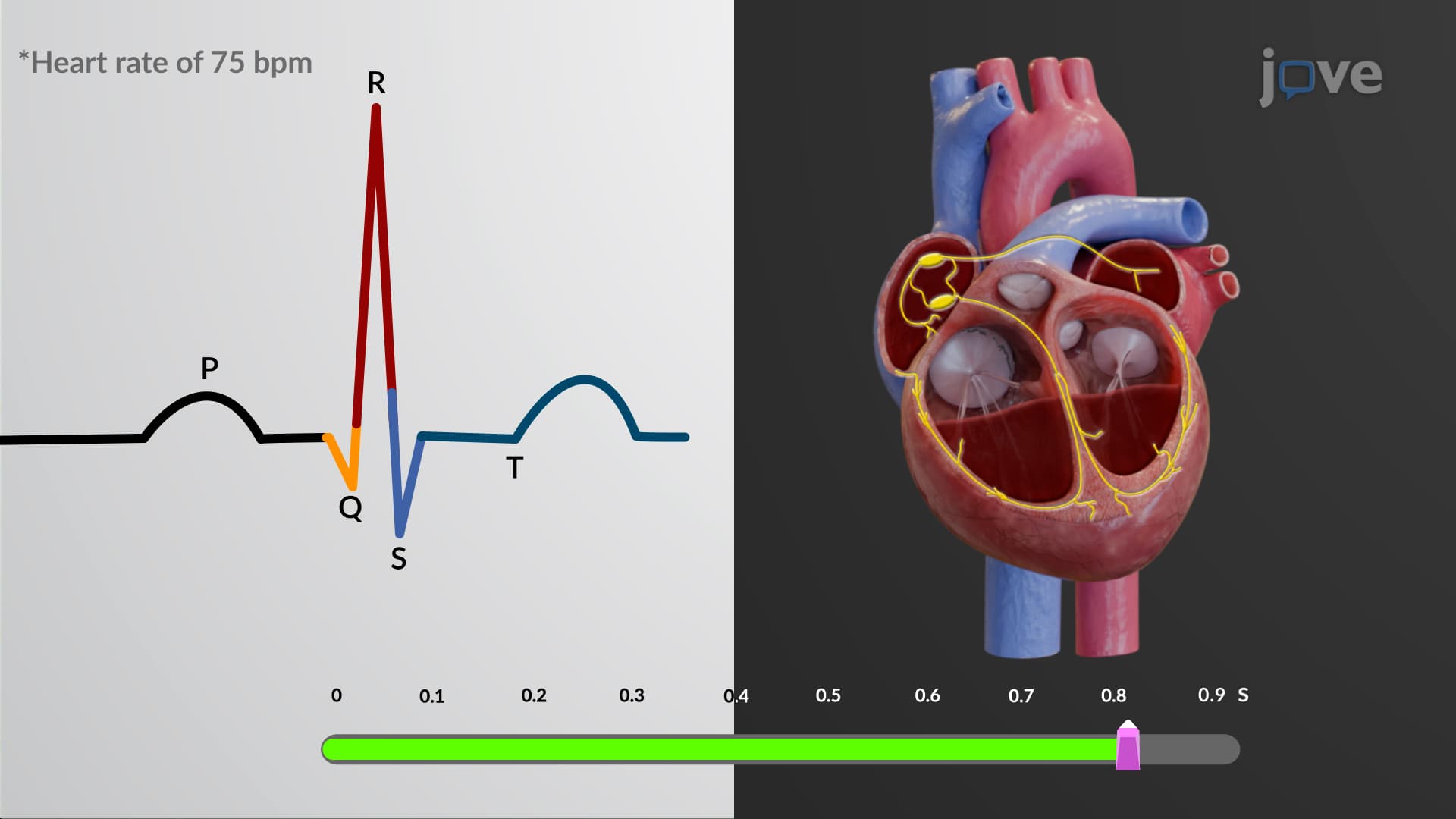
Understanding myocardial blood supply proves essential for healthcare professionals interpreting electrocardiograms (ECGs) and cardiac catheterization results. For instance, ST-elevation in leads II, III, and aVF typically indicates RCA occlusion affecting the inferior wall, while changes in leads V1-V4 suggest LAD involvement. This knowledge directly applies to USMLE Step 1 questions and NCLEX-RN cardiac scenarios that nursing students encounter in their board examinations.
Related Micro-courses