14 Concepts
6 Concepts
21 Concepts
23 Concepts
21 Concepts
15 Concepts
16 Concepts
26 Concepts
18 Concepts
19 Concepts
11 Concepts
6 Concepts
10 Concepts
22 Concepts
20 Concepts
26 Concepts
26 Concepts
14 Concepts
10 Concepts
12 Concepts
23 Concepts
29 Concepts
19 Concepts
38 Concepts
38 Concepts
25 Concepts
40 Concepts
25 Concepts
25 Concepts
20 Concepts
37 Concepts
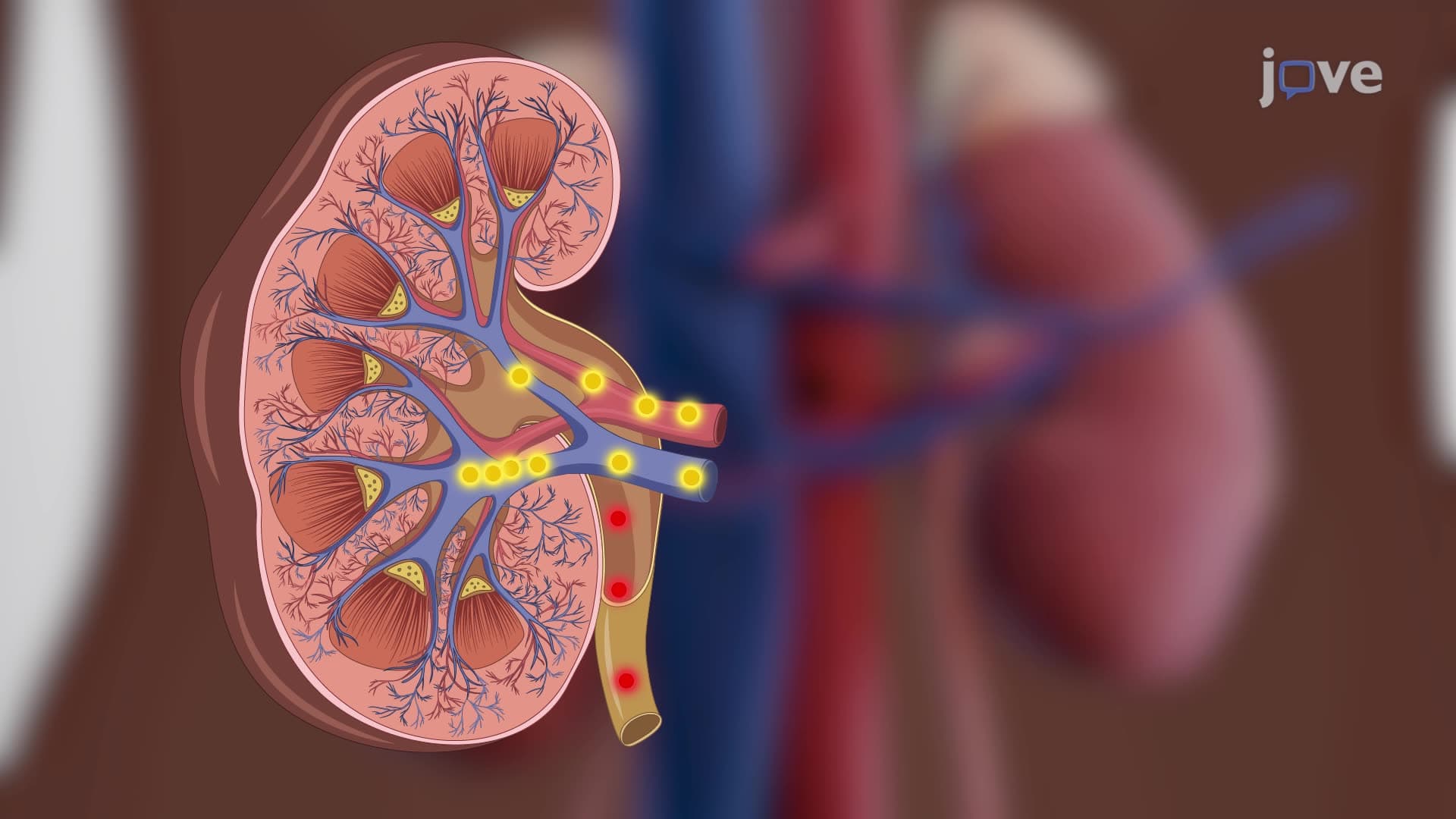
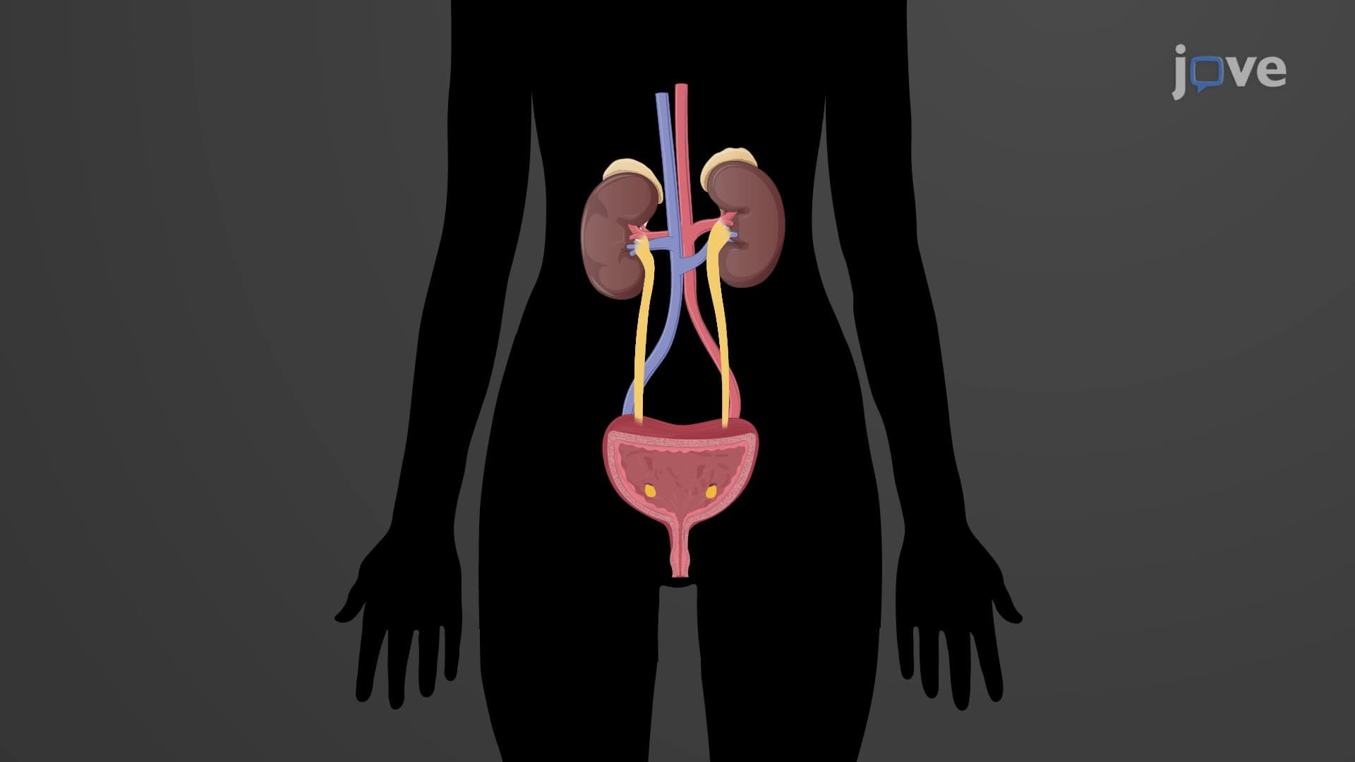
The urinary system comprises the kidneys, ureters, bladder, and urethra, working together to filter blood, regulate fluid balance, and eliminate waste through urine formation. Understanding kidney anatomy physiology and the structure and function of the urinary system is essential for students pursuing healthcare careers in the United States. This comprehensive course covers everything from nephron filtration mechanisms to clinical applications like dialysis, making complex renal physiology accessible through JoVE Coach's expert instruction.
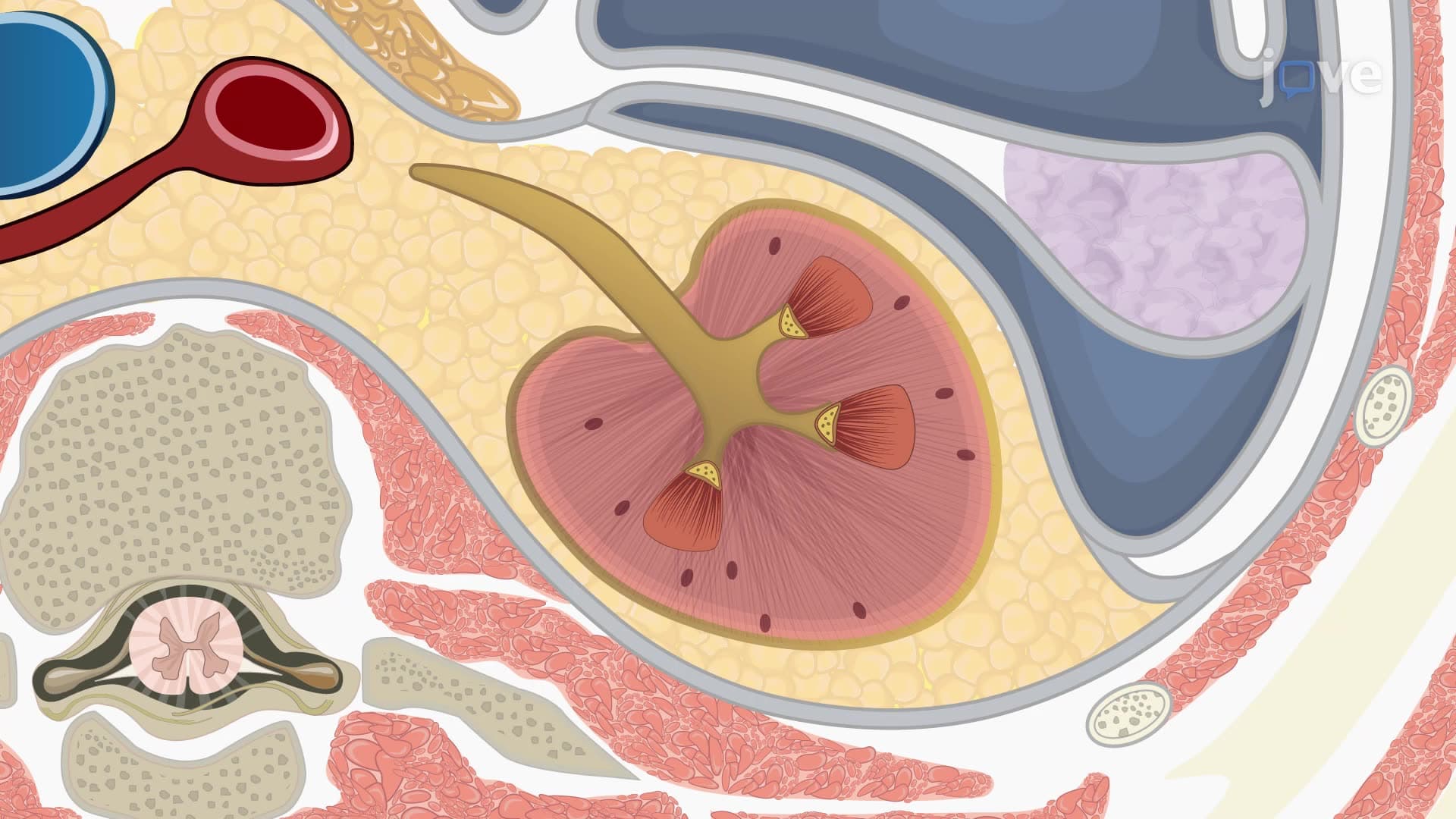
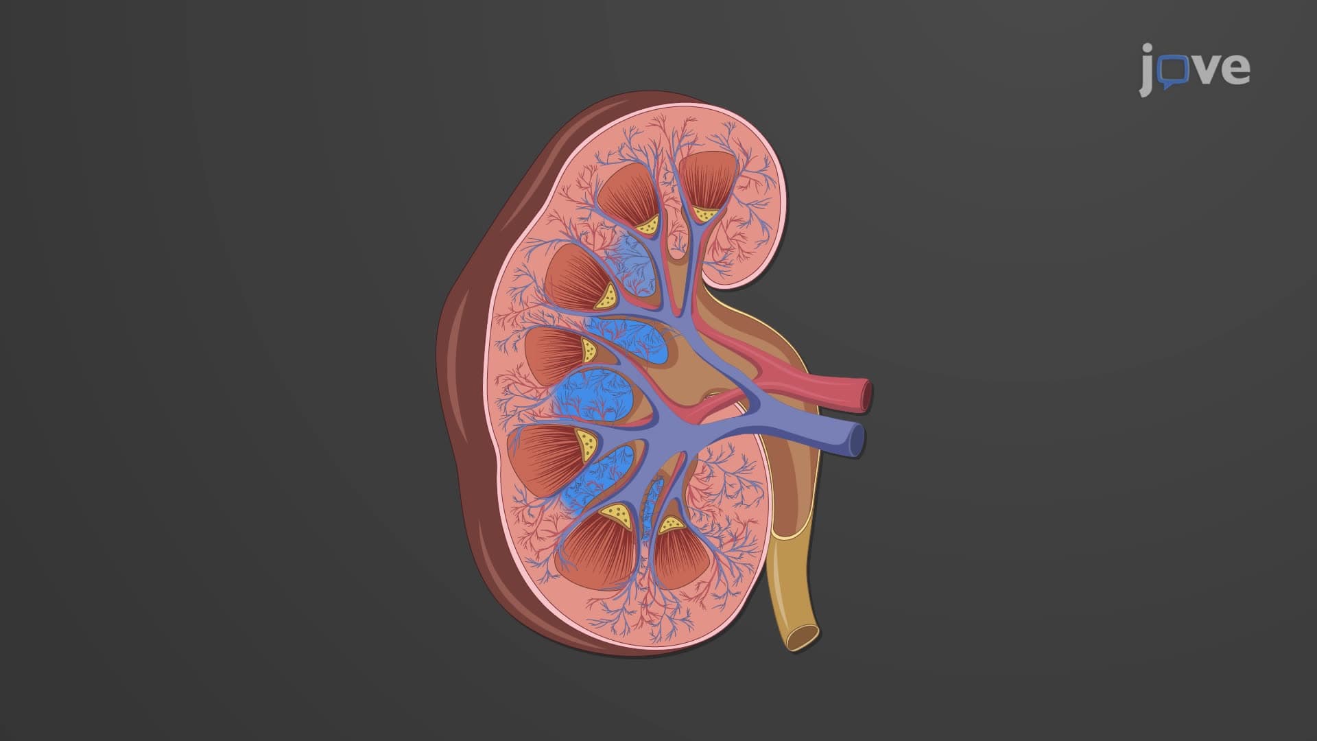
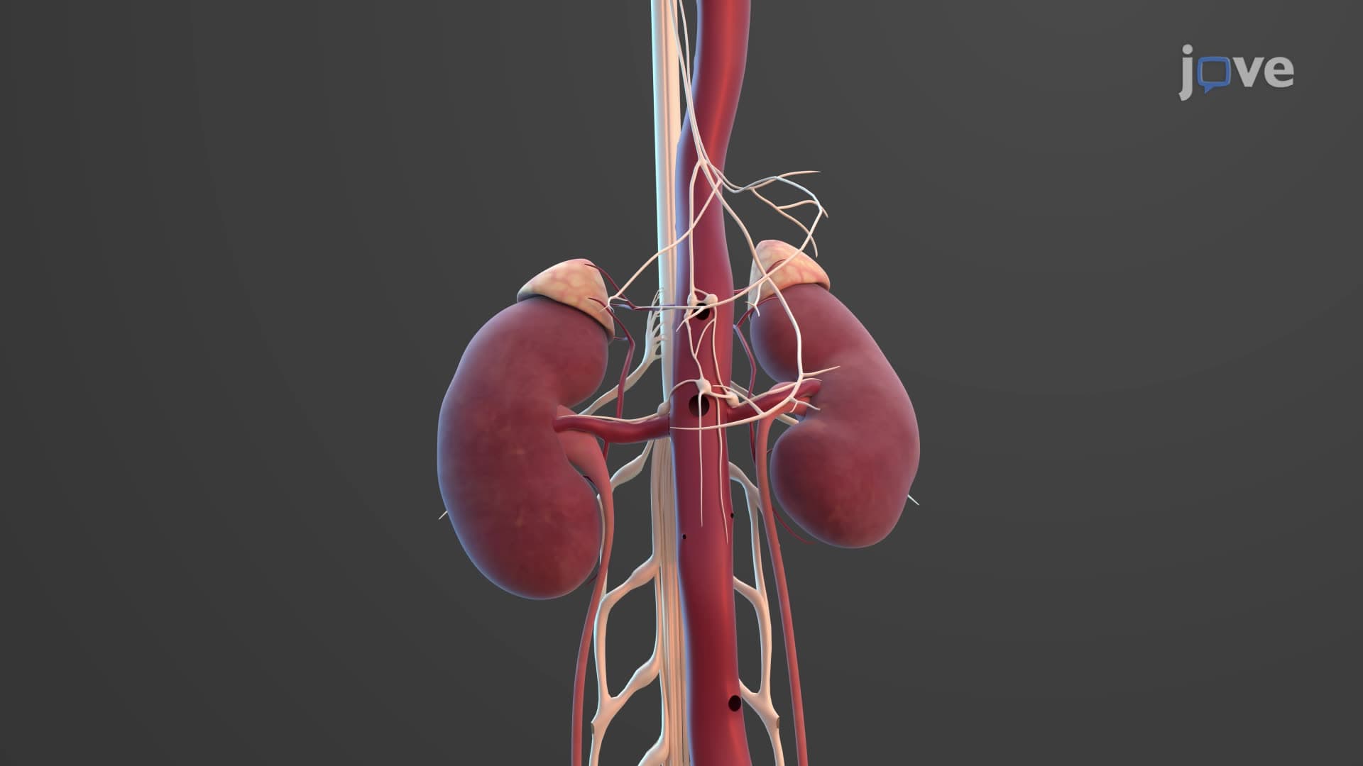
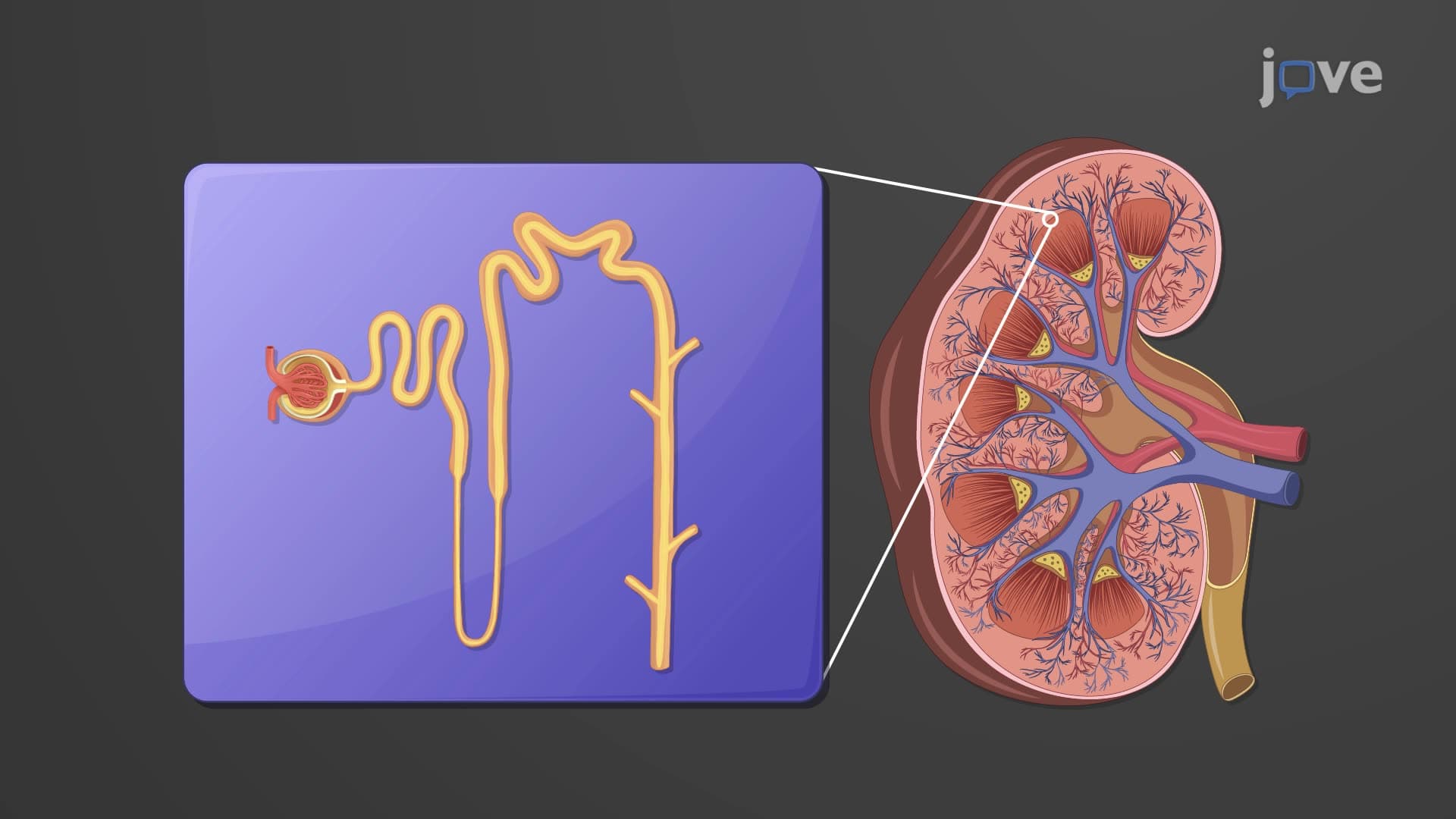
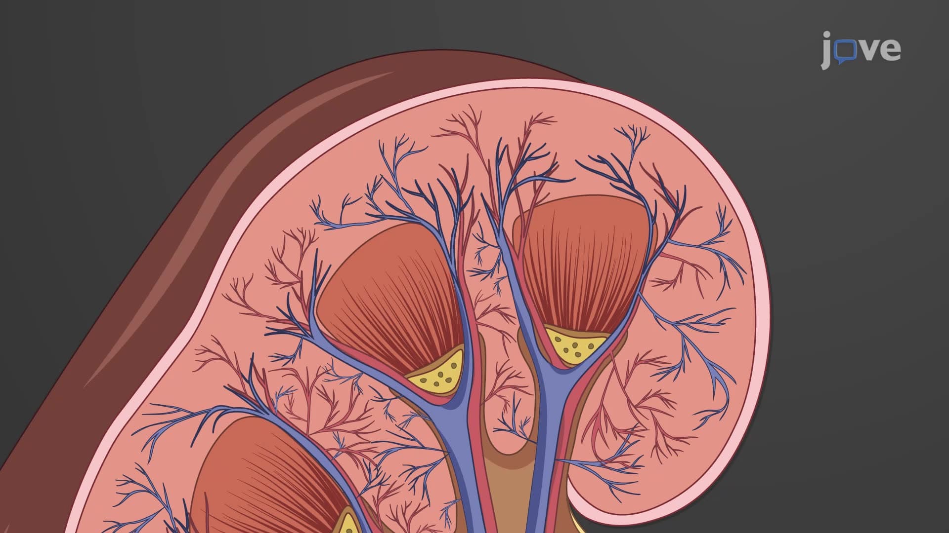
1. Urinary System Architecture and Kidney Anatomy: The urinary system consists of paired kidneys positioned at the T12-L3 vertebrae, protected by ribs and surrounded by three tissue layers. Each kidney measures approximately 10-12 cm long and contains distinct regions including the outer renal cortex, inner medulla with cone-shaped pyramids, and the renal pelvis drainage system. The renal hilum serves as the entry point for blood vessels, nerves, and the ureter. Understanding this structural organization is crucial for medical students preparing for USMLE Step 1, as kidney anatomy frequently appears in nephrology questions.
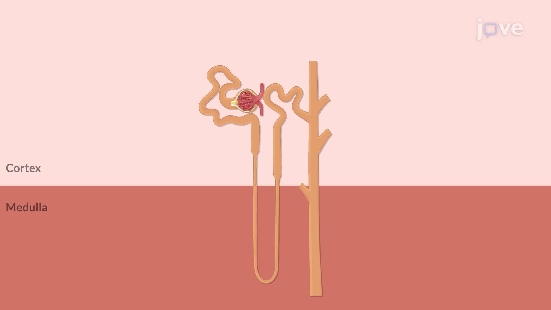
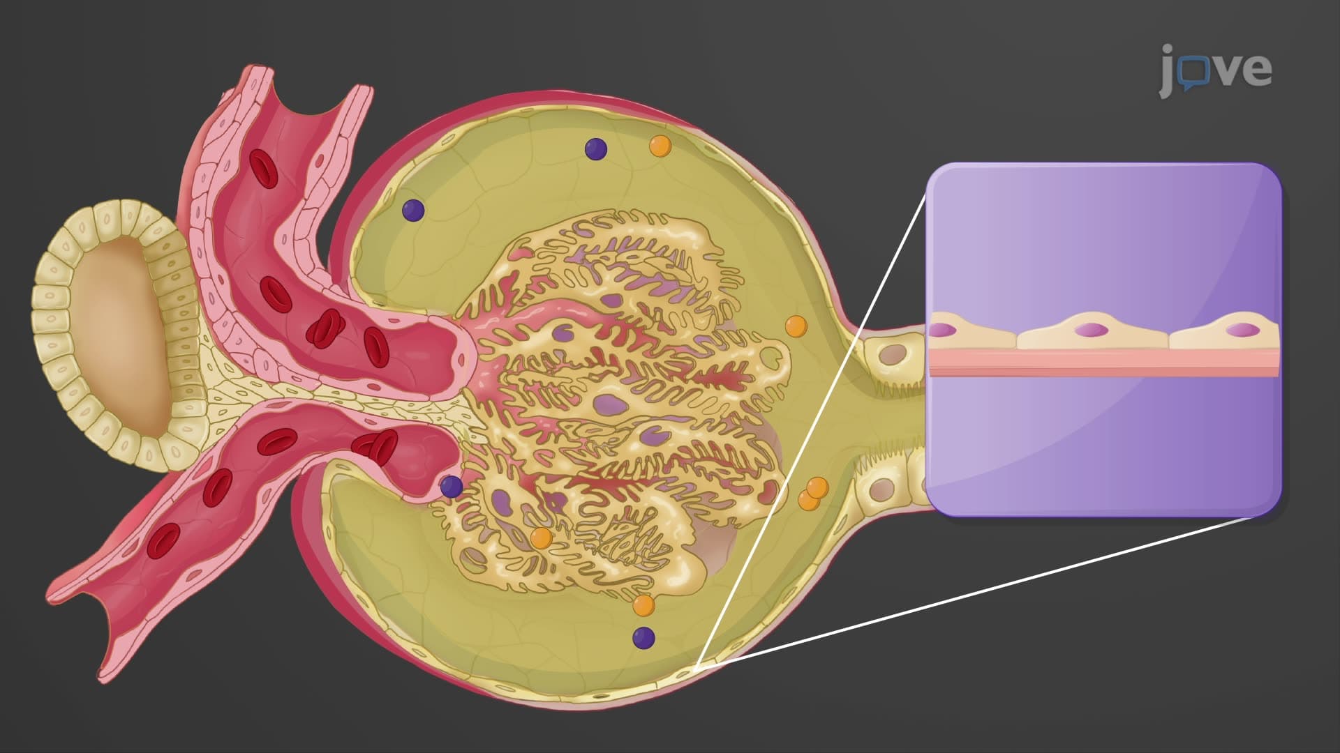
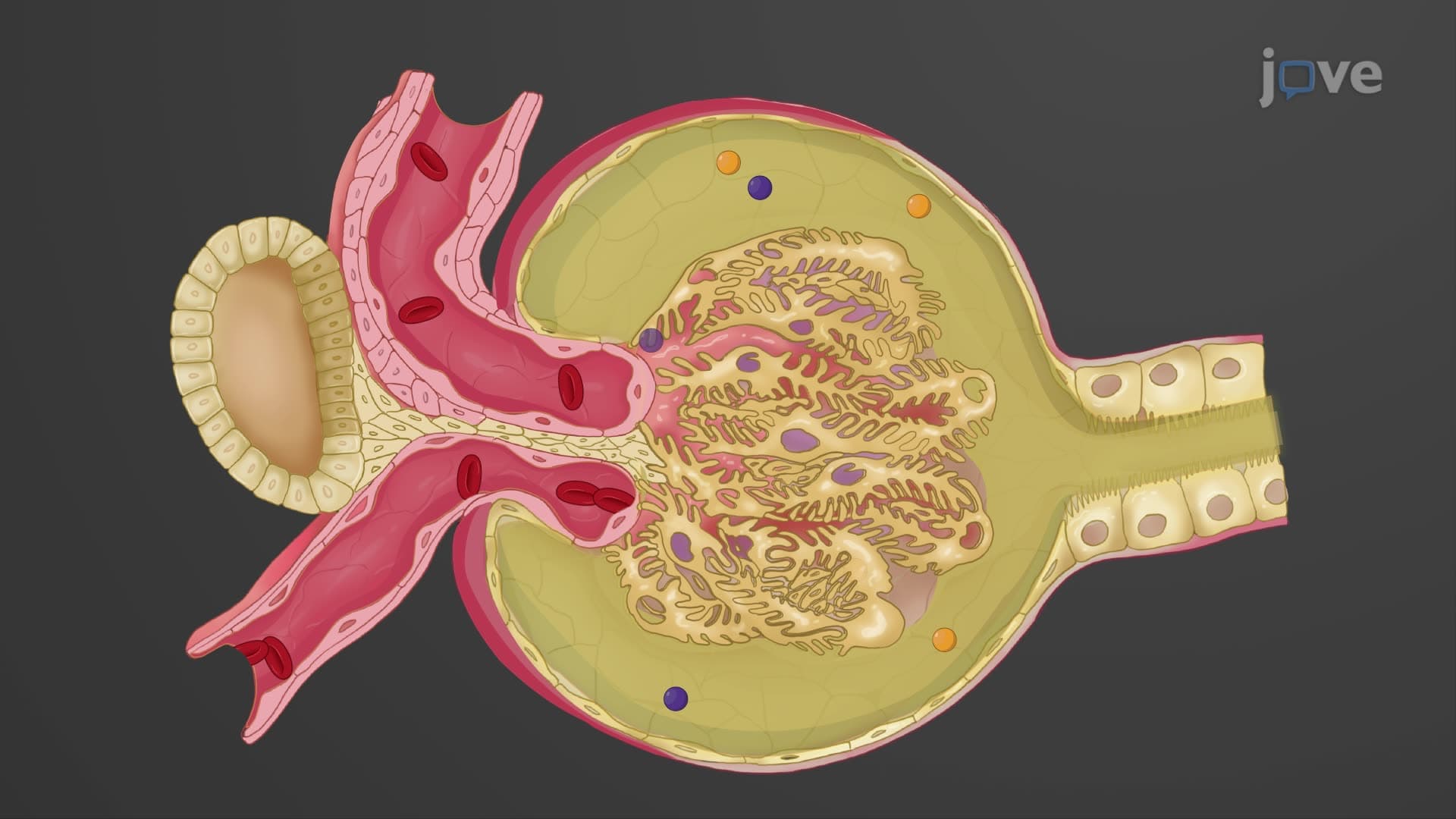
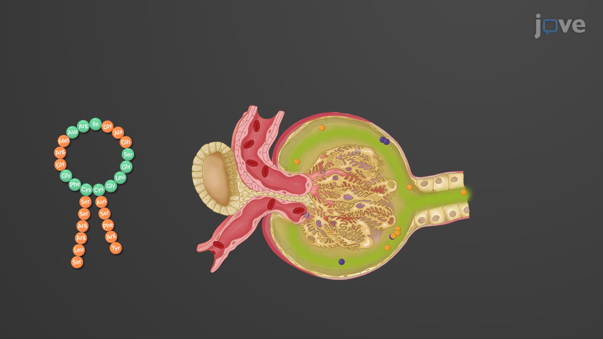
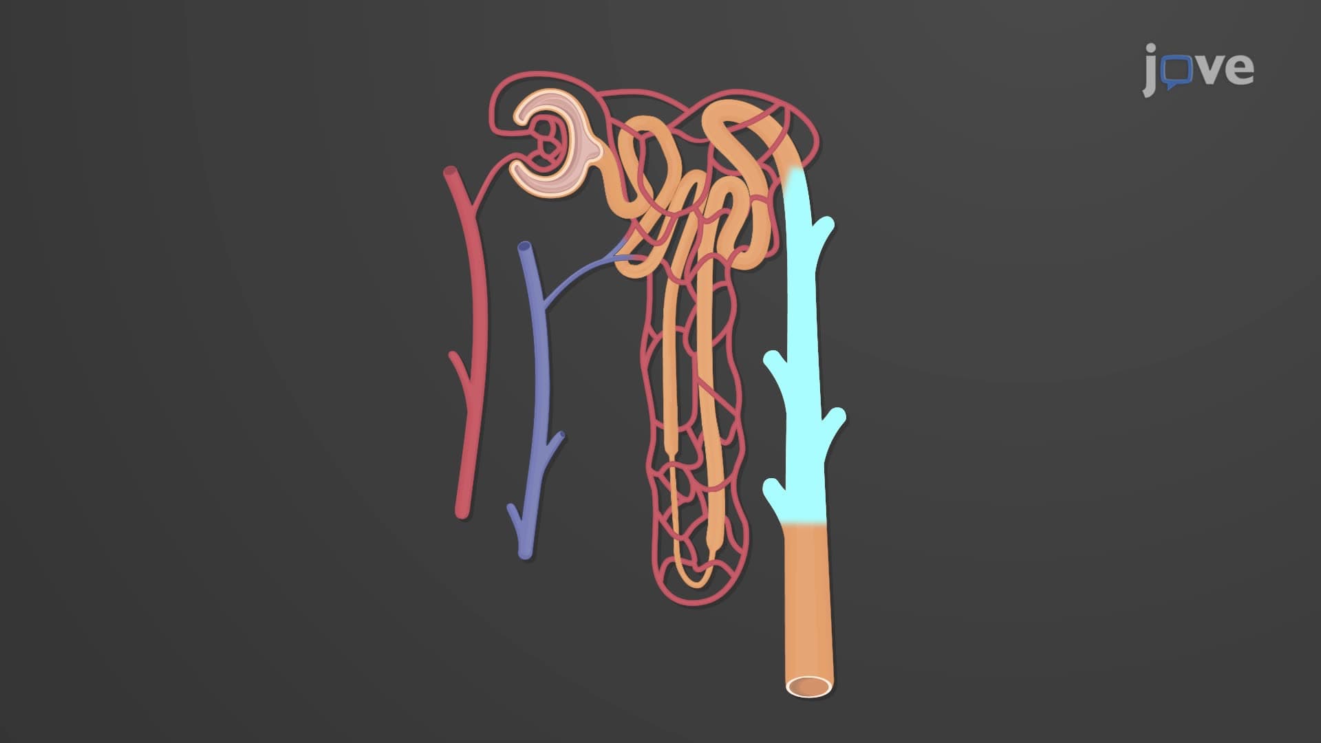
2. Nephron Structure and Specialized Cell Types: Each kidney contains about one million nephrons, the functional units responsible for filtration and urine formation. Cortical nephrons (80-85%) have shorter loops of Henle, while juxtamedullary nephrons (15-20%) extend deep into the medulla with longer loops essential for concentrating urine. The nephron includes the renal corpuscle with its glomerulus and Bowman's capsule, followed by the proximal convoluted tubule, loop of Henle, and distal convoluted tubule. Specialized cells like podocytes create filtration slits, while principal and intercalated cells in collecting ducts regulate sodium balance and pH.
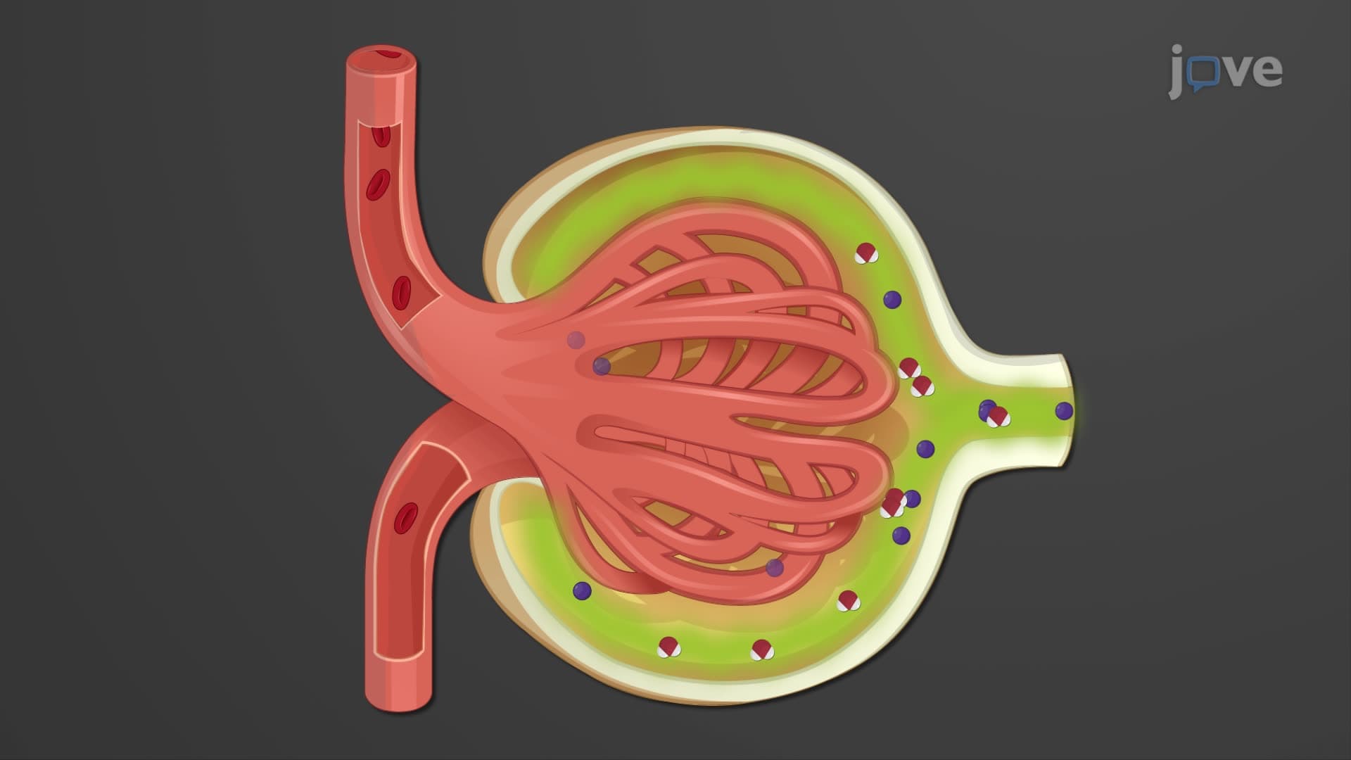
3. Glomerular Filtration Mechanisms and Pressure Dynamics: Blood filtration occurs at the glomerular filtration barrier, composed of fenestrated endothelial cells, basement membrane, and podocyte filtration slits. Net filtration pressure (NFP) results from glomerular blood hydrostatic pressure (55 mmHg) minus capsular hydrostatic pressure (15 mmHg) and blood colloid osmotic pressure (30 mmHg), yielding approximately 10 mmHg. This process produces 180 liters of filtrate daily, with 99% reabsorbed. MCAT students must understand how changes in these pressures affect filtration rates and kidney disease progression.
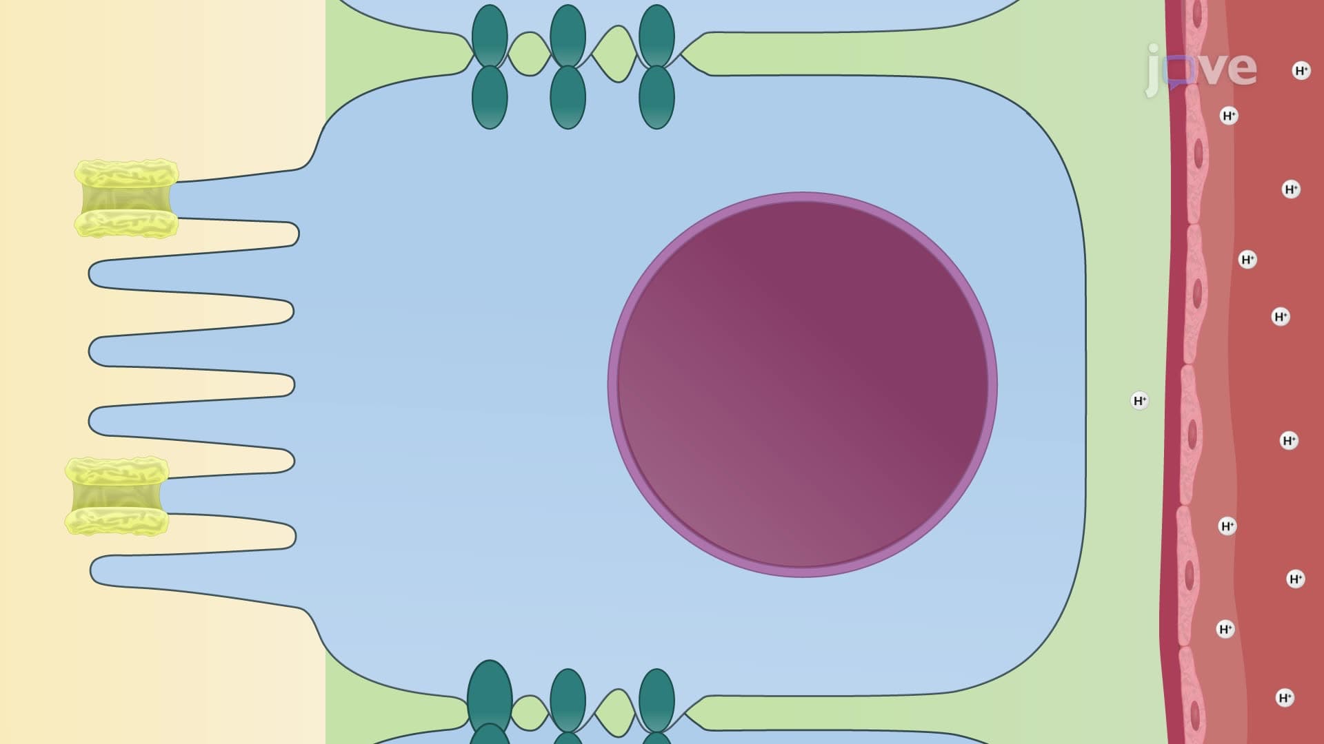
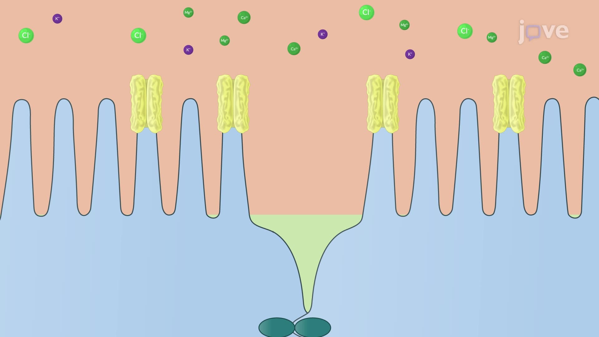
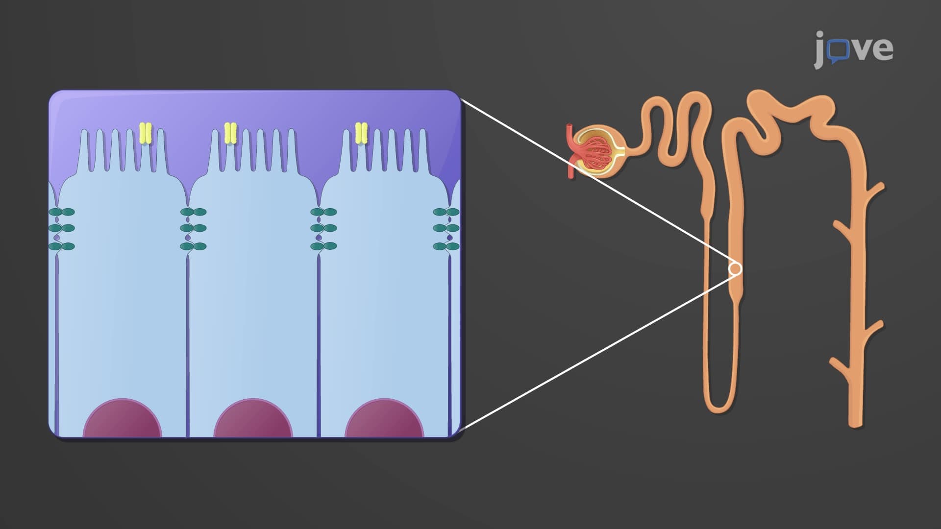
4. Tubular Reabsorption and Secretion Throughout the Nephron: The proximal convoluted tubule reabsorbs 65% of filtered sodium and water through active transport mechanisms including Na+/glucose symporters and Na+/H+ antiporters. The thick ascending limb of Henle uses Na+/K+/2Cl- symporters to reabsorb electrolytes while remaining impermeable to water. The distal convoluted tubule and collecting duct fine-tune electrolyte balance under hormonal control, with aldosterone increasing sodium reabsorption and ADH enhancing water permeability. These processes are frequently tested on nursing exams like NCLEX and TEAS.
5. Urine Concentration and Dilution Mechanisms: The kidney's ability to produce concentrated or dilute urine depends on the medullary osmotic gradient established by juxtamedullary nephrons. Countercurrent multiplication in the loop of Henle creates increasing osmolarity from cortex to medulla, while countercurrent exchange in vasa recta maintains this gradient. During dehydration, ADH release increases water reabsorption in collecting ducts, producing concentrated urine up to four times more concentrated than plasma. Conversely, overhydration suppresses ADH, leading to dilute urine production through continued ion reabsorption without water recovery.
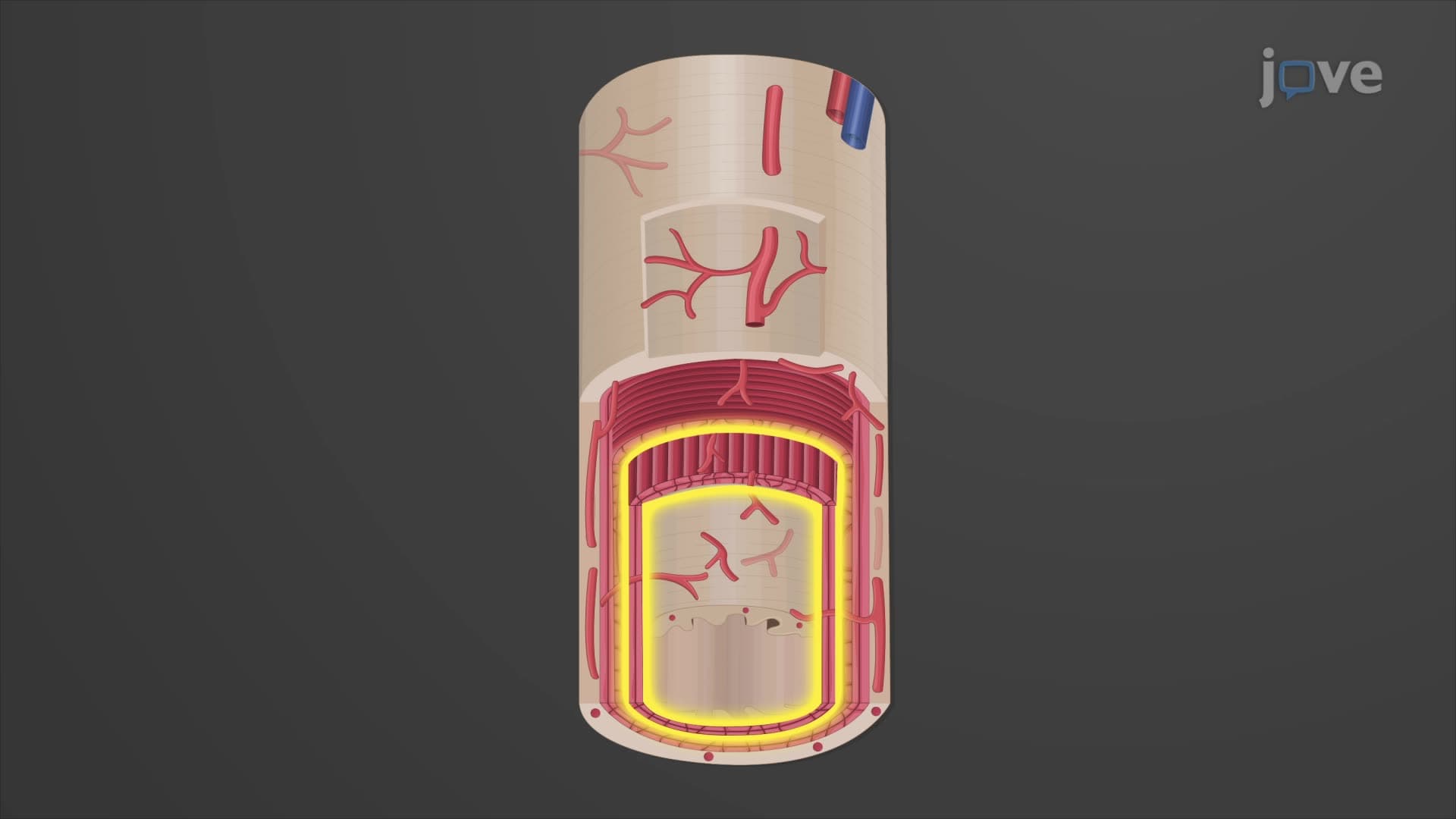
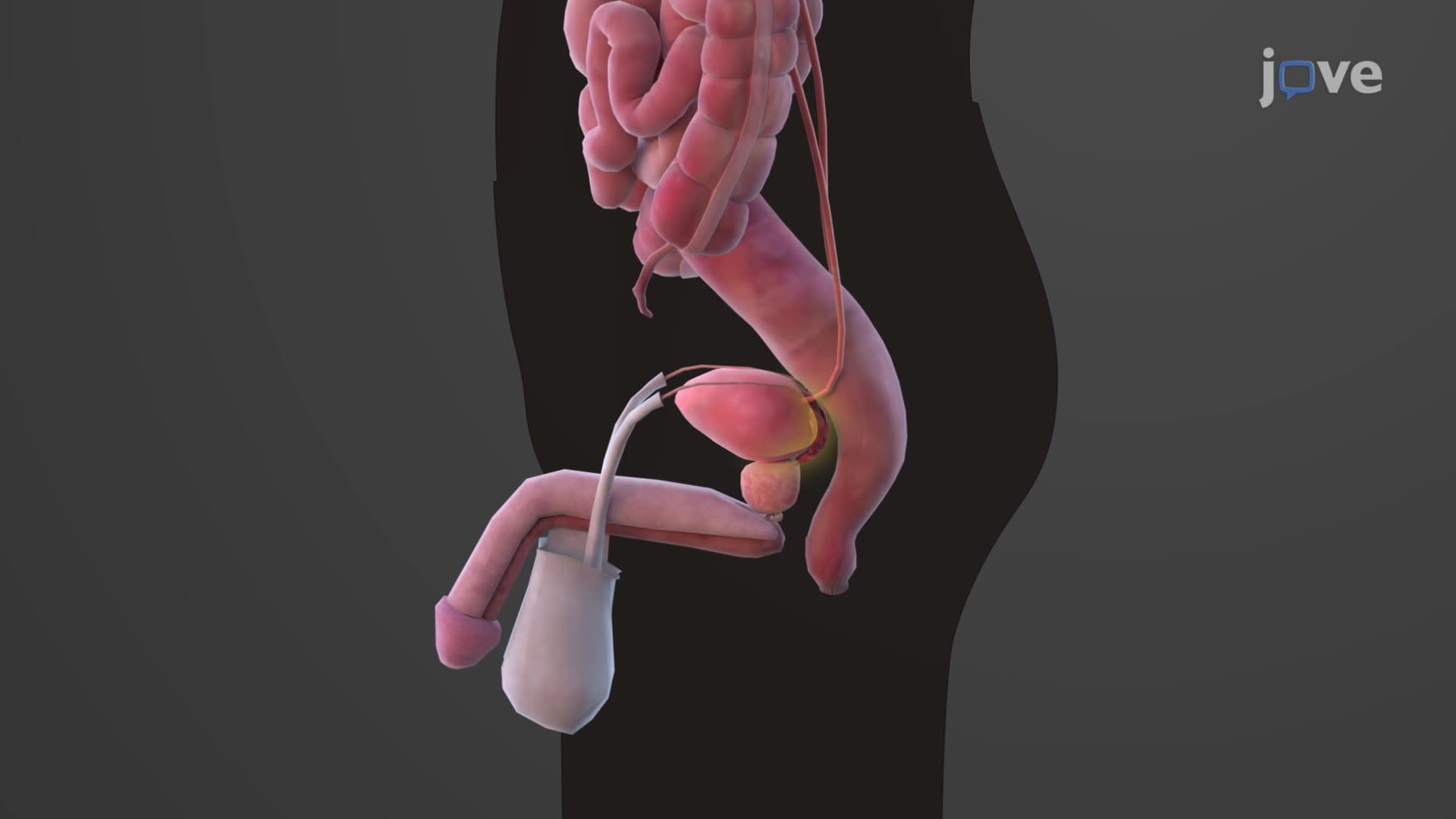
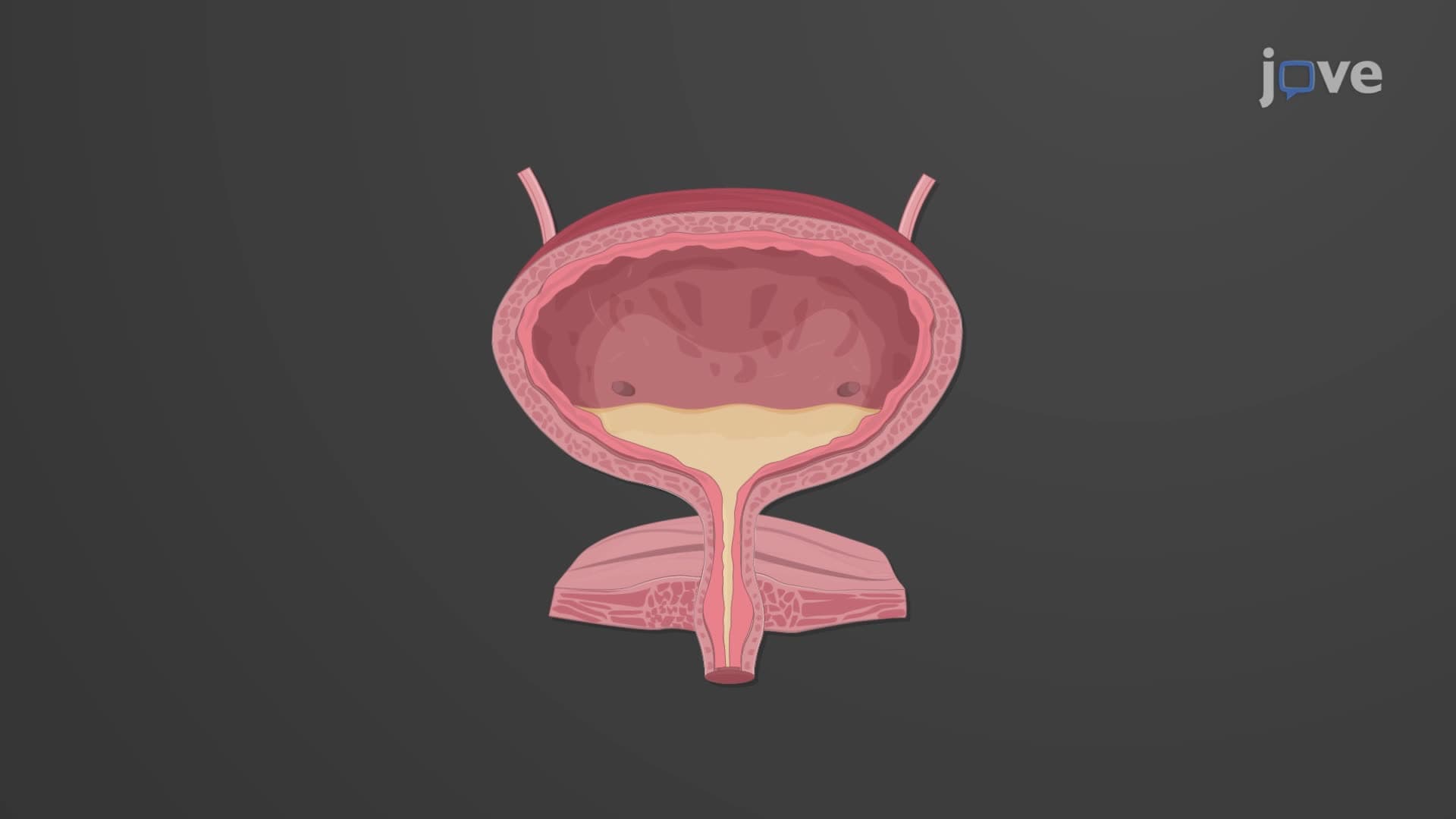
6. Lower Urinary Tract Function and Micturition Control: Ureters transport urine via peristaltic contractions through their three-layered walls containing transitional epithelium, smooth muscle, and connective tissue. The bladder stores up to 600 mL of urine in its distensible wall composed of detrusor muscle and rugae-containing mucosa. Micturition involves complex neural control through storage and voiding reflexes coordinated by sacral spinal segments and pontine centers. The internal urethral sphincter operates under autonomic control, while voluntary control of the external sphincter develops by age two, making this topic relevant for pediatric nursing students.
7. Clinical Applications: Renal Clearance and Diagnostic Testing: Renal clearance measurements assess kidney function by calculating the volume of plasma cleared of specific substances per minute. Creatinine clearance approximates glomerular filtration rate since creatinine is filtered but not reabsorbed or secreted. Inulin provides the gold standard for GFR measurement due to its complete filtration without reabsorption or secretion. These concepts are essential for pre-med students preparing for clinical rotations and understanding how healthcare providers monitor kidney function in conditions like diabetes, hypertension, and chronic kidney disease affecting millions of Americans.