Loading video…


 25 Concepts
 78.1K views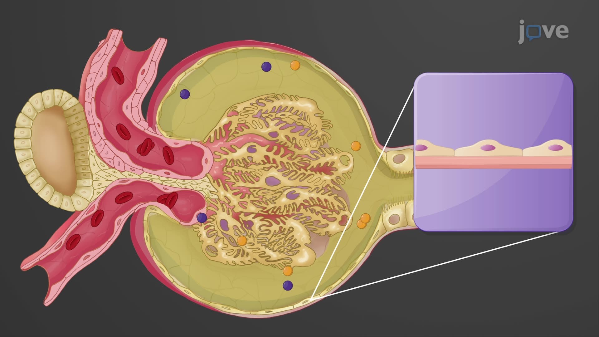

Video Summary: What Is Renal Corpuscle
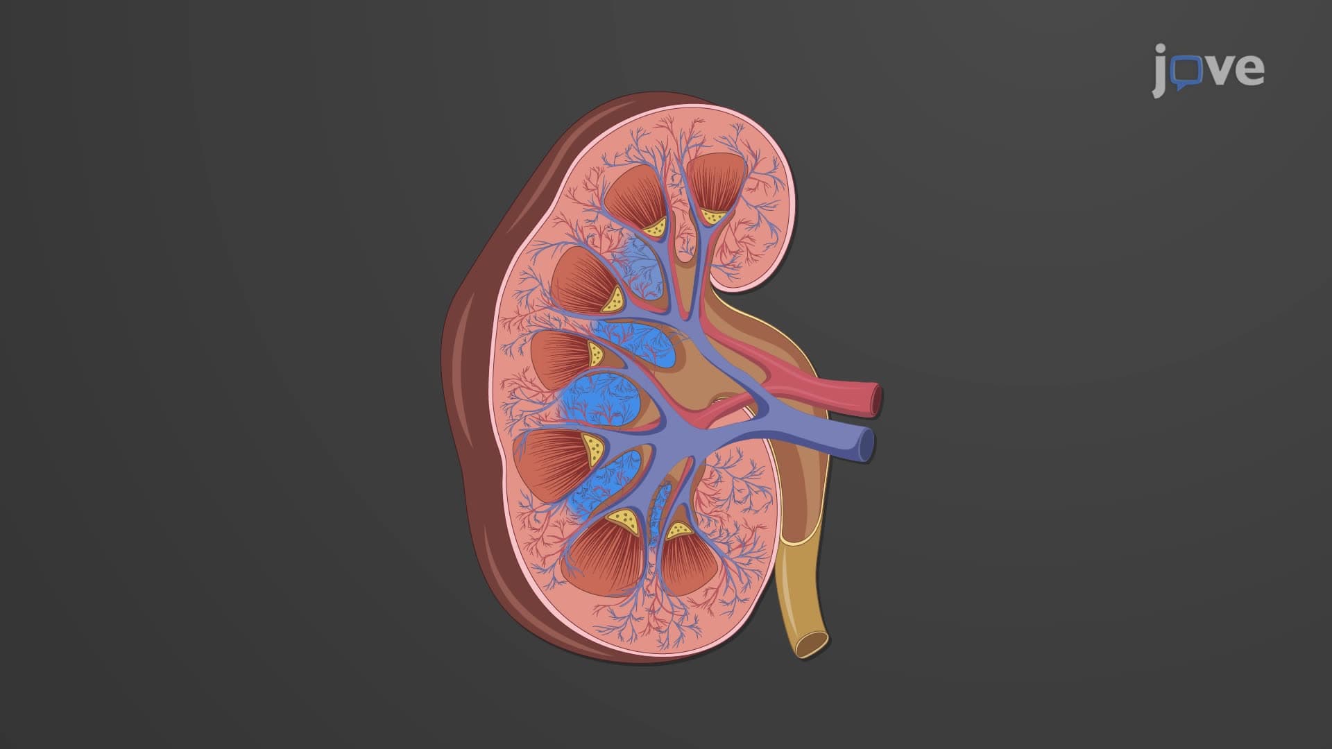
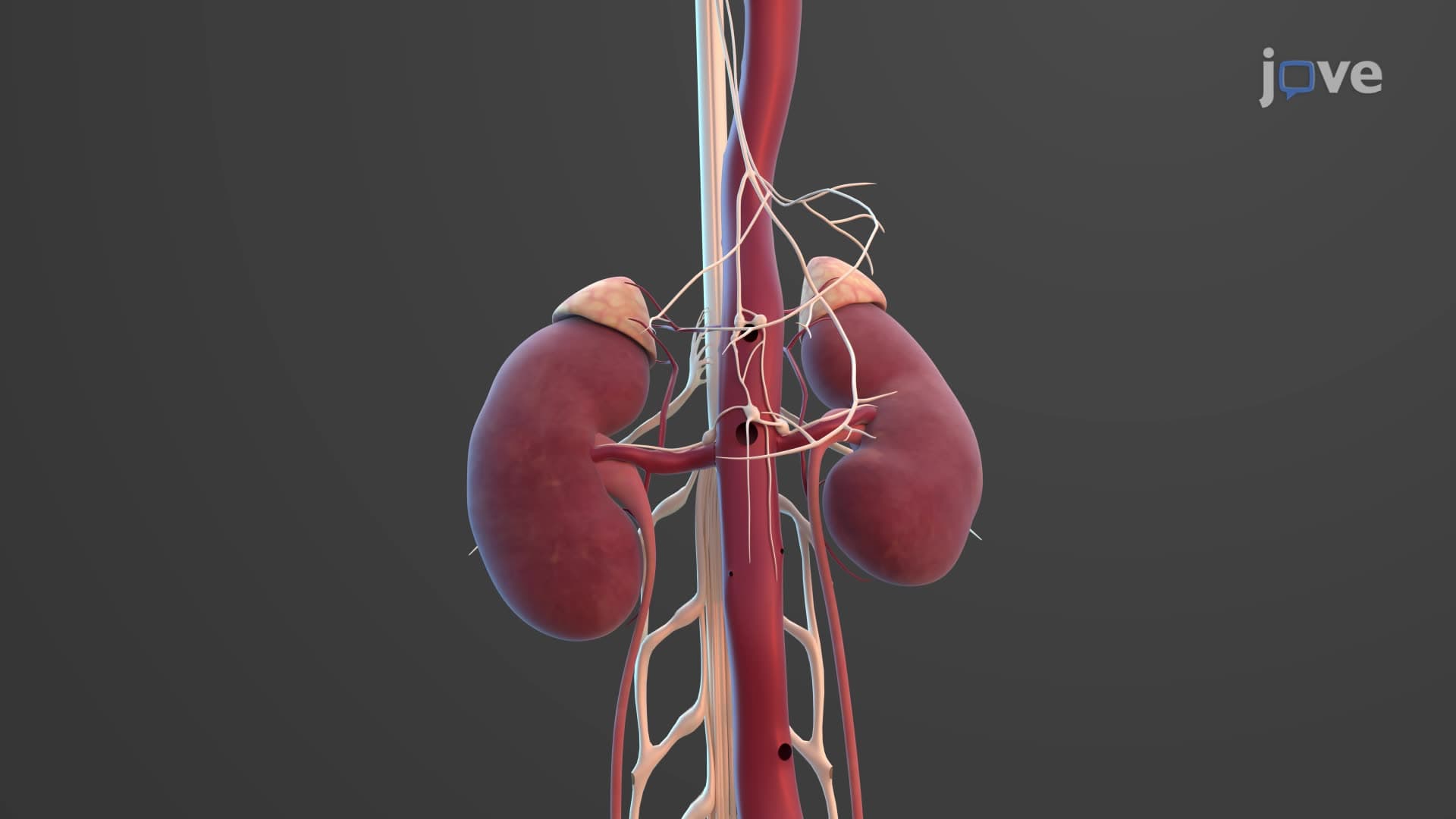
Did you know that your kidneys filter approximately 180 liters of blood daily-equivalent to filling a standard bathtub? Renal corpuscle anatomy biology reveals the microscopic filtration unit responsible for this remarkable process. At Johns Hopkins Medical School, students learn how each kidney contains about one million of these tiny filtering structures. The renal corpuscle consists of a specialized capillary cluster called the glomerulus surrounded by Bowman's capsule, creating the initial filtration system that removes waste from blood. Watch the full video on JoVE Coach to master this concept with expert-led visuals and step-by-step explanations.
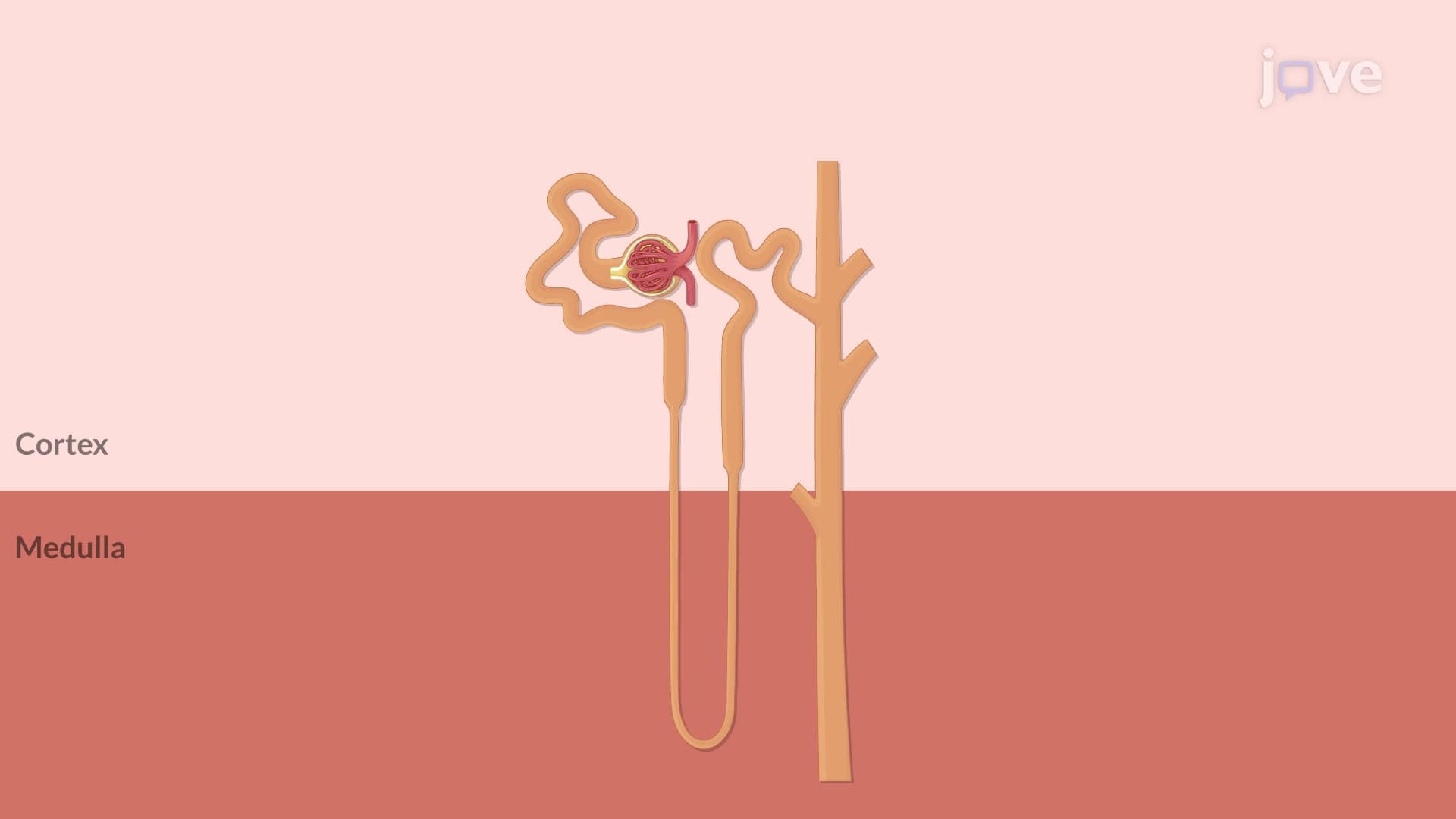
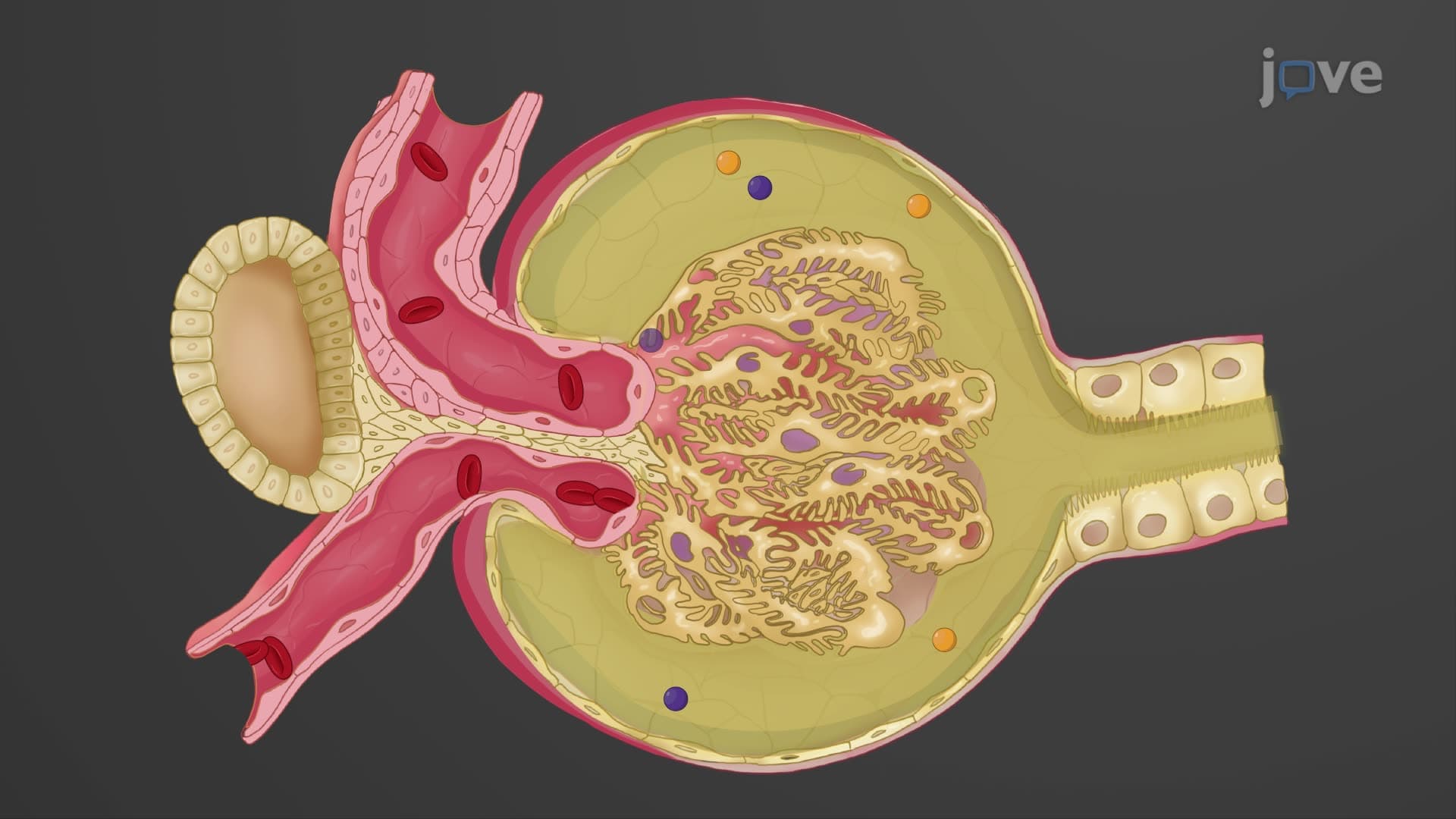
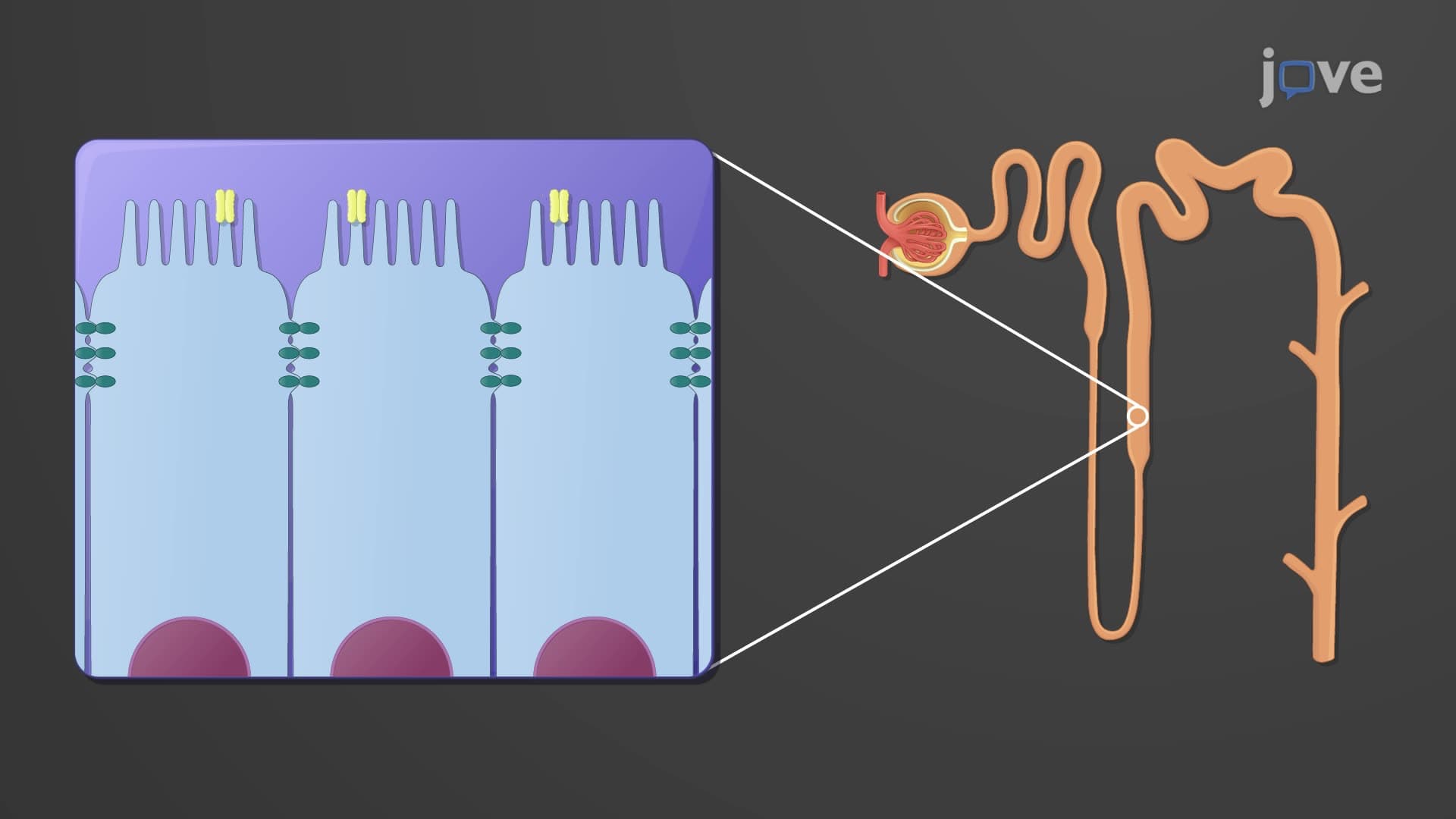
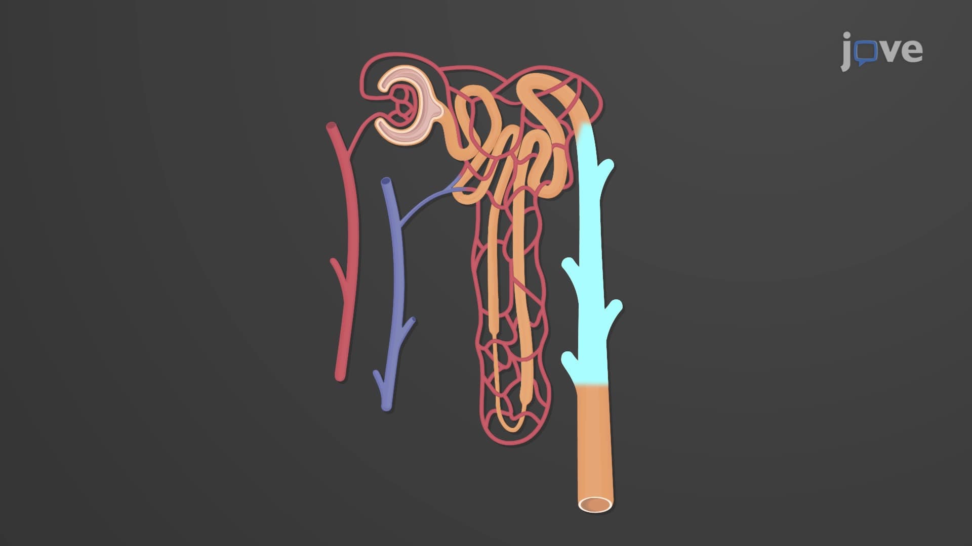
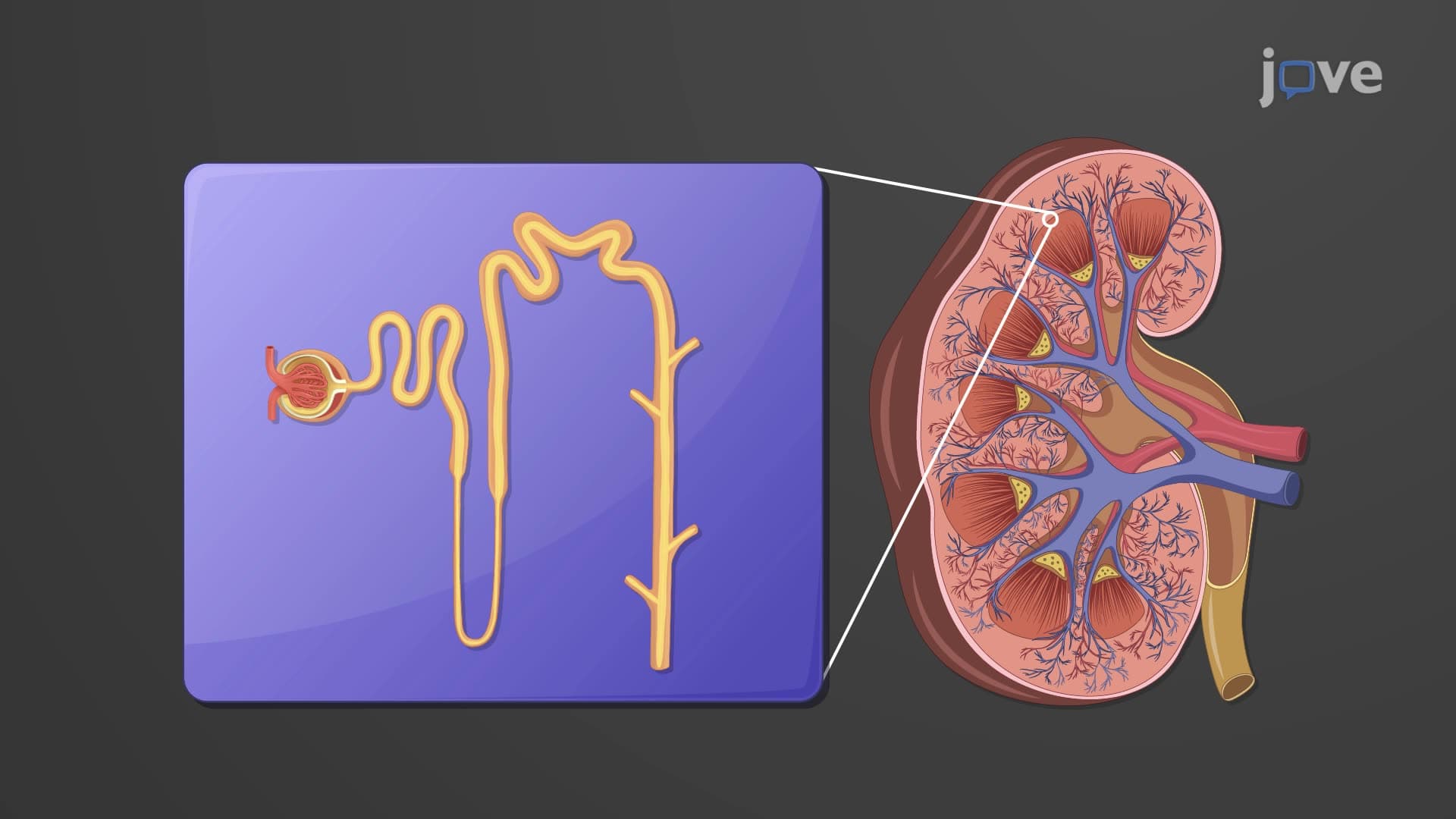
The renal corpuscle anatomy biology forms the foundation of kidney function, representing the initial filtration unit in each nephron. This microscopic structure, measuring only about 200 micrometers in diameter, performs the critical first step in urine formation by filtering blood plasma. Medical students at Harvard Medical School and other top US institutions spend considerable time mastering this concept because understanding renal corpuscles is essential for comprehending kidney physiology and pathology.
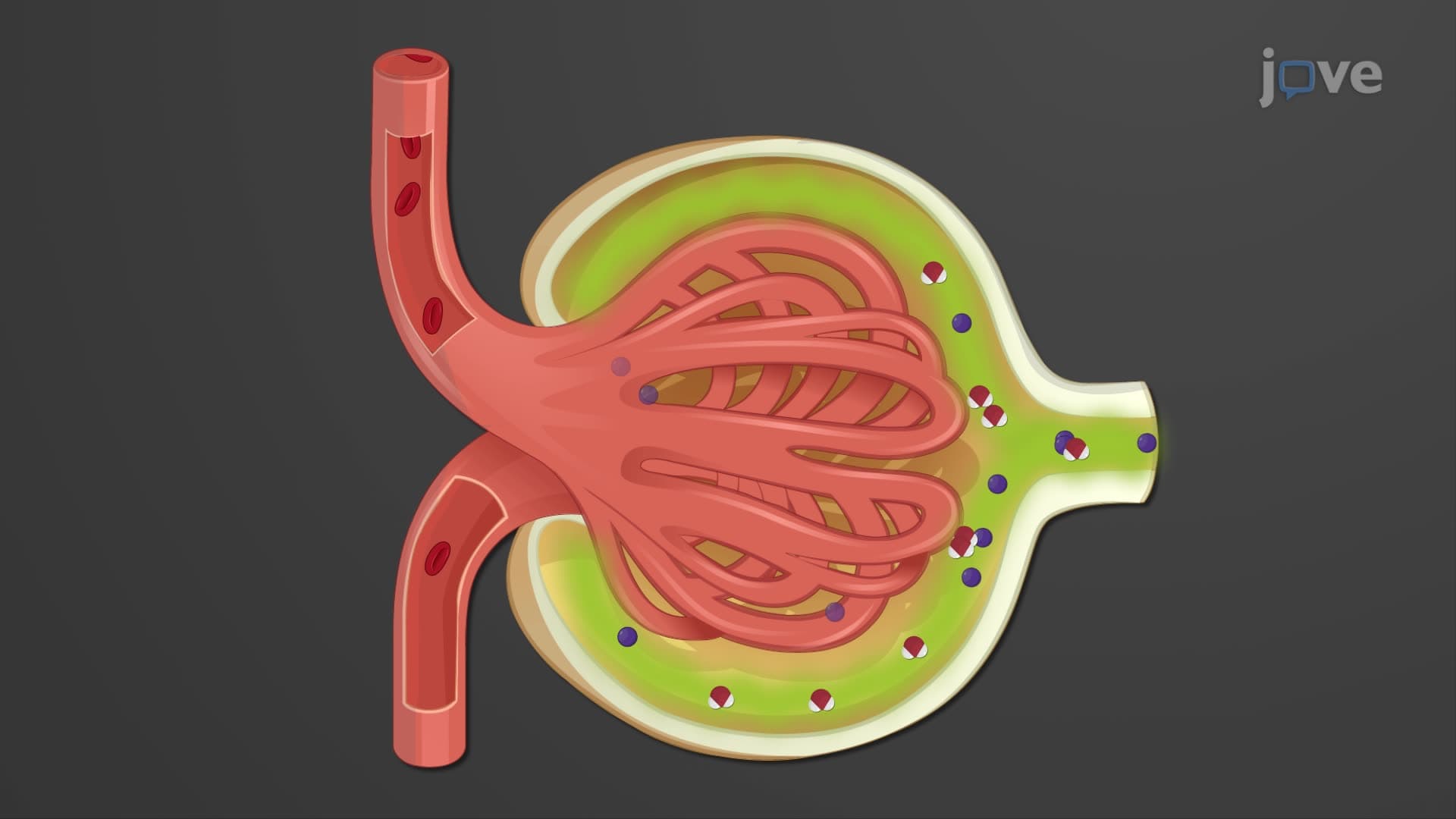
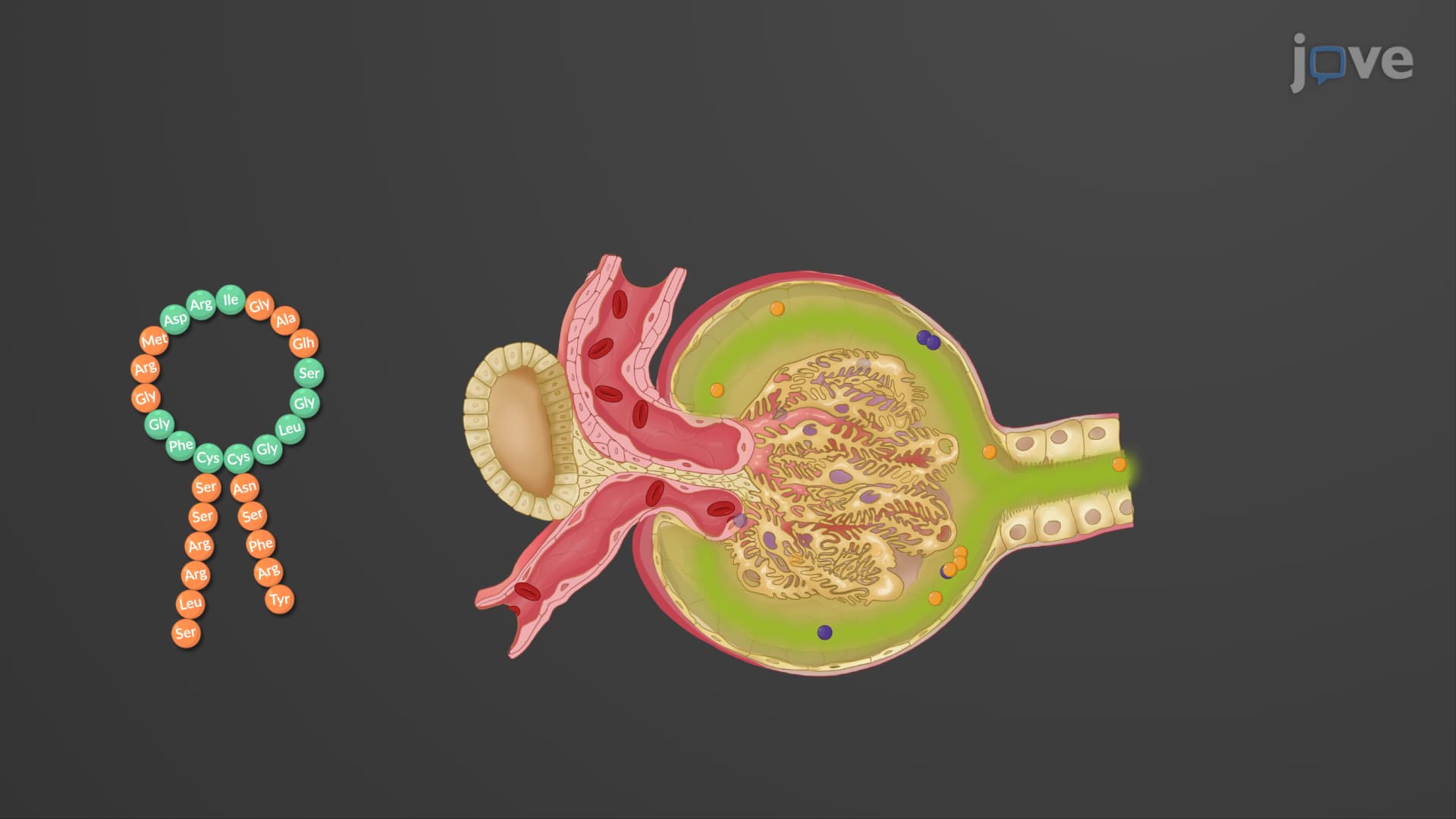
The glomerulus Bowman capsule explained begins with understanding the glomerulus as a unique capillary tuft. Unlike typical capillaries found elsewhere in the body, glomerular capillaries feature fenestrated endothelium-cells with numerous pores that dramatically increase permeability. This specialized structure allows water, electrolytes, glucose, amino acids, and small proteins to pass through while retaining larger blood components like red blood cells and most plasma proteins. The glomerulus receives blood from the afferent arteriole and drains into the efferent arteriole, creating high pressure that drives filtration.
What is the renal corpuscle in kidney anatomy becomes clearer when examining Bowman's capsule structure. This double-walled chamber surrounds the glomerulus and consists of two distinct layers. The outer parietal layer, composed of simple squamous epithelium, provides structural support but doesn't participate in filtration. The inner visceral layer contains highly specialized cells called podocytes that directly contact glomerular capillaries.
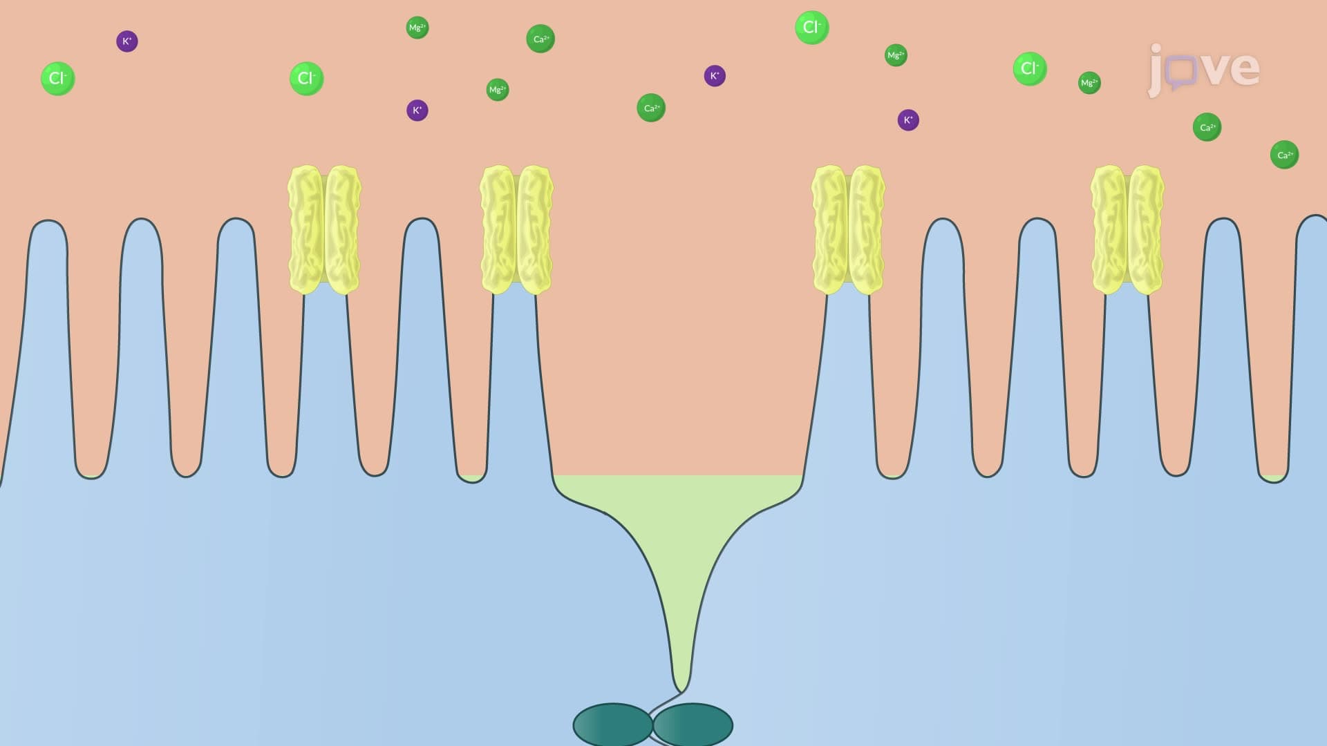
Podocytes represent one of the most fascinating cellular adaptations in human anatomy. These modified epithelial cells extend foot processes (pedicels) that interdigitate around glomerular capillaries. The spaces between these foot processes, called filtration slits, are bridged by a thin diaphragm that serves as the final filtration barrier. This intricate arrangement ensures that the filtration membrane corpuscle maintains selectivity based on size and charge. Students preparing for the MCAT or AP Biology exams should understand that this three-layer filtration barrier (fenestrated endothelium, basement membrane, and podocyte filtration slits) determines what substances enter the capsular space to become primary urine.
Related Micro-courses