Loading video…


 25 Concepts
 78.1K views

Video Summary: What Is Urethra
Did you know that urethral length differs dramatically between males and females-with males having a urethra roughly four times longer? Understanding urethra anatomy biology reveals how this critical tube connects your bladder to the outside world, enabling controlled urination. In US medical schools, students study how urethral dysfunction affects millions of Americans with conditions like incontinence or urinary retention. The urethra's dual role in males for both urine and reproductive function makes it essential knowledge for anatomy courses. Watch the full video on JoVE Coach to master this concept with expert-led visuals and step-by-step explanations.
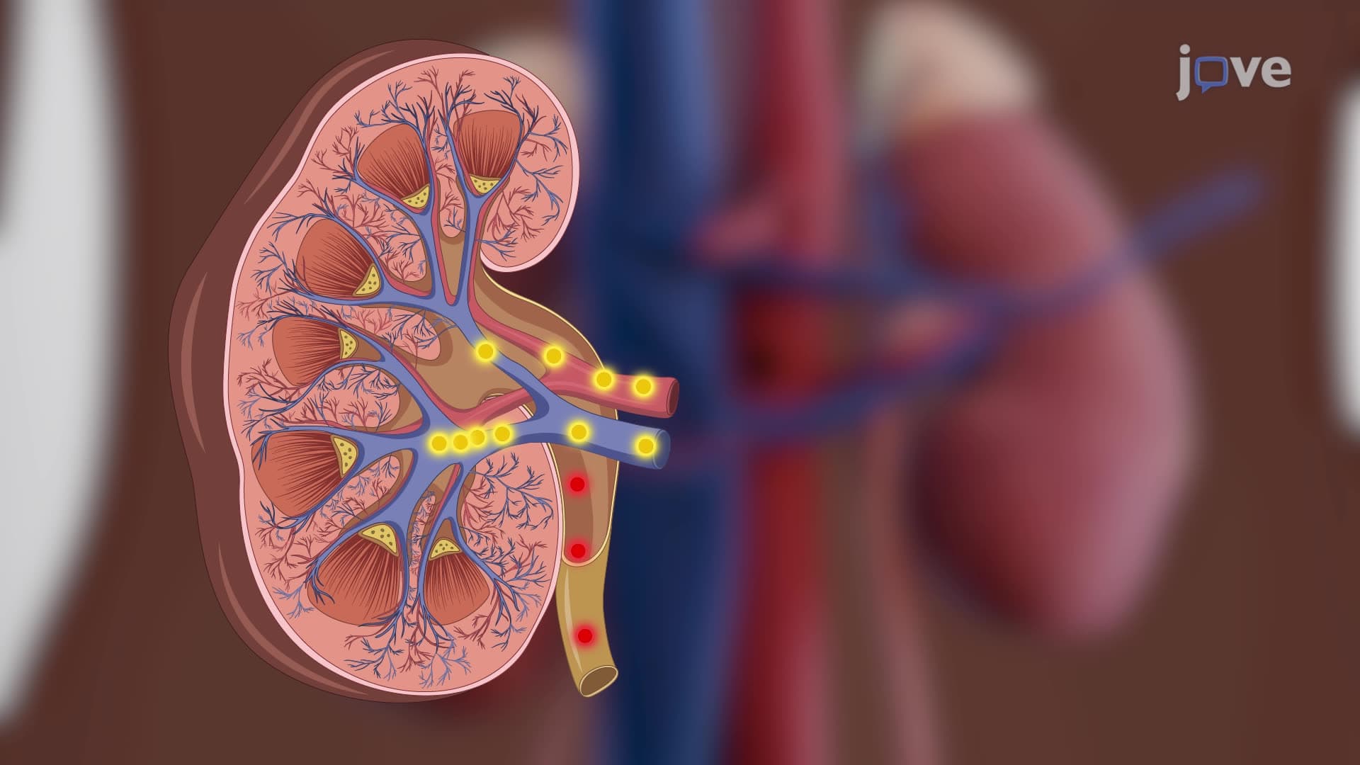
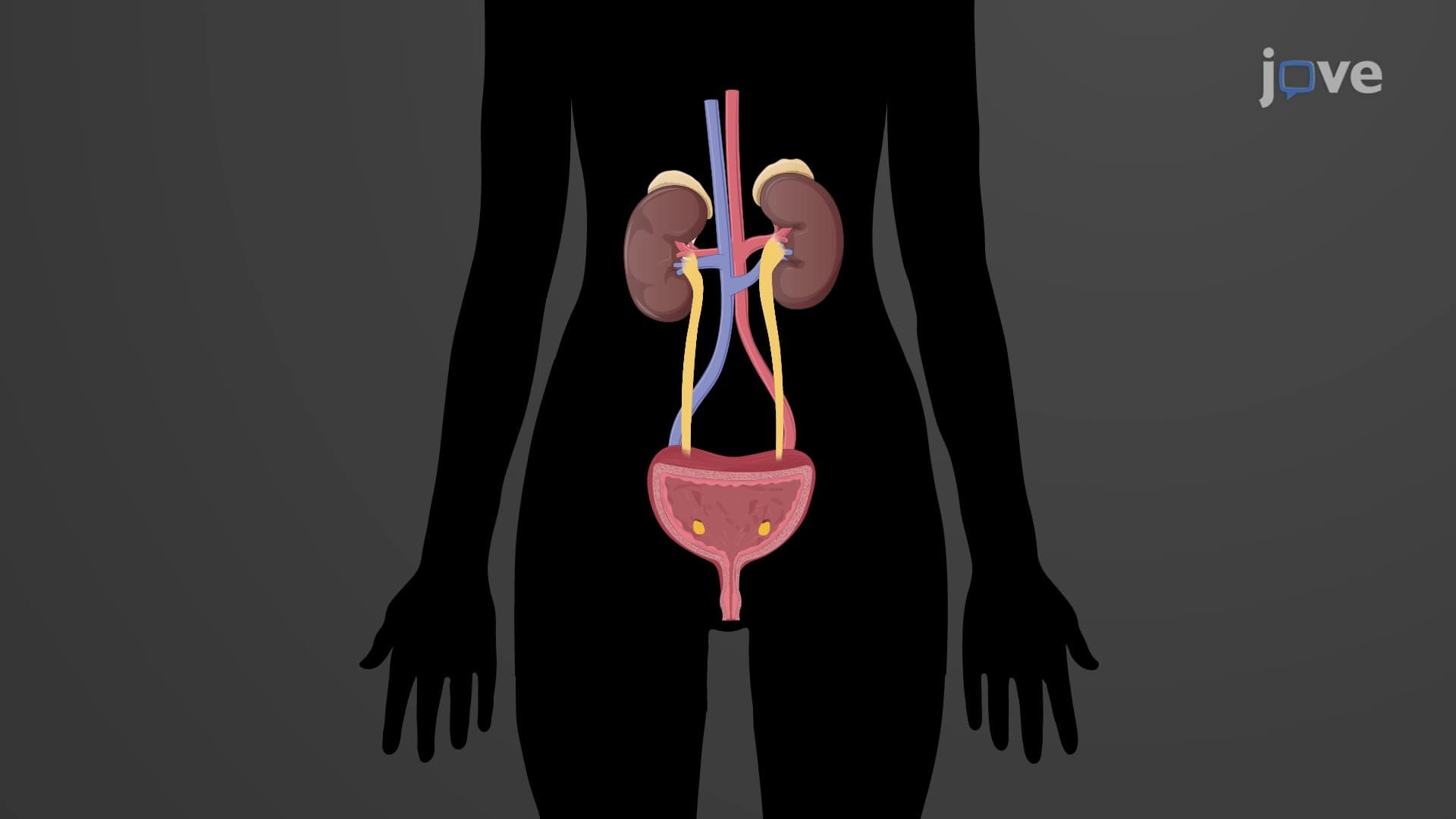
The urethra anatomy biology represents one of the most functionally important yet structurally simple components of the urinary system. This tubular conduit serves as the final pathway for urine elimination, connecting the bladder's internal environment to the external world. For students preparing for AP Biology or college anatomy courses, understanding urethral structure provides crucial insight into how the body maintains homeostasis through waste elimination.
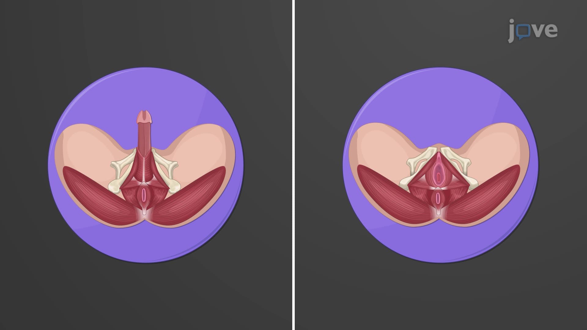
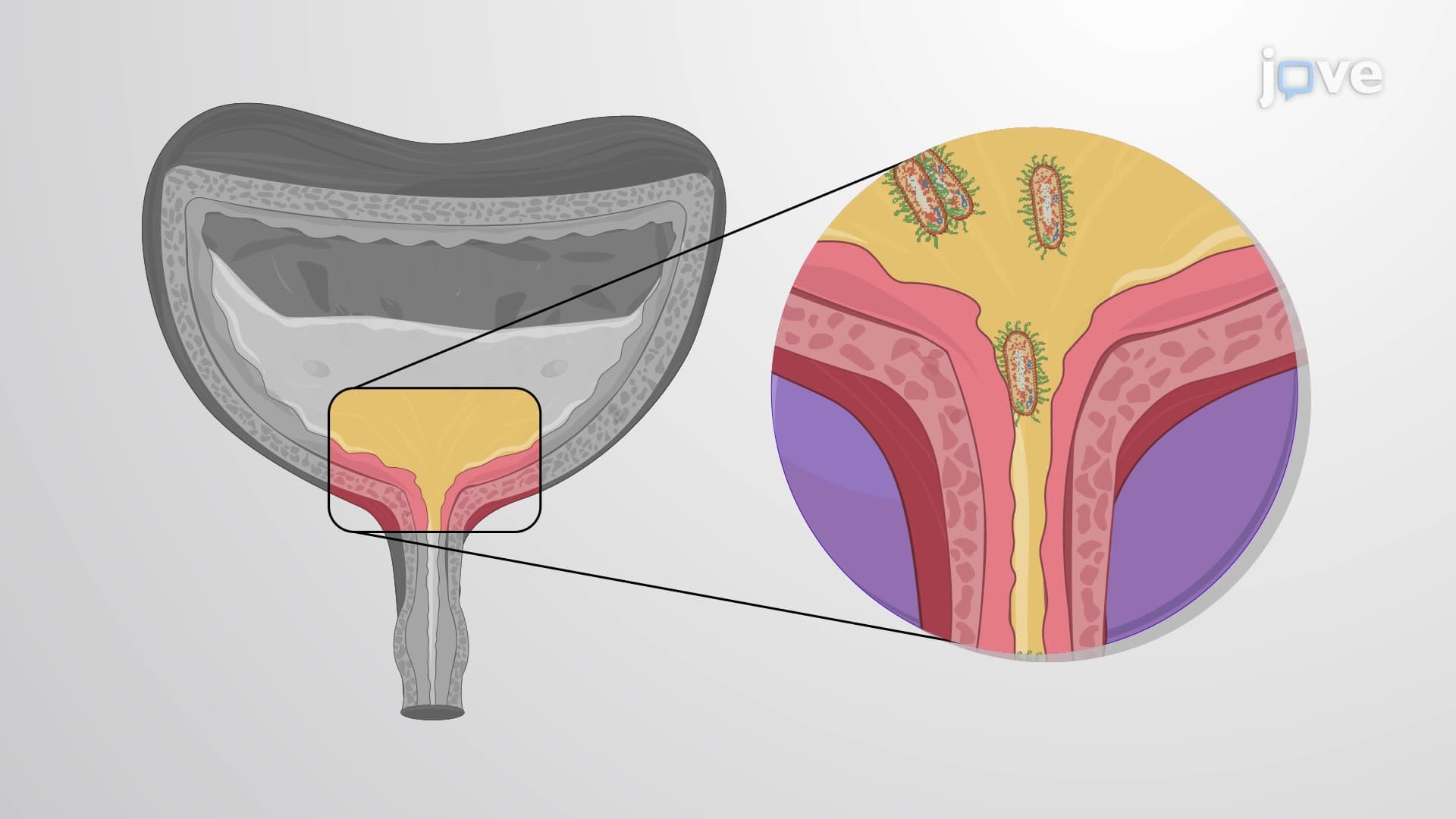
The most striking aspect of what is the urethra in urinary anatomy lies in its dramatic sexual dimorphism. Female urethras measure approximately 1.5 inches (4 cm) in length, opening just above the vaginal entrance. This shorter pathway explains why women experience higher rates of urinary tract infections-bacteria have less distance to travel from the external environment to the bladder.
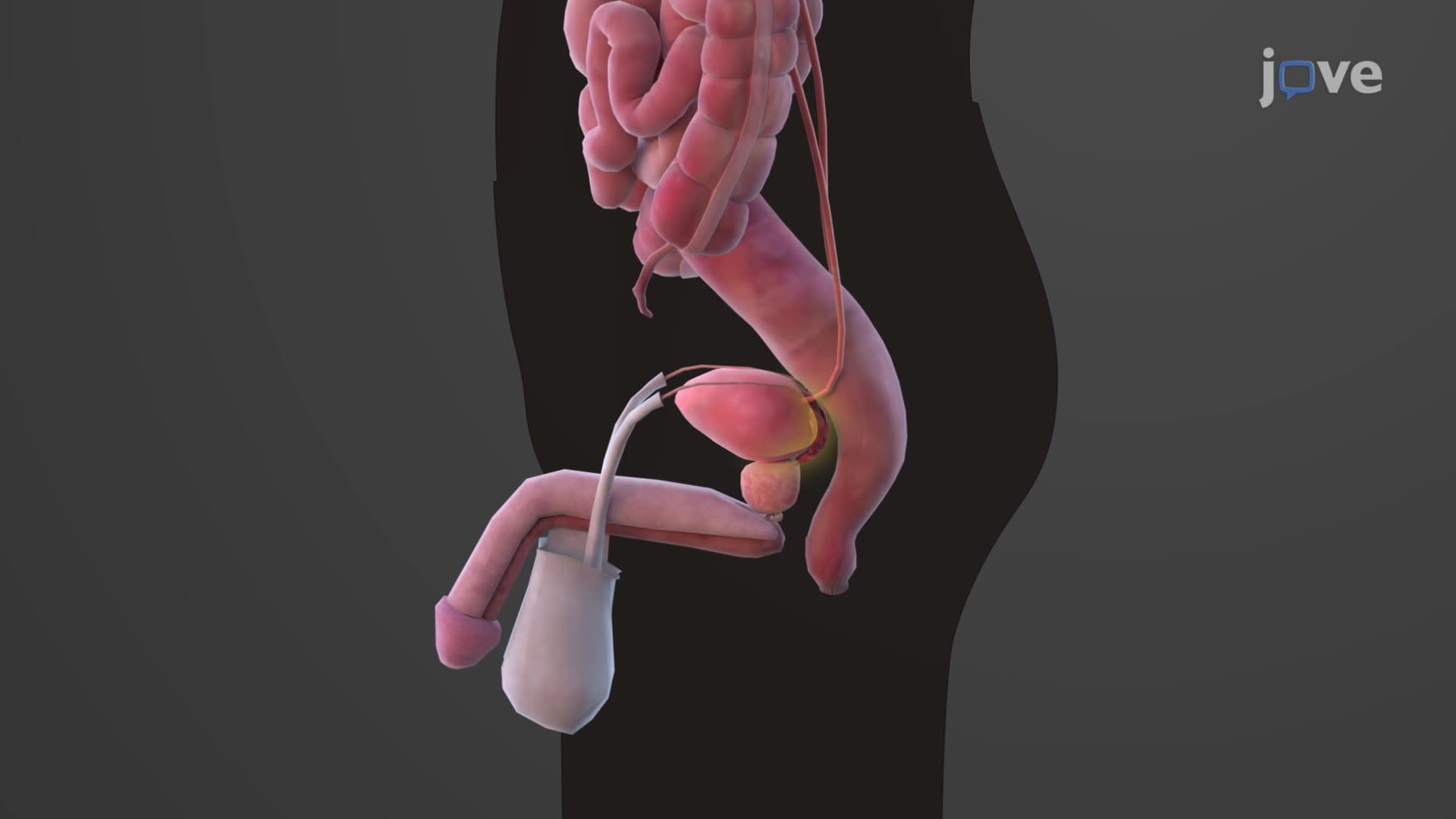
Male urethras, conversely, extend roughly 8 inches (20 cm) through three distinct regions. The prostatic urethra, the widest segment, passes through the prostate gland where seminal fluid joins urine during ejaculation. The membranous urethra represents the shortest, most vulnerable section as it traverses the urogenital diaphragm. Finally, the spongy (penile) urethra extends through erectile tissue, explaining why urination becomes difficult during sexual arousal.
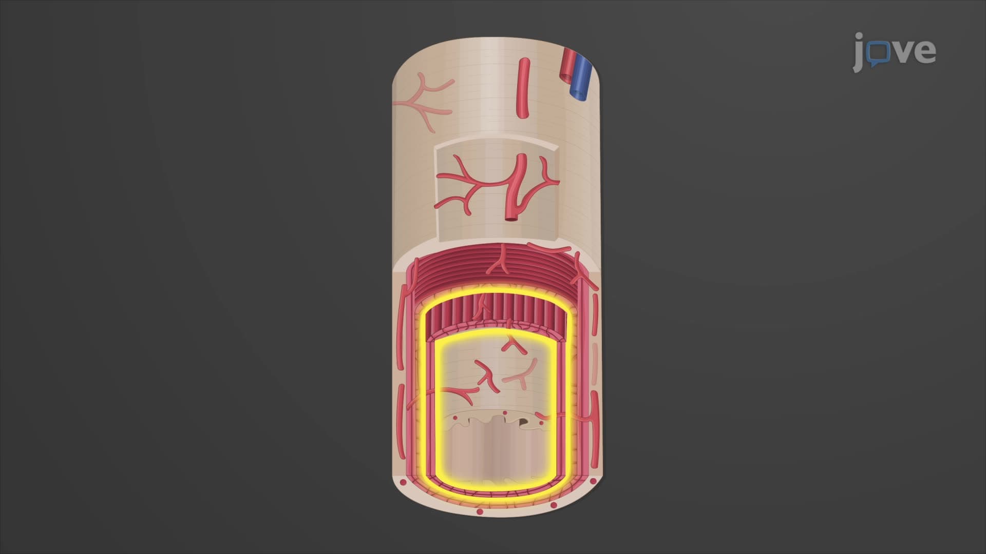
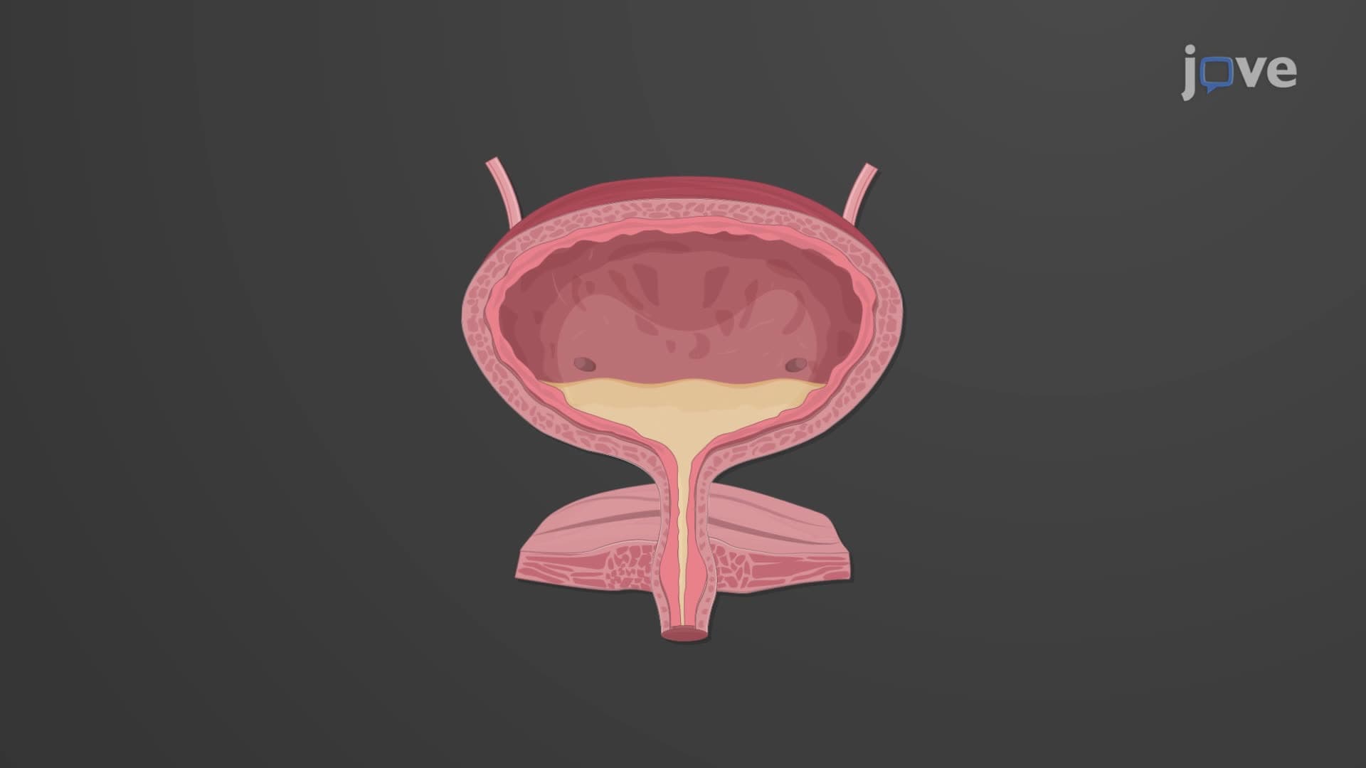
The external urethral sphincter and its internal counterpart work in coordinated opposition to maintain continence. The internal sphincter, composed of smooth muscle, operates unconsciously through autonomic nervous system control. This involuntary mechanism prevents constant urine leakage, maintaining baseline bladder pressure.
The external sphincter, however, consists of skeletal muscle under conscious control. This voluntary mechanism allows deliberate urination initiation or suppression-a skill developed during childhood toilet training. MCAT students should recognize this as a classic example of dual innervation, where both voluntary and involuntary systems coordinate for optimal function.
For pre-med students tackling MCAT passages, urethral anatomy frequently appears in kidney physiology contexts. Understanding male female urethra length differences helps explain infection patterns, surgical complications, and catheterization procedures. Medical schools across the US, from Harvard to Stanford, emphasize how anatomical knowledge directly impacts clinical decision-making in urology and emergency medicine.
Related Micro-courses