Loading video…


 25 Concepts
 78.1K views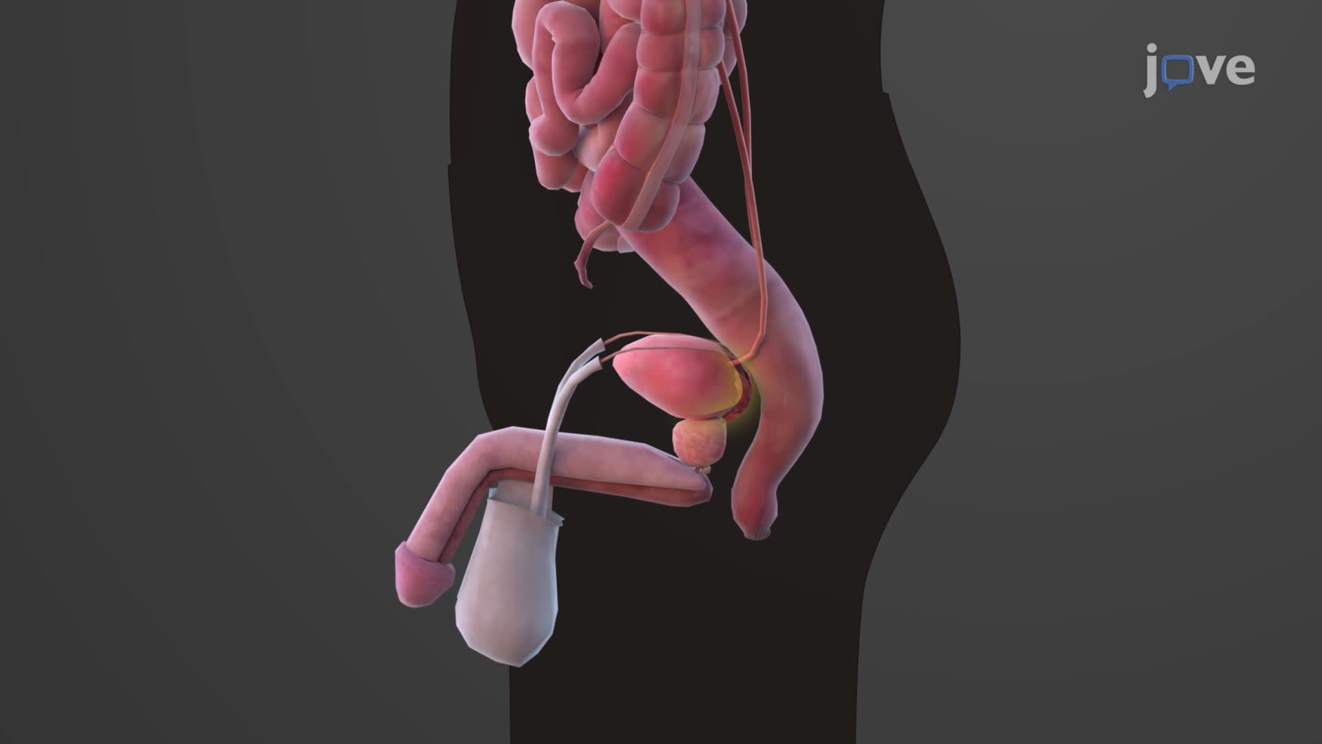

Video Summary: What Is Urinary Bladder
Did you know your urinary bladder anatomy biology allows this remarkable organ to expand from ping-pong ball size to hold nearly two and a half cups of fluid? The What is Urinary Bladder reveals a sophisticated hollow organ positioned behind your pubic bone, featuring specialized layers including the detrusor muscle that contracts during urination. Students at Johns Hopkins University study this essential component of the urinary system to understand how our bodies efficiently store and eliminate waste. Watch the full video on JoVE Coach to master this concept with expert-led visuals and step-by-step explanations.
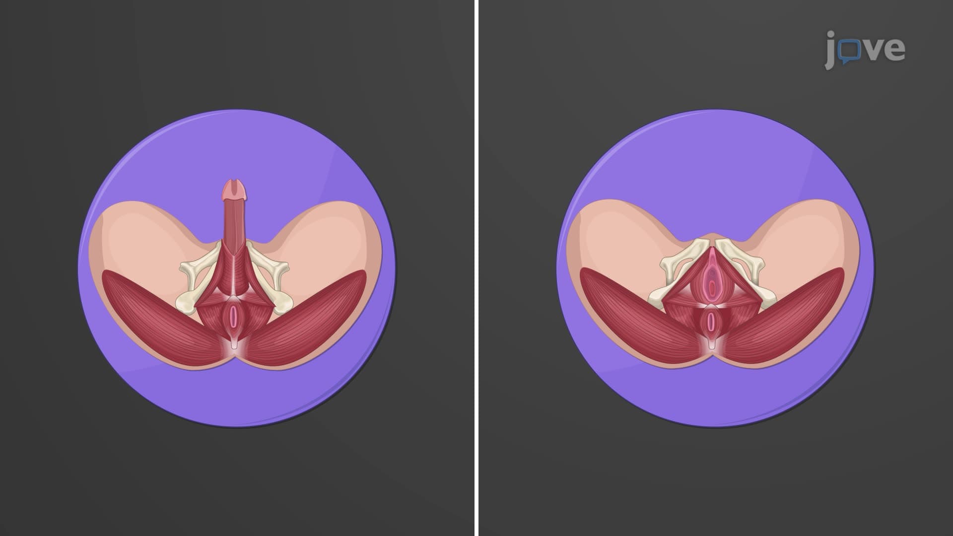
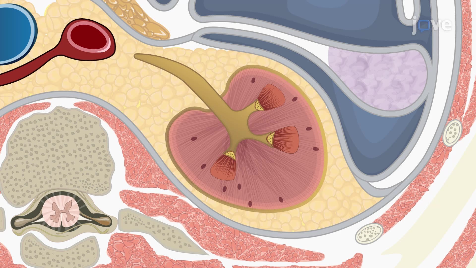
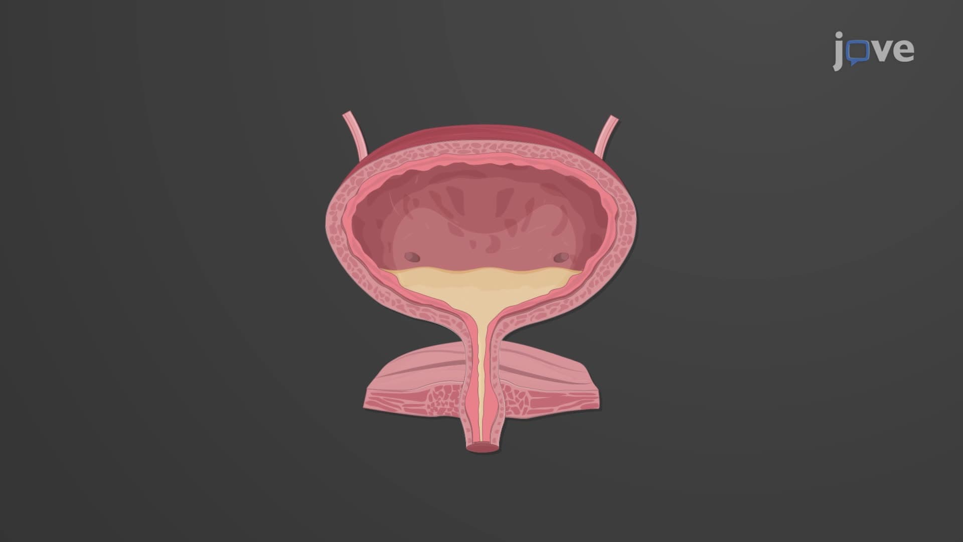
The urinary bladder anatomy biology represents one of the most adaptable organs in the human body. This hollow, muscular sac sits strategically in the pelvis, positioned retroperitoneally behind the pubic symphysis. The bladder structure function explained becomes clearer when examining its precise anatomical relationships. In males, the bladder rests directly anterior to the rectum, while in females, it's positioned anterior to the vagina and inferior to the uterus. This positioning is crucial for medical professionals performing procedures like catheterization at hospitals such as Mayo Clinic or Cleveland Clinic.
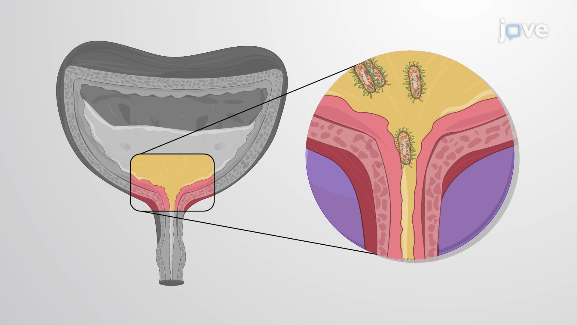
The trigone bladder anatomy forms a distinctive triangular region on the bladder floor, created by two ureteric orifices and the internal urethral orifice. This smooth, non-distensible area contrasts sharply with the rest of the bladder wall and serves as a crucial landmark during cystoscopic procedures. Medical students preparing for the USMLE Step 1 must understand that the trigone's unique structure prevents backflow of urine into the ureters during bladder contraction. The internal urethral sphincter at the trigone's apex plays a vital role in maintaining continence.
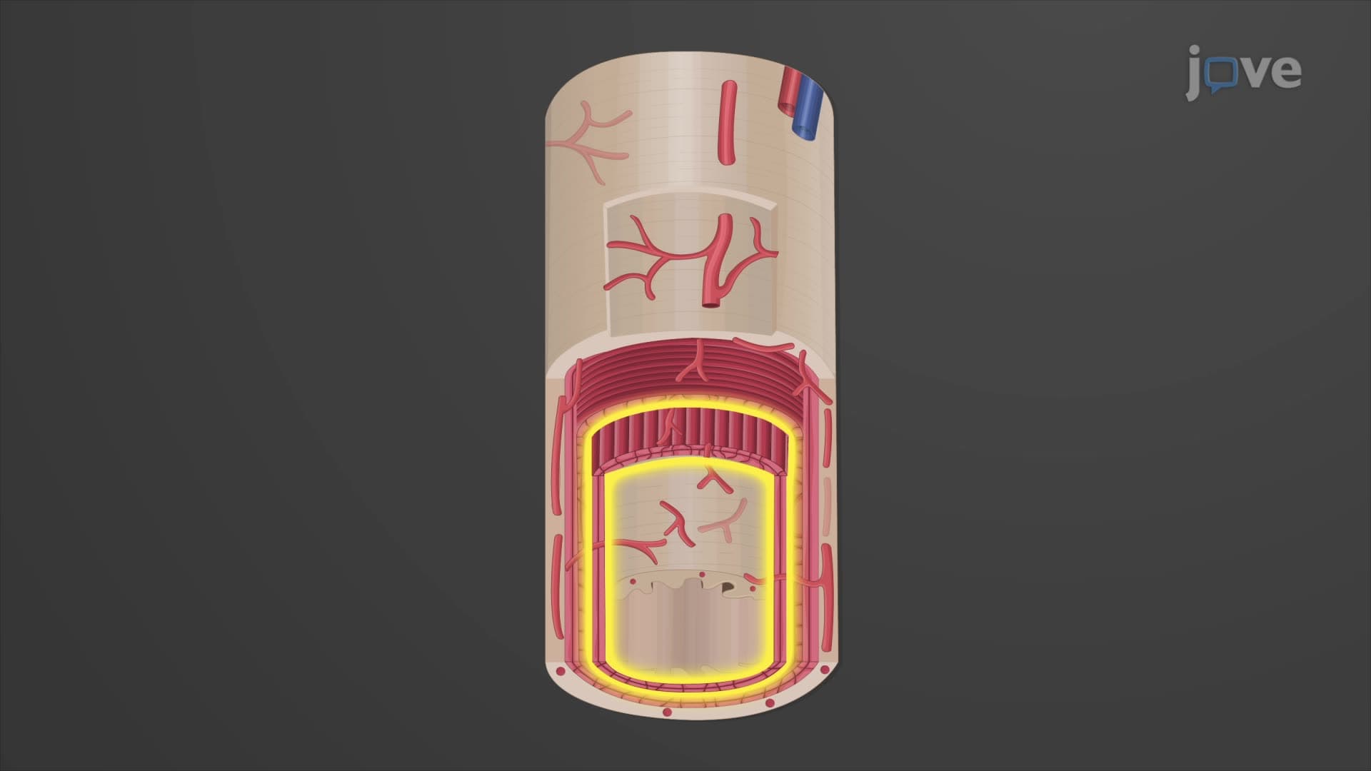
The bladder wall transitional epithelium represents a marvel of biological engineering. The innermost mucosa contains specialized transitional epithelium arranged in rugae, allowing dramatic expansion without structural damage. Students taking AP Biology or college anatomy courses should note how this transitional epithelium can stretch from 6-8 cell layers when empty to just 2-3 layers when full. The middle muscularis layer houses the detrusor muscle bladder, composed of smooth muscle fibers arranged in inner and outer longitudinal layers surrounding a circular layer. This arrangement enables coordinated contraction during micturition.
Understanding bladder capacity function becomes essential when studying urological disorders common in American healthcare. The normal bladder capacity of approximately 600 mL represents the balance between storage efficiency and anatomical constraints. Pre-med students studying for the MCAT should recognize how conditions like neurogenic bladder or detrusor instability can disrupt this delicate balance. The adventitia, continuous with ureteral walls, provides structural support while allowing necessary mobility during filling and emptying cycles.
Related Micro-courses