Loading subject page…

Micro-course: The Urinary System
78,053 views
Learn key concepts and earn your certificate.



Ureters

00:00
01:22
Video Summary: What Is Ureters
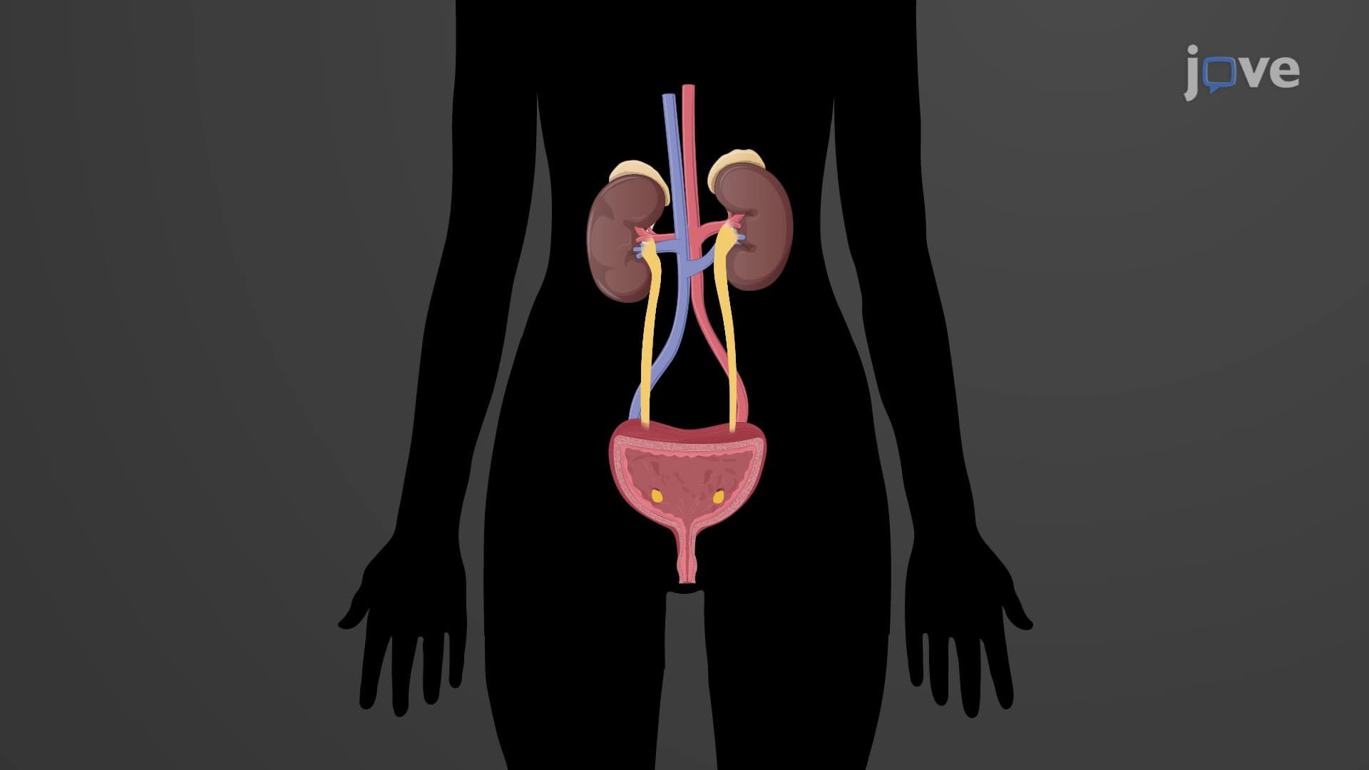
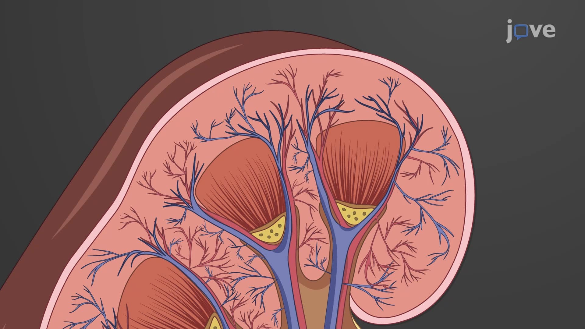
Ever wonder how your kidneys efficiently transport nearly 50 gallons of filtered urine to your bladder daily without any backflow? The ureters anatomy biology involves sophisticated tubular structures that act like one-way highways in your urinary system. These remarkable 10-12 inch tubes use muscular contractions similar to how your esophagus moves food to your stomach, ensuring urine flows only toward the bladder. Watch the full video on JoVE Coach to master this concept with expert-led visuals and step-by-step explanations.
- Understand what is ureters and their essential role as retroperitoneal transport tubes connecting kidneys to the urinary bladder
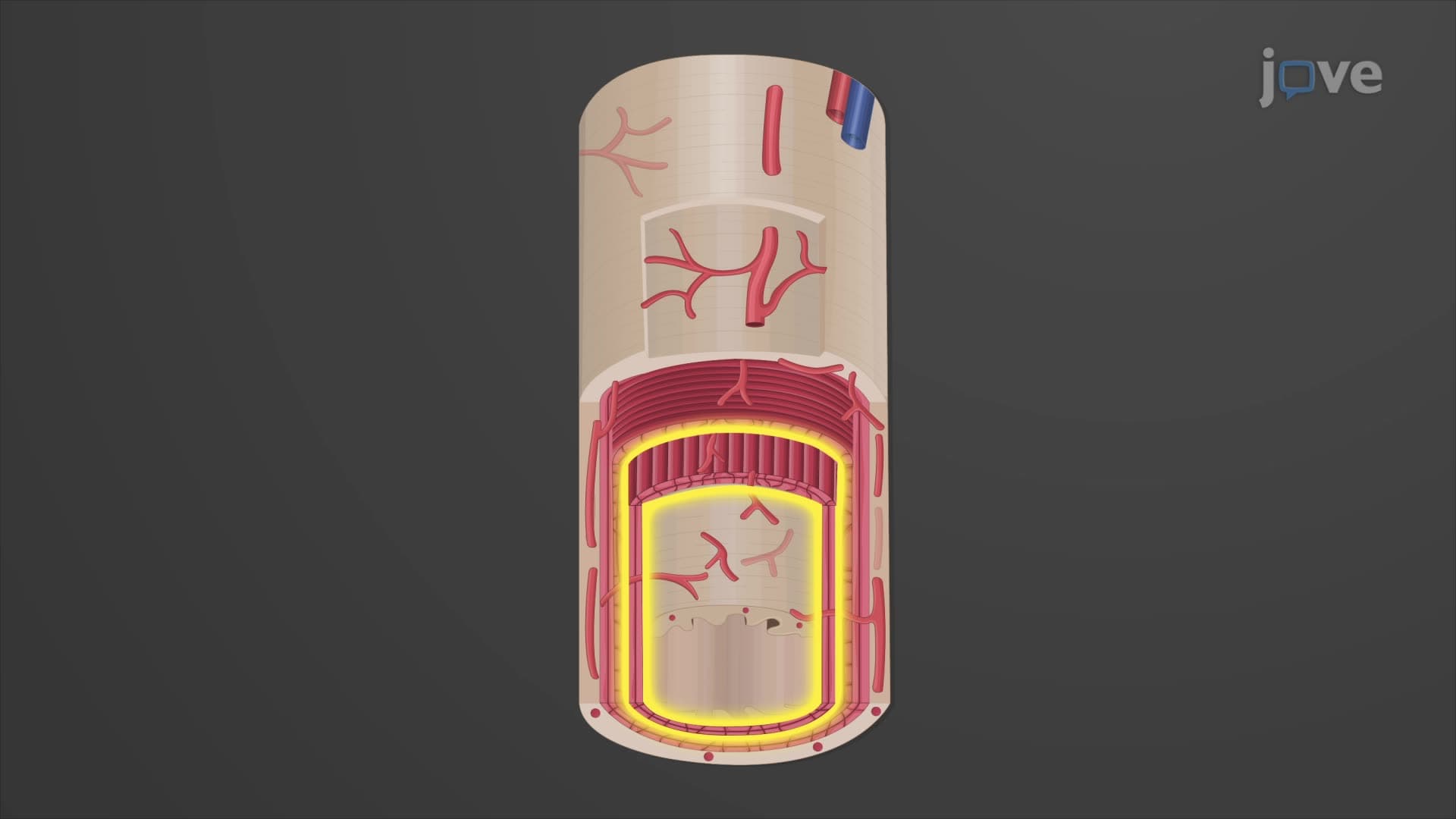
- Identify the three distinct histological layers of ureter structure: mucosa, muscularis, and adventitia
- Analyze how ureter peristalsis creates unidirectional urine flow through coordinated smooth muscle contractions
- Compare the varying ureter diameter from 10mm at the renal pelvis to 1mm at the bladder entry point
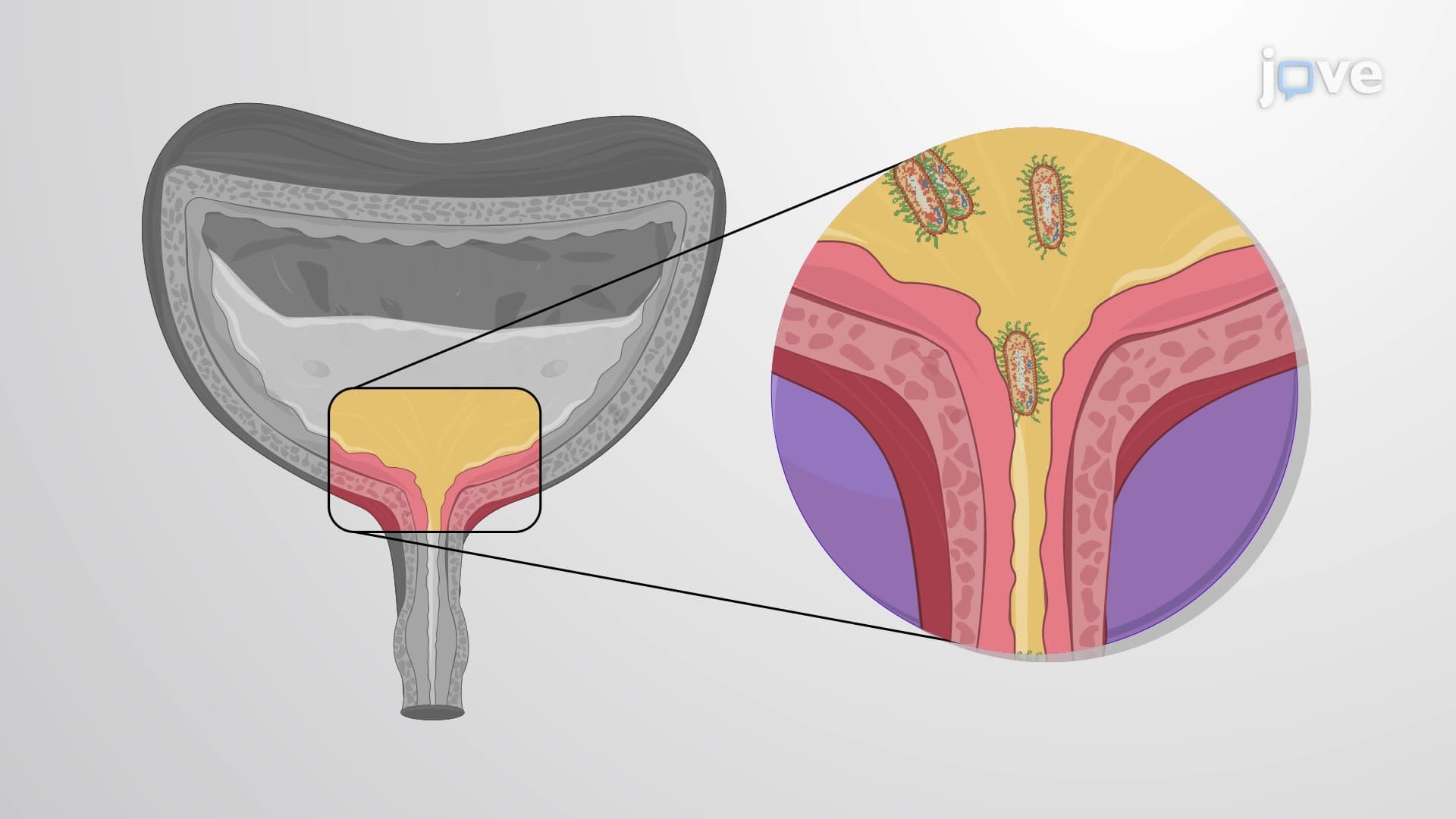
- Evaluate the anti-reflux mechanism created by the oblique entry angle of ureters into the bladder wall
- Apply knowledge of transitional epithelium's role in allowing ureter expansion during high urine volume
- Recognize the clinical significance of ureter anatomy in preventing kidney damage from urine backflow
- Explore the relationship between ureter length (25-30 cm) and efficient gravitational urine transport
What is Ureters: Essential Urinary System Components
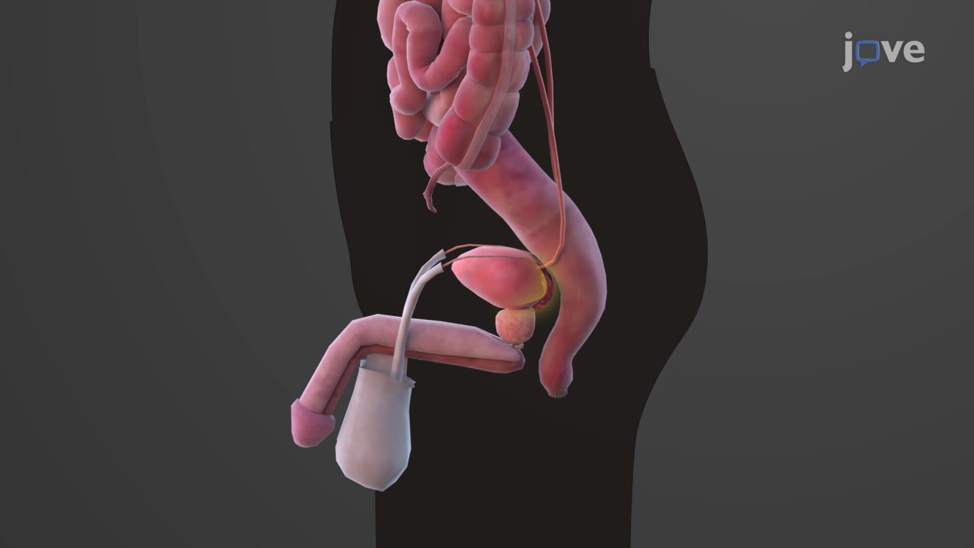
The ureters anatomy biology represents one of the most elegantly designed transport systems in human physiology. These paired retroperitoneal tubes serve as critical conduits in the urinary system, ensuring efficient urine transport while preventing potentially dangerous backflow that could damage the kidneys. Understanding ureter structure function explained is fundamental for students preparing for advanced biology courses and medical school prerequisites.
Structural Architecture and Dimensional Specifications
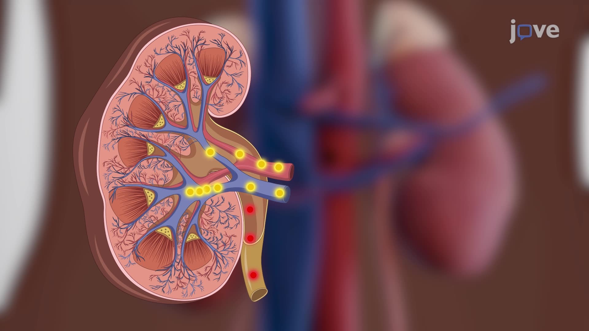
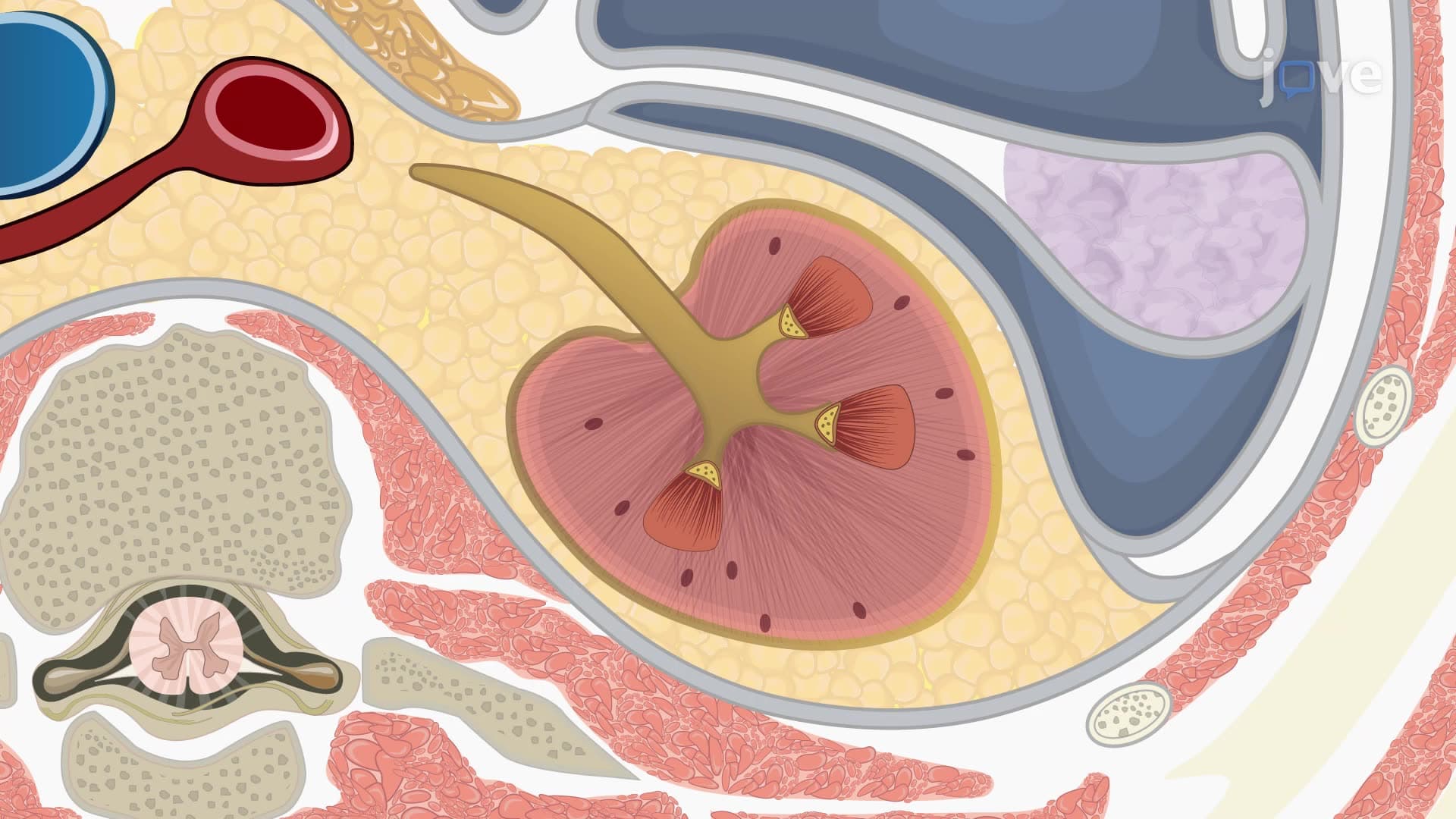
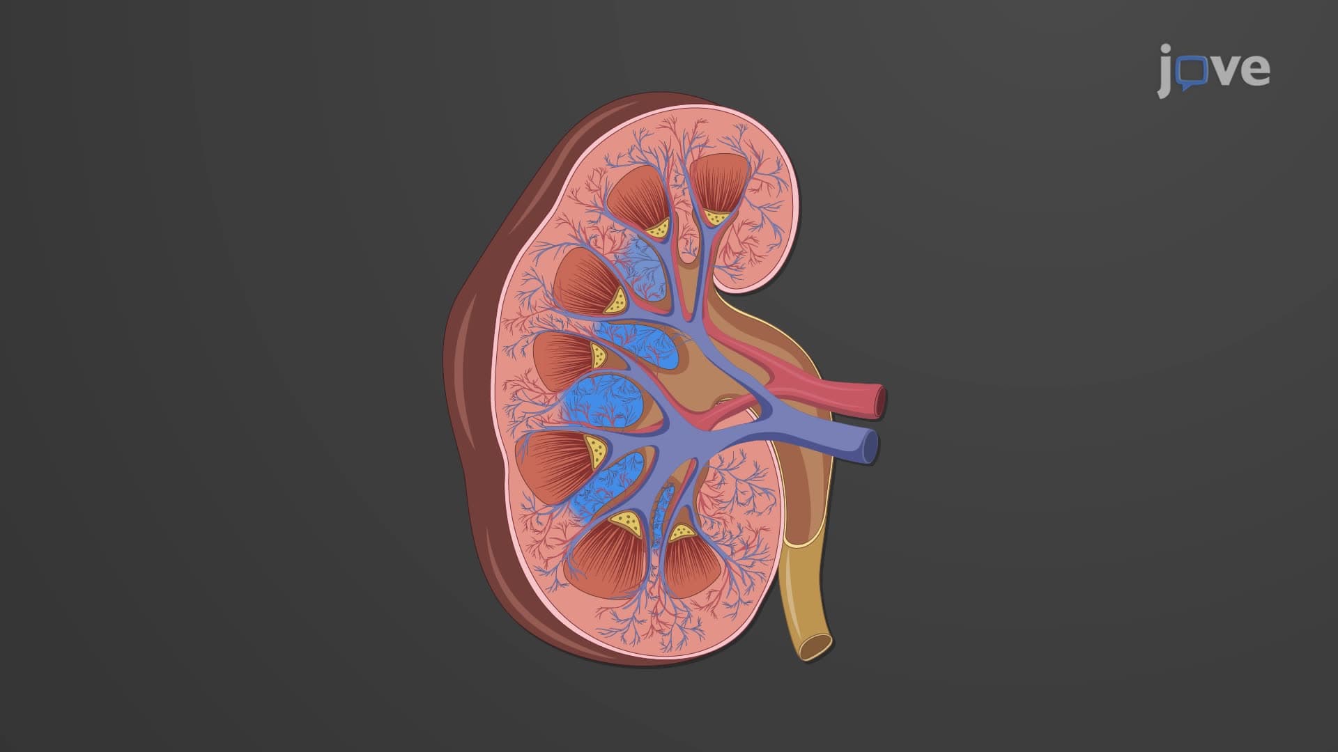
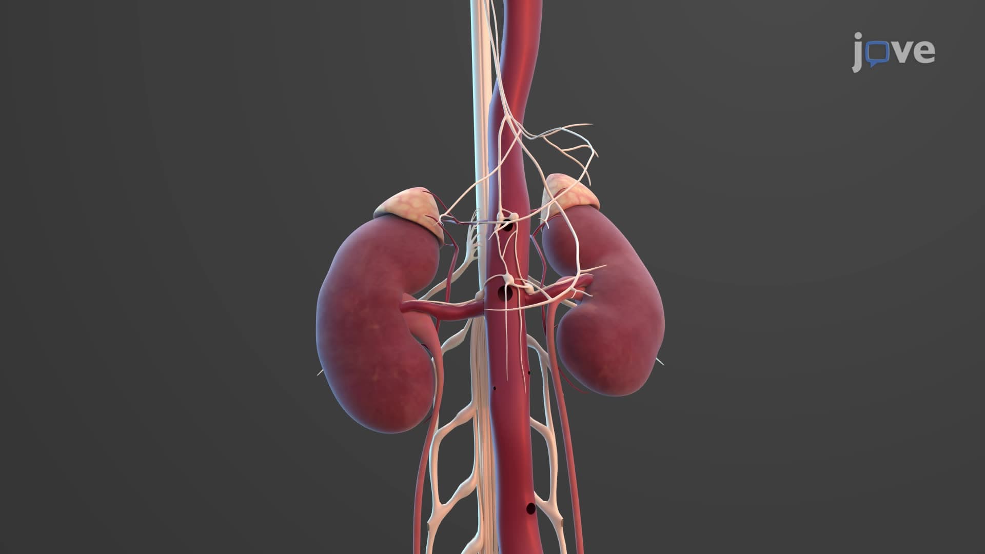
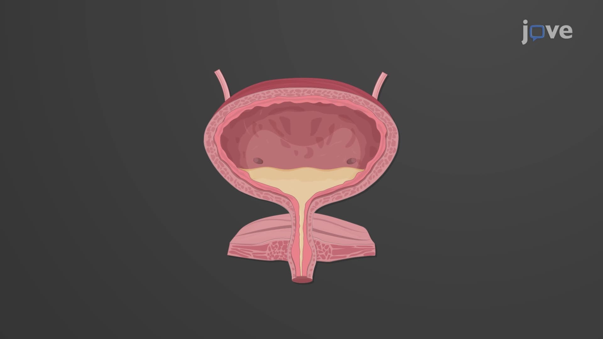
The ureter length anatomy spans approximately 25-30 centimeters (10-12 inches) on each side of the vertebral column. This dimension is crucial because it allows gravity to assist in urine transport while providing sufficient length for the muscular contractions that drive peristalsis. The what are the ureters in urinary anatomy question often focuses on their variable diameter-starting at 10mm where they connect to the renal pelvis and narrowing dramatically to just 1mm at their oblique entry point into the posterior bladder wall.
This tapering design serves multiple functions. The wider proximal diameter accommodates the initial urine collection from the kidney, while the narrow distal opening creates a natural valve mechanism. Students studying for the MCAT or AP Biology exams should note that this anatomical feature prevents urine reflux, which could cause serious complications like pyelonephritis or kidney damage.
Three-Layer Histological Organization
The ureteral wall three layer structure demonstrates sophisticated biological engineering. The innermost mucosa contains transitional epithelium-the same specialized tissue found in the bladder-allowing dramatic expansion during peak urine flow periods. This adaptability is crucial during situations like rapid hydration or diuretic medication use.
The middle muscularis layer drives ureter peristalsis urine transport through coordinated smooth muscle contractions. The inner longitudinal fibers work with outer circular fibers to create wave-like contractions occurring 1-5 times per minute. This mechanism is similar to esophageal peristalsis, making it an excellent comparison point for students learning multiple body systems.
Clinical Relevance and Functional Integration
The outermost adventitia provides structural support and protection, anchoring the ureters in their retroperitoneal position. This positioning protects them from abdominal trauma while maintaining their essential ureter kidney bladder connection. Medical students studying for the USMLE Step 1 should understand that ureter obstruction-whether from kidney stones, tumors, or anatomical abnormalities-can rapidly lead to hydronephrosis and permanent kidney damage, making ureter anatomy knowledge clinically critical.
Frequently Asked Questions
Ureters are paired tubes (you have two) that transport urine from each kidney to the bladder, measuring 25-30 cm long. The urethra is a single tube that carries urine from the bladder to outside the body. This distinction is frequently tested on MCAT and AP Biology exams.
MCAT questions often focus on ureter peristalsis mechanisms, the anti-reflux valve function, and clinical scenarios involving kidney stones. USMLE Step 1 emphasizes embryological development, anatomical relationships, and pathological conditions like hydronephrosis. Expect integration with endocrine and cardiovascular systems.
Ureter anatomy appears in AP Bio Unit 5 (Heredity) when studying excretory system evolution, and Unit 7 (Natural Selection) regarding mammalian adaptations. Students should understand how smooth muscle peristalsis relates to cell communication and homeostasis maintenance. The three-layer histological structure exemplifies tissue organization principles.
The ureter diameter narrows from 10mm to just 1mm at three anatomical constriction points. When stones attempt to pass through these narrow areas, they stretch the ureter wall and trigger intense pain signals. Emergency departments across the US see thousands of these cases annually, making this clinically relevant knowledge.
Not at all-ureter anatomy is quite straightforward compared to other organ systems. The three-layer structure follows the same pattern as the digestive tract, and the transport function resembles how your esophagus moves food. Students with basic knowledge of smooth muscle and epithelial tissue can easily master this concept.
Create comparison charts linking ureters to familiar structures like the esophagus (peristalsis), blood vessels (three layers), and garden hoses (diameter changes). Use mnemonics like "Mucosa, Muscularis, aDventitia" (M-M-A) for the layers. Practice drawing the kidney-ureter-bladder pathway repeatedly.
Ureter problems directly impact cardiovascular health through blood pressure regulation and fluid balance. Endocrine connections include ADH and aldosterone effects on urine production. Nervous system involvement includes pain pathways and autonomic control of peristalsis, making ureters excellent for integrated learning approaches.
Progress to ureter embryological development, studying how these structures form from the ureteric bud. Explore comparative anatomy across mammals, and investigate current research on bioengineered ureter replacements for congenital abnormalities. These topics prepare you for upper-level physiology and potential medical research.
Related Micro-courses

20 Concepts

37 Concepts

14 Concepts