Loading subject page…


57,806 views


Video Summary: Vertebral Column Regions and Curvature Explained
Ever wonder why humans can walk upright while most animals can't? The vertebral column regions curvatures create an S-shaped spine that's perfectly engineered for bipedal locomotion. Consider how NFL quarterbacks like Tom Brady maintain perfect posture under pressure-their spine's four distinct curves distribute forces efficiently from cervical to lumbar regions. This Vertebral Column Regions And Curvature Explained content reveals how 33 infant vertebrae fuse into 26 adult bones, creating the thoracic kyphosis and lumbar lordosis that define human anatomy. Watch the full video on JoVE Coach to master this concept with expert-led visuals and step-by-step explanations.
The human spine represents one of evolution's most remarkable engineering achievements. Unlike our quadrupedal ancestors, the human vertebral column regions curvatures create an S-shaped structure that enables efficient bipedal locomotion while protecting the delicate spinal cord. This complex architecture involves precise regional specialization and carefully timed developmental changes.
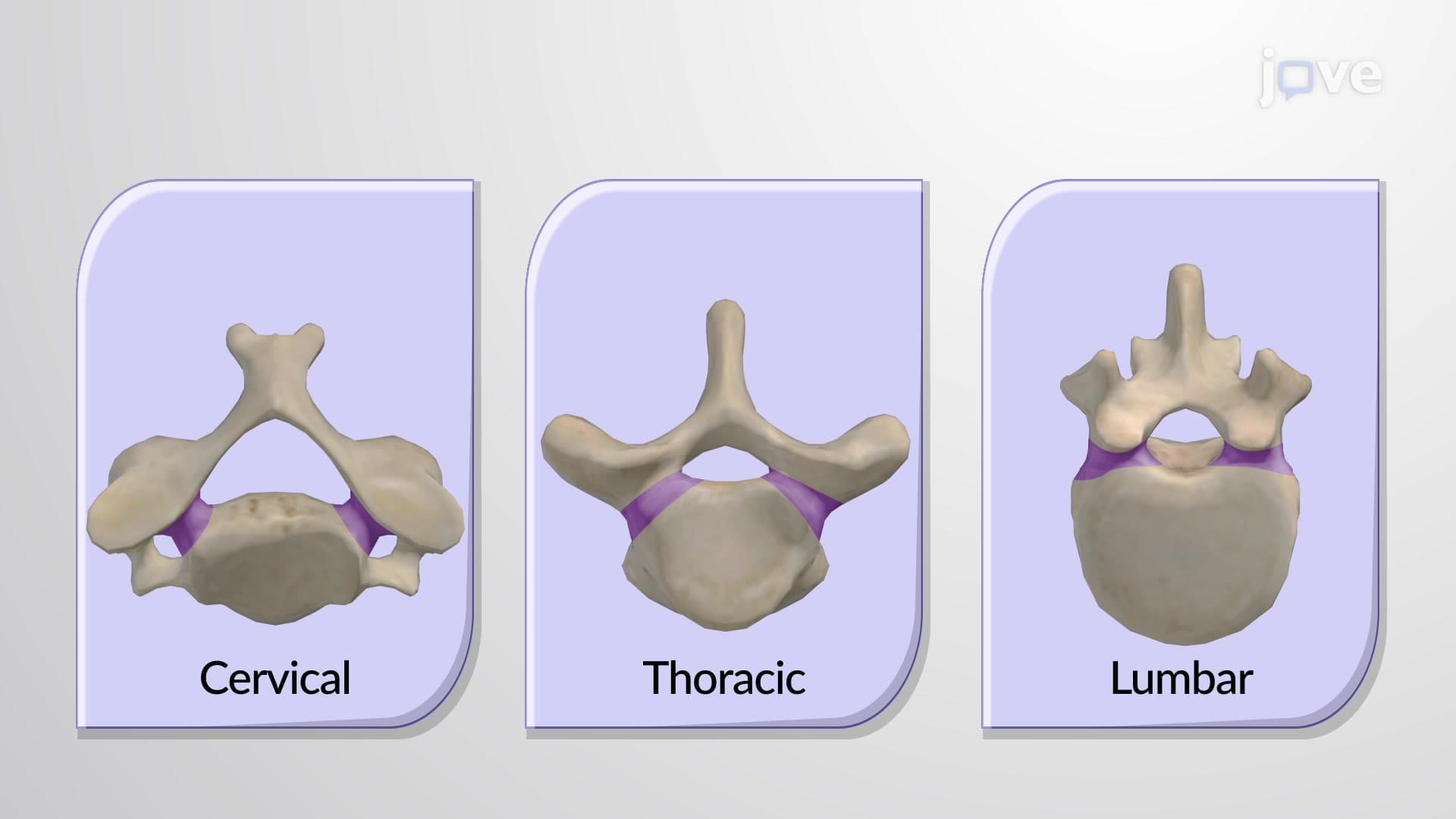
The spine regions and curves anatomy divides into five functionally distinct areas. The cervical region (C1-C7) supports the head's weight-approximately 10-12 pounds in adults-while providing maximum mobility for head rotation and flexion. The atlas (C1) and axis (C2) vertebrae create a specialized joint allowing 50% of neck rotation, crucial for activities like driving or sports.
The thoracic region (T1-T12) forms the longest spinal section, with each vertebra connecting to ribs that protect vital organs. These vertebrae are intermediate in size and less mobile due to rib attachments. The lumbar region (L1-L5) contains the spine's largest vertebrae, designed to bear the body's full weight during activities like weightlifting or jumping-forces that can exceed 10 times body weight in elite athletes.
Understanding what are the vertebral column regions and curvatures requires examining both structural and developmental aspects. Infants begin with 33 separate vertebrae, but the five sacral vertebrae fuse by age 26 to form the sacrum, while the four coccygeal vertebrae fuse into the coccyx. This fusion process creates the stable pelvic attachment necessary for upright walking.
The spine's characteristic curvatures develop through a fascinating timeline. Primary curvatures-thoracic sacral kyphosis-exist at birth, reflecting our fetal position. Secondary curvatures-cervical lumbar lordosis-develop as infants gain motor control. The cervical lordosis emerges when babies lift their heads around 3-4 months, while lumbar lordosis develops during walking at 12-15 months.
This spine curvature anatomy knowledge proves essential for various US examinations. MCAT students encounter spinal anatomy in biology sections, while nursing students preparing for NCLEX-RN must understand spinal assessment techniques. Physical therapy students learn how abnormal curvatures-excessive kyphosis (hunchback) or lordosis (swayback)-impact patient mobility and treatment planning.
Healthcare professionals regularly assess spinal curvatures using standardized measurements. The normal cervical lordosis measures 20-40 degrees, thoracic kyphosis 20-45 degrees, and lumbar lordosis 30-50 degrees. Deviations outside these ranges often indicate pathological conditions requiring intervention, making this knowledge crucial for clinical practice and board examinations.
Related Micro-courses