Loading subject page…


57,806 views


Video Summary: What are the Thoracic Cage Ribs
Ever wonder why your chest automatically expands when you breathe? The thoracic cage ribs anatomy reveals a sophisticated 24-rib system that protects your heart and lungs while enabling respiration. Consider how a baseball player's protective chest gear mimics this natural armor-our ribs form a flexible cage with 12 pairs working in perfect harmony. Watch the full video on JoVE Coach to master this concept with expert-led visuals and step-by-step explanations.
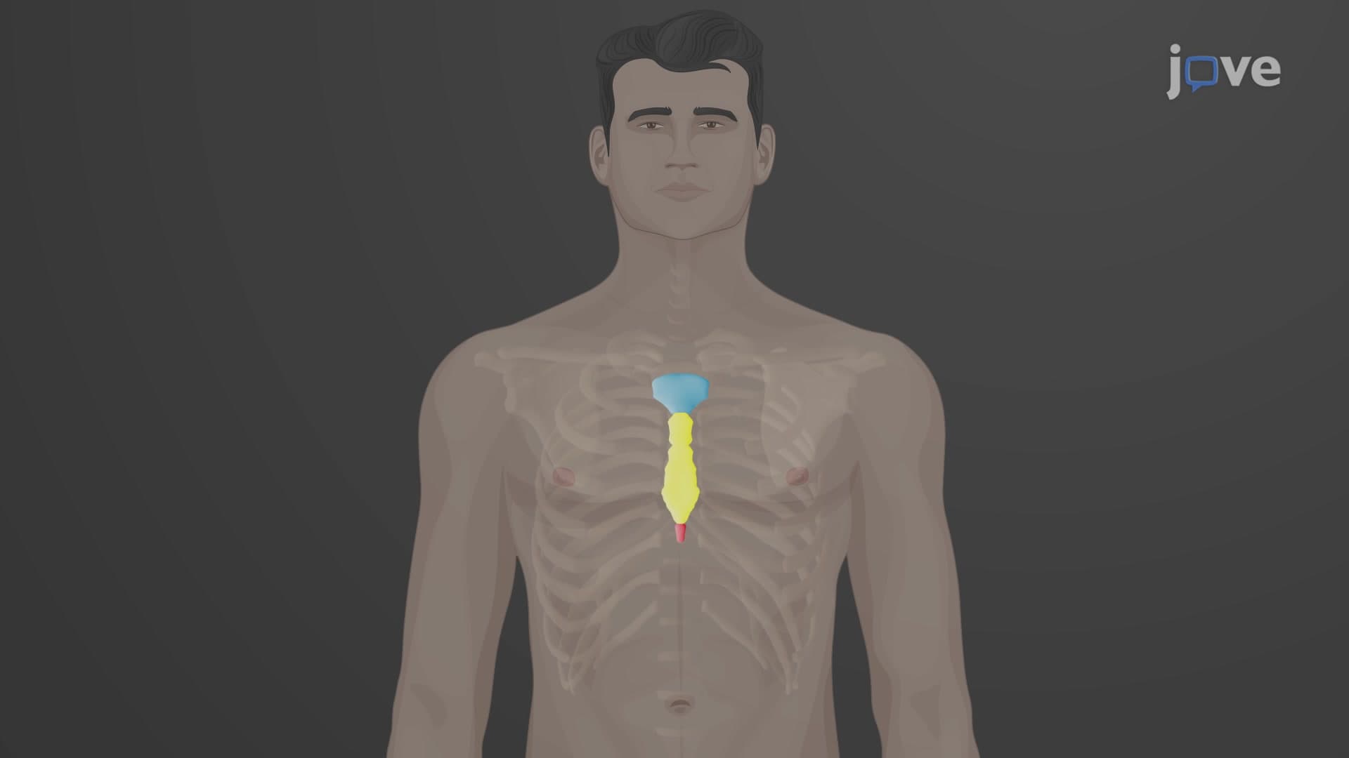
The thoracic cage ribs anatomy represents one of the most elegant engineering solutions in human biology. This protective framework consists of 12 pairs of curved bones that form the lateral walls of the thoracic cavity, working alongside the sternum and thoracic vertebrae to create a flexible yet sturdy enclosure for vital organs. Unlike rigid protective gear, these ribs must balance protection with mobility, expanding and contracting approximately 20,000 times daily during normal breathing.
The rib bones thorax explained reveals a hierarchical classification system. The first seven pairs, termed true ribs or vertebrosternal ribs, establish direct connections to the sternum through their individual costal cartilages-think of these as the "premium seats" with direct access. Ribs 8-10, known as false ribs or vertebrochondral ribs, take an indirect route, connecting to the costal cartilage of the seventh rib above them. This design allows for greater flexibility in the lower thoracic region while maintaining structural integrity.
The final two pairs (ribs 11-12) earn the designation "floating ribs" because they lack any anterior attachment, instead embedding their costal cartilages directly into the chest wall musculature. This arrangement provides maximum flexibility for the lower thoracic cage, accommodating the dynamic movements required during deep breathing or physical exertion-crucial for athletes like swimmers or runners who demand maximum lung capacity.
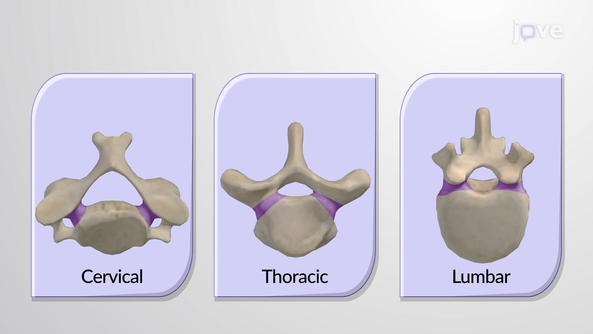
Each individual rib showcases sophisticated anatomical engineering. The rib anatomy head neck relationship begins at the posterior end, where the rib head contains articular facets that connect with costal facets on two adjacent thoracic vertebrae. This dual articulation-with both the same-numbered vertebra and the one above-creates exceptional stability while allowing rotational movement during breathing.
The tubercle, projecting from the short neck region, provides an additional point of articulation with the transverse process of the corresponding vertebra. This three-point attachment system (head to two vertebral bodies, tubercle to transverse process) creates what biomechanical engineers call a "kinetic chain"-allowing coordinated movement while preventing individual rib displacement.
The rib body features a distinctive costal groove along its internal surface, serving as a protected highway for intercostal nerves, arteries, and veins. Medical students studying for the MCAT or USMLE examinations must understand this groove's clinical significance, as improper chest tube placement can damage these neurovascular bundles, leading to complications.
Understanding thoracic rib articulation proves essential for various US medical examinations. MCAT questions frequently test knowledge of rib classification and respiratory mechanics, while nursing students preparing for NCLEX or HESI A2 exams encounter scenarios involving chest tube insertions and rib fracture assessments. AP Biology students explore how intercostal muscles utilize rib flexibility to alter thoracic volume, demonstrating the relationship between anatomy and physiology that appears on standardized tests and college midterm examinations.
Related Micro-courses