Loading subject page…

- Anatomy and Physiology
- The Axial Skeleton
- Cranial Bones: Lateral View
Micro-course: The Axial Skeleton
57,801 views
Learn key concepts and earn your certificate.




Cranial Bones: Lateral View

00:00
01:27
Video Summary: What Is Cranial Bones Lateral View
When emergency room physicians at Johns Hopkins examine head trauma patients, they rely heavily on understanding the cranial bones lateral view to assess potential fractures. This side perspective reveals critical structures like the temporal bone's zygomatic arch and the sphenoid's pterygoid processes-features invisible from other angles. The lateral skull view provides essential anatomical landmarks that medical professionals use daily for diagnosis and treatment planning. Watch the full video on JoVE Coach to master this concept with expert-led visuals and step-by-step explanations.
- Understand what cranial bones lateral view reveals and why this anatomical perspective is clinically significant
- Identify the temporal bone's key features including the squamous part, zygomatic process, and mastoid process
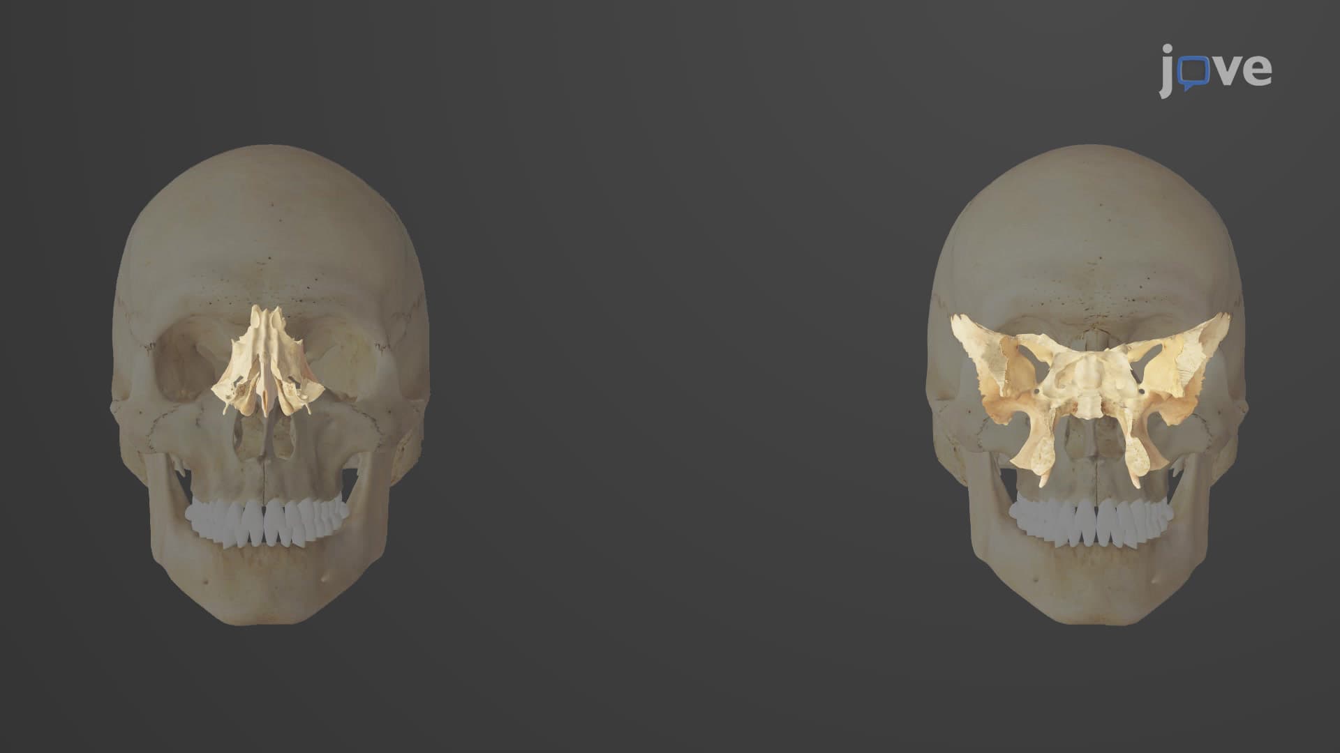
- Analyze the sphenoid bone's complex structure, focusing on the sella turcica, greater wings, and pterygoid processes
- Recognize the ethmoid bone's cribriform plate and crista galli in lateral perspective
- Compare how different cranial bones articulate with each other when viewed from the side
- Evaluate the functional significance of foramina visible in lateral skull anatomy
- Apply knowledge of lateral cranial anatomy to understand clinical presentations of head injuries
- Explore the relationship between skull bone positioning and protection of underlying neural structures
Understanding Cranial Bones Lateral View Fundamentals
The cranial bones lateral view provides one of the most comprehensive perspectives for studying skull anatomy, revealing structural relationships invisible from anterior or posterior angles. This side perspective showcases how the eight cranial bones-frontal, parietal, temporal, occipital, sphenoid, ethmoid, lacrimal, and nasal-interconnect to form the protective cranial vault. Medical students at institutions like Harvard Medical School spend considerable time mastering this view because it's essential for understanding both normal anatomy and pathological conditions.
Temporal Bone Architecture in Lateral Perspective
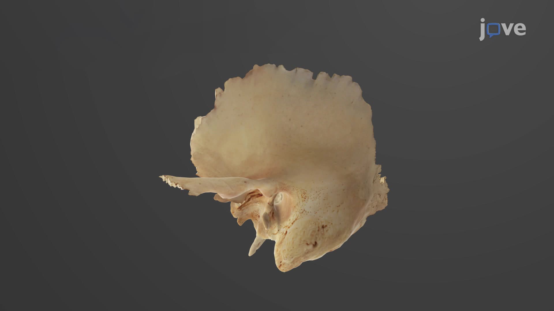
The temporal bone dominates much of the lateral skull view, displaying its squamous portion that forms part of the middle cranial fossa. The zygomatic process extends anteriorly to meet the zygomatic bone, creating the prominent zygomatic arch that you can feel just below your eye. This arch serves as an attachment site for the powerful masseter muscle used in chewing. The mastoid process, visible as a rounded projection behind the ear, anchors several neck muscles including the sternocleidomastoid. Students preparing for the MCAT often encounter questions about temporal bone fractures, which can damage the facial nerve running through the stylomastoid foramen.
Sphenoid Bone Complexity and Clinical Significance
The sphenoid bone appears remarkably complex in lateral view, earning its nickname as the "keystone" of the skull base. Its greater wing forms part of the middle cranial fossa and contains three crucial foramina: the foramen rotundum (maxillary nerve), foramen ovale (mandibular nerve), and foramen spinosum (middle meningeal artery). The pterygoid processes, projecting downward from the sphenoid body, serve as attachment points for muscles of mastication. Pre-med students at schools like UCLA often study sphenoid anatomy intensively because neurosurgeons must navigate around this bone during procedures involving the pituitary gland, which sits in the sella turcica.
Ethmoid Bone and Olfactory Function
Though partially obscured in lateral view, the ethmoid bone plays a crucial role in olfactory function. Its cribriform plate contains numerous perforations allowing olfactory nerve filaments to pass from the nasal cavity to the brain. The crista galli ("rooster's comb") projects upward, providing attachment for the falx cerebri, a membrane that separates the brain's hemispheres. Understanding ethmoid anatomy becomes particularly important for students studying conditions like anosmia (loss of smell), which has gained attention following COVID-19 research at institutions like the NIH.
Frequently Asked Questions
The cranial bones lateral view is a side perspective of the skull that reveals the temporal, sphenoid, and ethmoid bones along with their articulations and foramina. This view is crucial because it shows anatomical relationships invisible from other angles, making it essential for medical diagnosis, surgical planning, and understanding how cranial bones protect vital structures like the brain and cranial nerves.
The temporal bone is most prominent, showing its squamous part, zygomatic process, and mastoid process. The sphenoid bone displays its greater wing, pterygoid processes, and various foramina. The ethmoid bone appears between the sphenoid and nasal bones, while portions of the frontal, parietal, and zygomatic bones are also clearly visible in this perspective.
MCAT questions frequently test knowledge of cranial foramina, bone articulations, and clinical correlations visible in lateral view. Students must identify structures like the zygomatic arch, pterion (where four bones meet), and various foramina that allow nerve and vessel passage. Understanding these relationships helps answer questions about head trauma, cranial nerve pathways, and developmental abnormalities.
Temporal bone fractures can damage hearing and facial nerve function, while sphenoid bone involvement occurs in pituitary tumors and certain types of skull base fractures. Ethmoid bone damage can cause anosmia or cerebrospinal fluid leaks. Emergency physicians at trauma centers regularly assess these bones when evaluating head injuries using CT scans that provide detailed lateral perspectives.
While initially challenging due to the three-dimensional relationships involved, lateral skull anatomy becomes manageable with systematic study and hands-on practice. Starting with major landmarks like the zygomatic arch and mastoid process, then progressing to smaller details like individual foramina, allows students to build understanding progressively without feeling overwhelmed.
Create memorable associations between each foramen and its contents-for example, "Rotundum for maXillary" (both have X sounds) or "Ovale for mandibular" (think "oval mouth"). Practice with unlabeled skull models and use clinical scenarios to understand why each opening exists, as this functional approach aids long-term retention better than pure memorization.
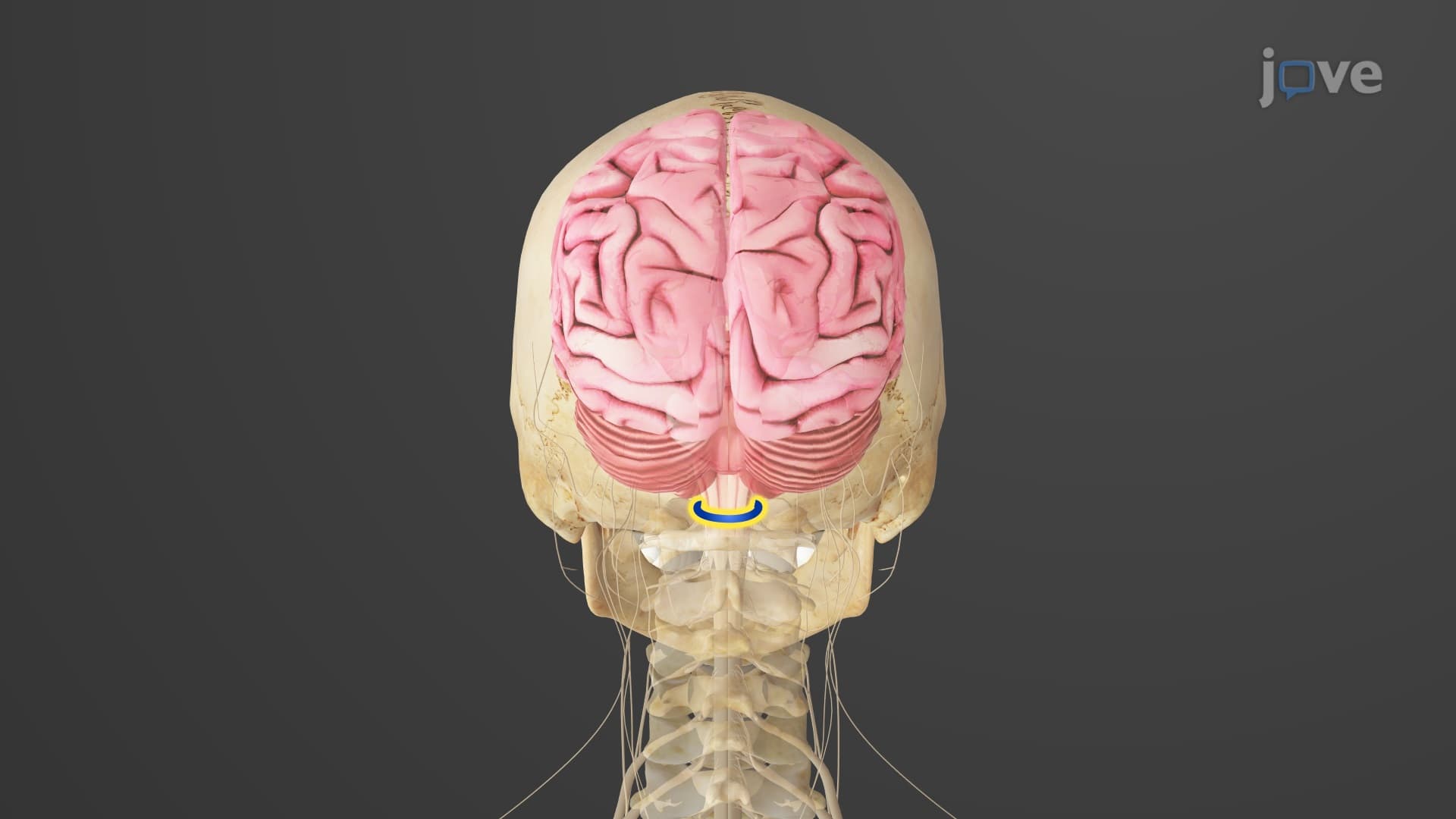
The lateral view demonstrates how multiple cranial bones work together to form a protective vault around the brain. The temporal bone's squamous part covers the temporal lobe, while the sphenoid's greater wing protects deeper brain structures. Understanding these relationships helps explain why certain impact patterns cause specific types of brain injuries and why neurosurgeons approach different brain regions through particular surgical corridors.
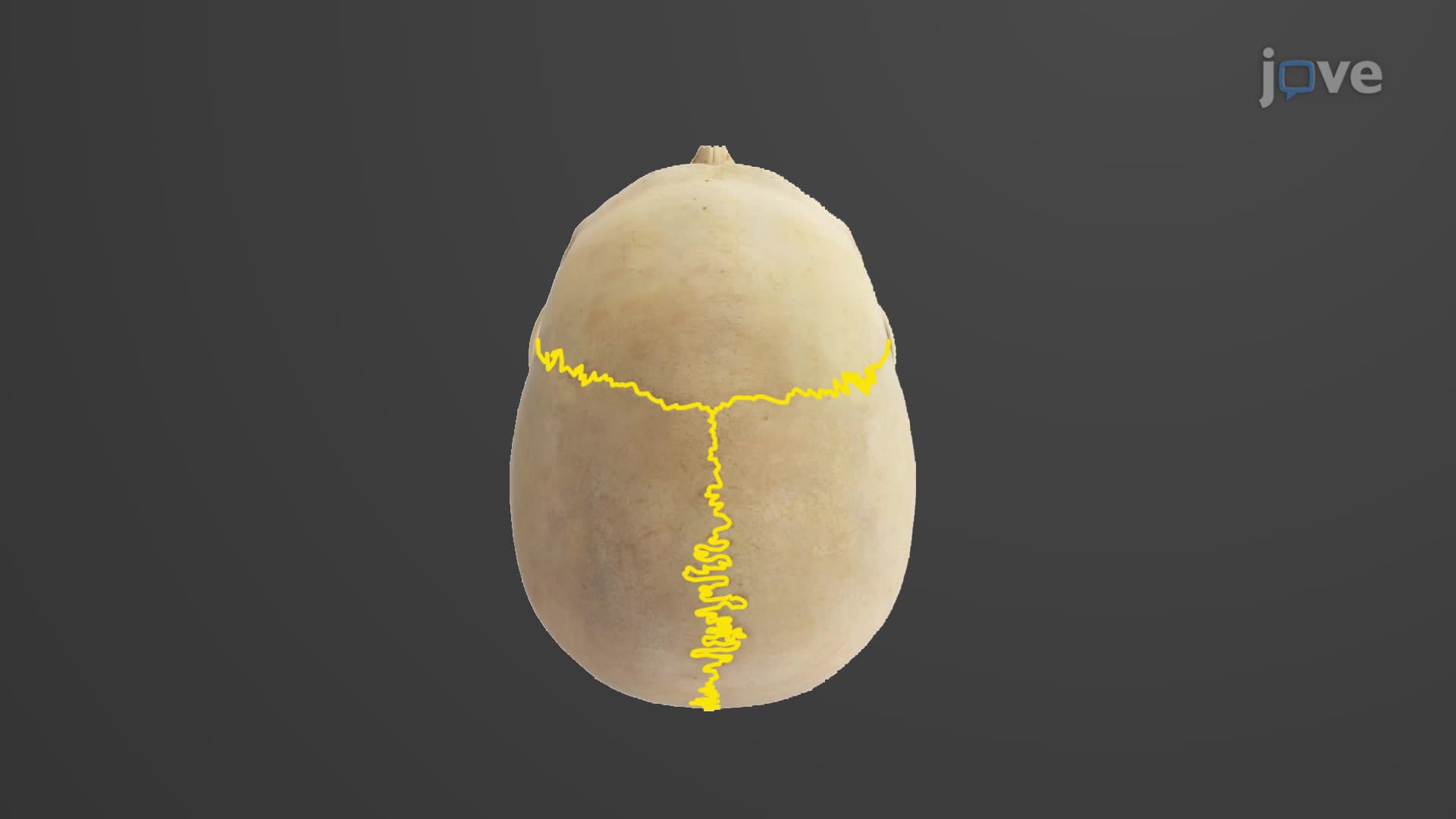
Students should progress to studying cranial sutures and their fusion patterns, detailed foraminal anatomy with specific nerve and vessel pathways, and radiological interpretation of lateral skull X-rays and CT scans. Advanced topics include skull base surgery approaches and developmental abnormalities affecting cranial bone formation, which become relevant for students pursuing neurosurgery or radiology specialties.
Related Micro-courses