Loading video…


 11 Concepts
 57.8K views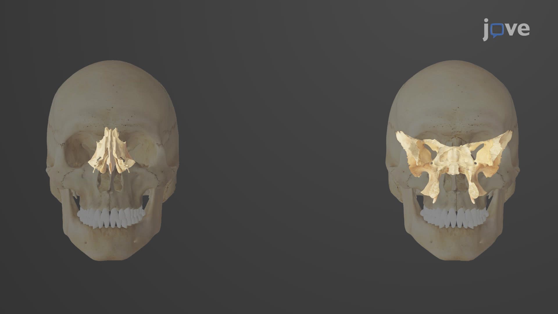

Video Summary: What Is Overview of the Skull
Did you know your skull contains 22 bones that work together like a protective helmet for your brain? Understanding the skull overview anatomy reveals how cranial and facial bones create this remarkable structure. In US medical schools like Johns Hopkins, students learn that the human skull structure overview divides into two main regions: the neurocranium protecting the brain and the viscerocranium forming your face. Watch the full video on JoVE Coach to master this concept with expert-led visuals and step-by-step explanations.
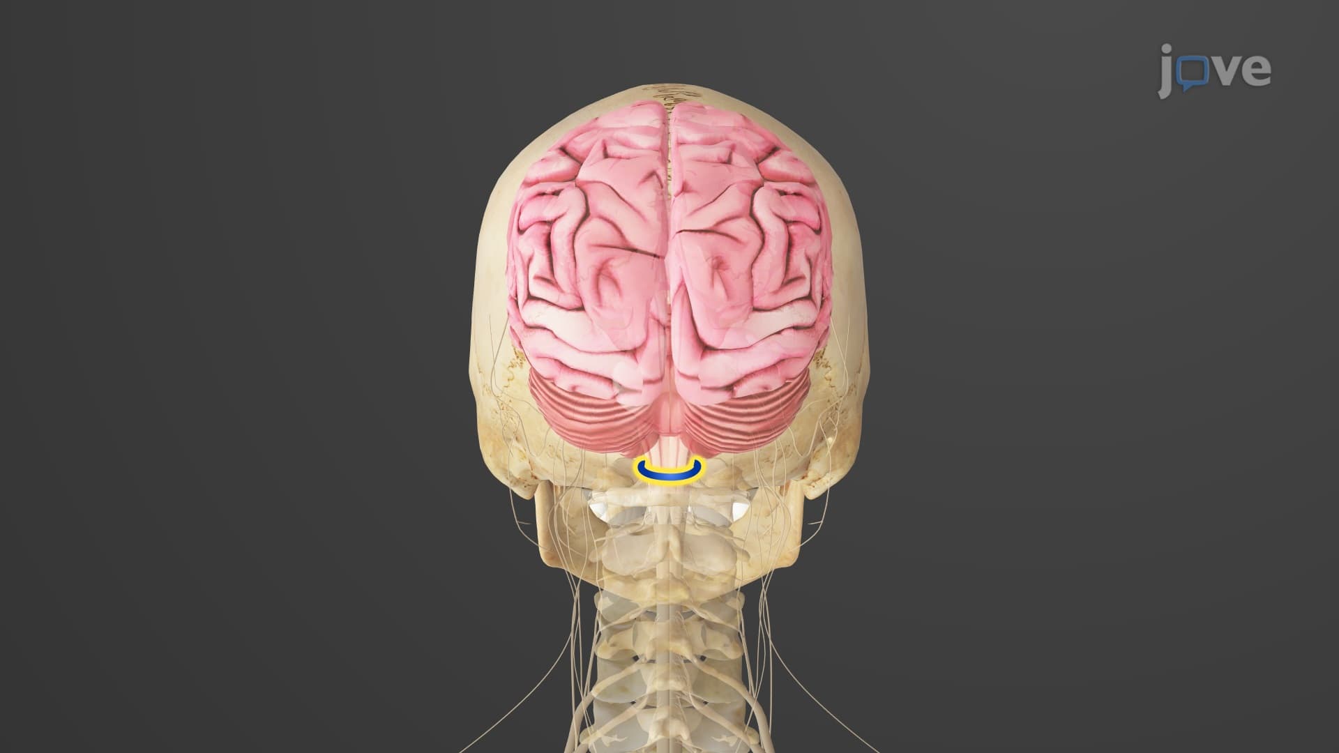
The skull overview anatomy encompasses 22 bones that form the head's bony framework, divided into two functional regions. The neurocranium consists of eight bones protecting the brain, while the viscerocranium contains fourteen bones forming the facial skeleton. This division reflects the skull's dual role: safeguarding vital neural structures while providing attachment sites for muscles of mastication and facial expression.
Medical students at institutions like UCLA and Harvard learn that understanding this human skull structure overview is crucial for clinical practice, as skull anatomy directly relates to neurological examinations, dental procedures, and trauma assessment.
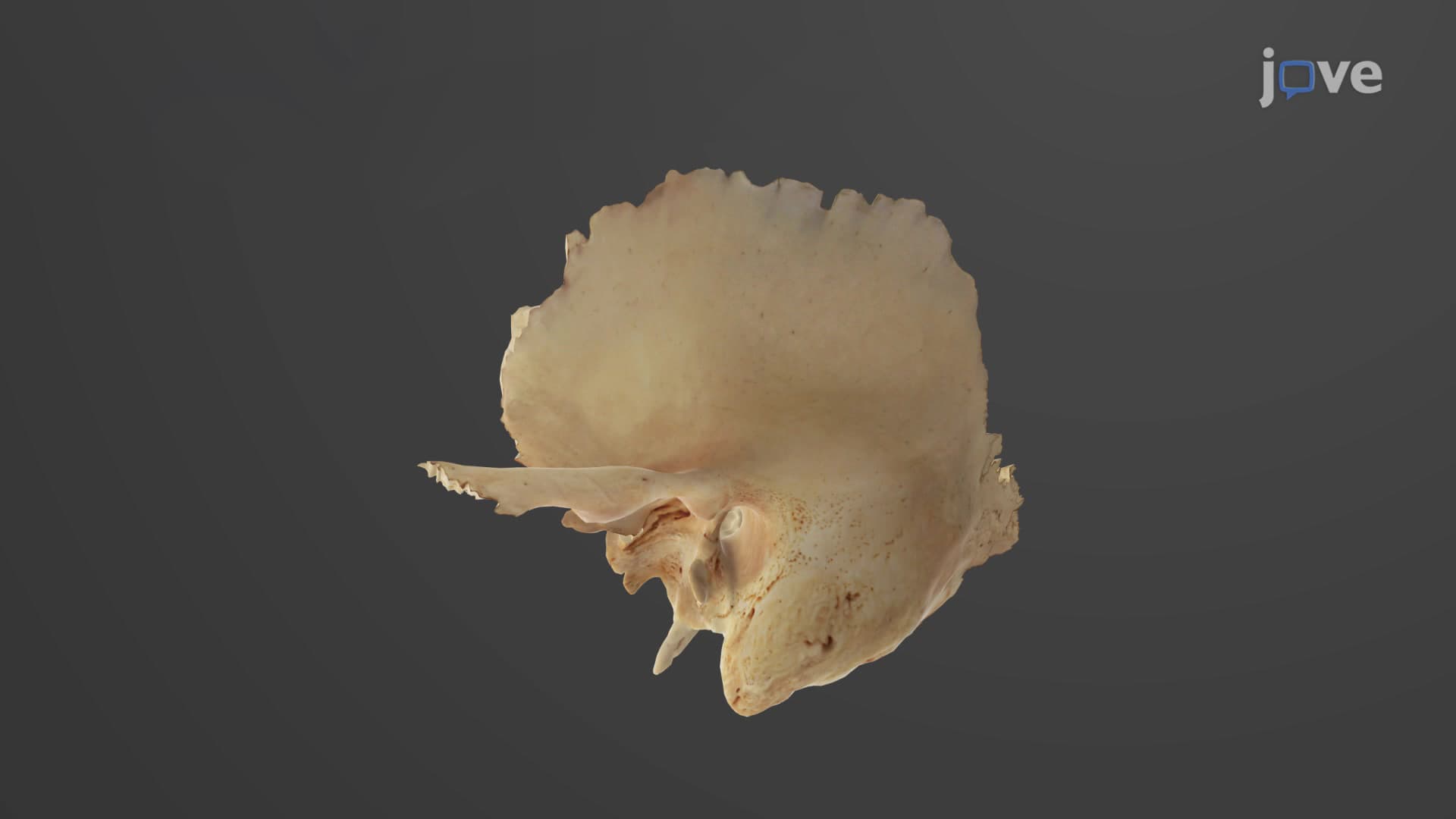
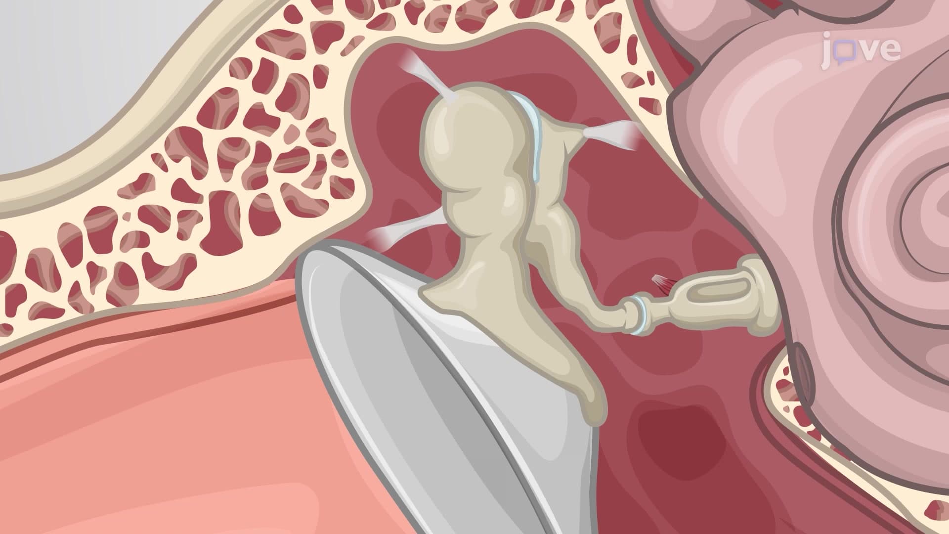
The neurocranium includes the frontal bone forming your forehead, paired parietal bones creating the skull's top and sides, and the occipital bone at the back. Temporal bones house your ear structures, while the ethmoid and sphenoid bones form the skull base's complex interior. These bones create the cranial cavity's 1,400-cubic-centimeter space housing the brain.
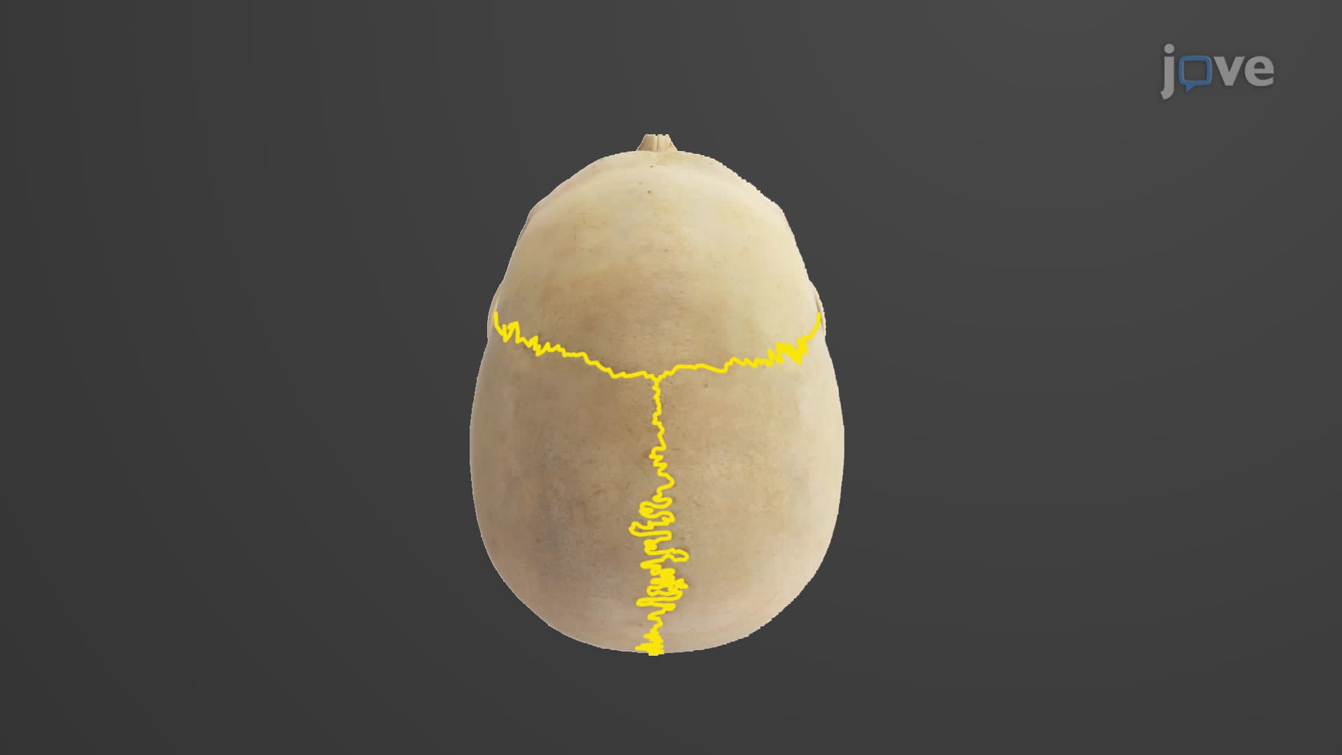
On the MCAT and AP Biology exams, students frequently encounter questions about how these bones develop through intramembranous ossification, with sutures allowing skull expansion during childhood growth. The coronal suture between frontal and parietal bones, sagittal suture between parietal bones, and lambdoid suture separating parietal and occipital bones are commonly tested landmarks.
What is an overview of the skull in anatomy wouldn't be complete without examining the viscerocranium. The mandible forms the movable lower jaw, while paired maxillae create the upper jaw and contain the maxillary sinuses. Nasal bones shape the nose bridge, zygomatic bones form cheekbones, and lacrimal bones house tear ducts.
Smaller bones like the palatine bones separate nasal and oral cavities, while the vomer contributes to the nasal septum. These intricate relationships explain why facial fractures, common in US emergency departments, often involve multiple bones simultaneously.
Understanding skull anatomy overview proves essential for healthcare careers. Nurses taking the NCLEX-RN encounter questions about skull assessment, while pre-med students face detailed skull questions on the MCAT. The concept appears regularly in college anatomy courses, particularly when studying head trauma patterns seen in American football injuries or motor vehicle accidents treated at trauma centers nationwide.
Related Micro-courses