Loading video…


 11 Concepts
 57.8K views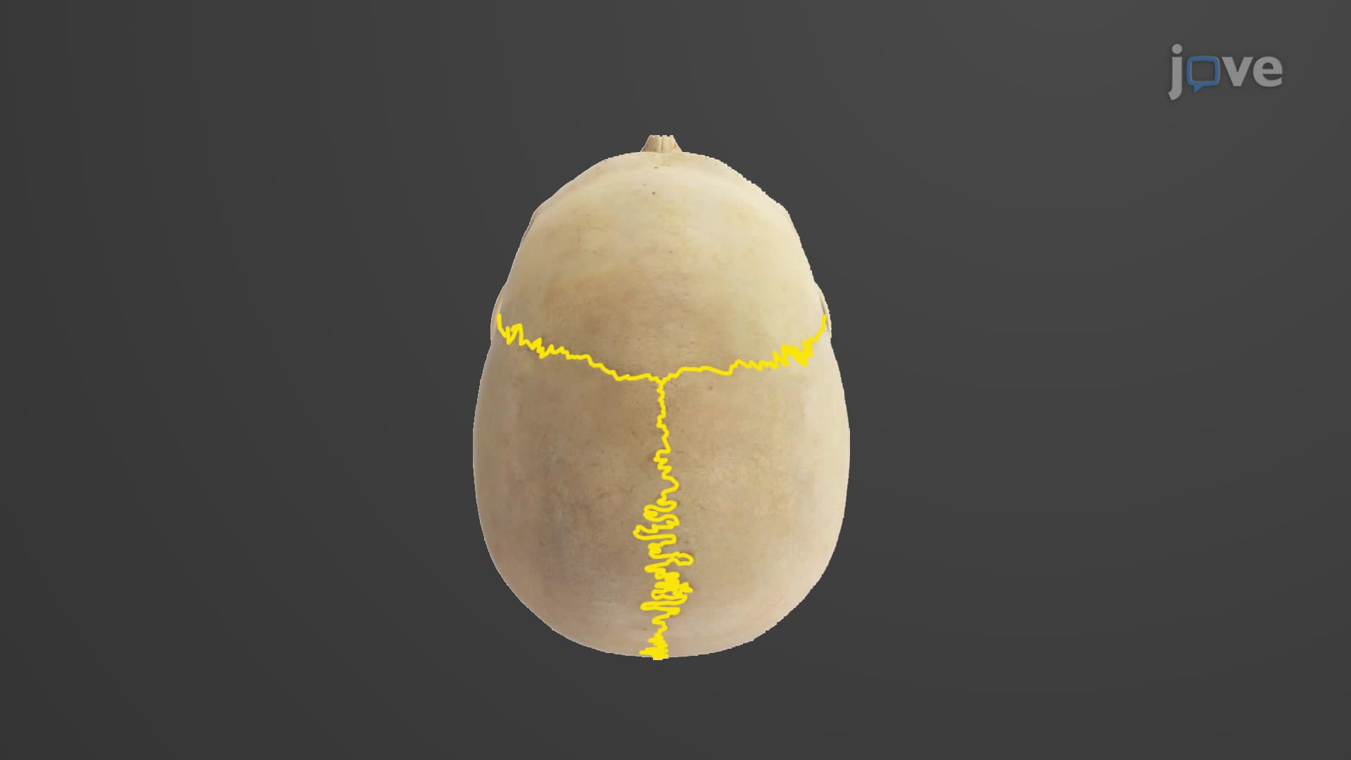

Video Summary: What Is Sutures of the Skull
Ever wonder why newborn babies can squeeze through the birth canal without skull fractures? Skull sutures anatomy reveals the fascinating design of cranial bone connections that make this possible. These immovable fibrous joints between skull bones start as flexible fontanelles in infants-the "soft spots" pediatricians examine during routine checkups at Children's Hospital of Philadelphia and other US medical centers. What is sutures of the skull becomes clear when we understand how these seam-like structures protect our brain while allowing for growth. Watch the full video on JoVE Coach to master this concept with expert-led visuals and step-by-step explanations.
Skull sutures anatomy represents one of the most elegant examples of form following function in human biology. These specialized joints serve as both protective barriers and developmental accommodations, making them crucial topics in AP Biology, college anatomy courses, and pre-medical studies across US universities.
The coronal sagittal lambdoid suture system forms the primary framework of cranial protection. The coronal suture creates the anterior boundary, connecting the frontal bone to both parietal bones in a crown-like pattern-hence its name from the Latin "corona" meaning crown. Students preparing for the MCAT often remember this by visualizing a royal crown sitting atop the forehead.
The sagittal suture runs along the skull's midline, joining the two parietal bones like an arrow's path (sagittal derives from "sagitta," meaning arrow). This squamous suture skull connection, along with the lambdoid suture at the skull's posterior, creates a three-dimensional puzzle that neurosurgeons at institutions like Johns Hopkins navigate during complex procedures.
Fibrous skull joint composition involves dense collagen fibers that provide immovable stability while maintaining slight flexibility during impact. This design prevents the catastrophic fracture patterns seen in rigid materials, explaining why football helmets and motorcycle gear incorporate similar flexible-joint principles.
During fetal development, fontanelles allow for brain growth rates of up to 15 cm in circumference during the first year. Pediatricians at Children's National Hospital monitor fontanelle closure timing, as premature fusion (craniosynostosis) or delayed closure can indicate developmental concerns requiring surgical intervention.
Understanding skull suture anatomy proves essential for students pursuing healthcare careers. Radiologists identify suture lines on CT scans to assess trauma patients, while plastic surgeons working on craniofacial reconstruction rely on suture knowledge for optimal surgical approaches. The naming convention-where sutures take names from connected bones-helps medical students systematically learn cranial anatomy for USMLE Step 1 examinations.
Related Micro-courses