Loading subject page…


57,806 views
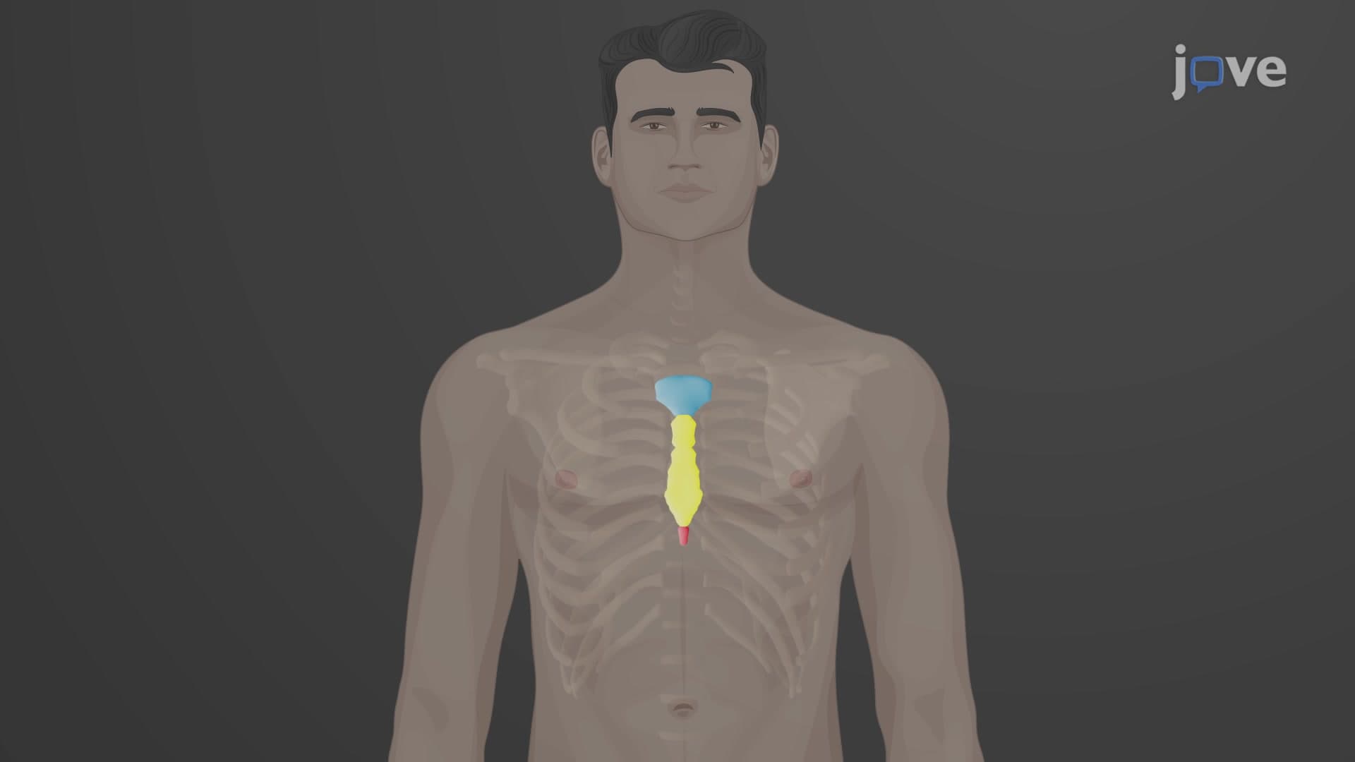

Video Summary: What Is the Thoracic Cage Sternum
Ever wondered how your chest stays protected during a baseball slide or football tackle? The sternum thoracic cage anatomy forms your body's natural armor, with the T-shaped sternum bone acting as the central shield for your heart and lungs. During CPR training at American Red Cross centers, instructors emphasize proper hand placement on the sternum to avoid injury while saving lives. What is The Thoracic Cage Sternum reveals how this remarkable bone connects your entire rib cage system. Watch the full video on JoVE Coach to master this concept with expert-led visuals and step-by-step explanations.
The sternum represents one of anatomy's most ingenious designs-a flat, T-shaped bone that serves as the central anchor point for your entire thoracic cage. This remarkable structure doesn't just protect your vital organs; it creates the mechanical foundation that allows your ribcage to expand and contract with every breath you take.
The sternum bone structure explained reveals a fascinating developmental story. What appears as a single bone actually forms through the fusion of three distinct components: the manubrium (top), body (middle), and xiphoid process (bottom). The manubrium creates the broad upper portion, featuring the distinctive jugular notch you can feel at the base of your throat-the same landmark nurses use when assessing your pulse. The sternal angle, where the manubrium meets the body, serves as a critical reference point that aligns with the second rib, making it invaluable for medical professionals during chest examinations.
The sternum's body forms the longest segment, providing attachment sites for ribs three through seven via their costal cartilages. This connection system allows for the flexibility needed during breathing while maintaining structural integrity. Students preparing for the MCAT or AP Biology exams should note how this arrangement facilitates the "bucket handle" motion of ribs during respiration.
Understanding sternum attachment to ribs becomes crucial in medical emergencies. When EMTs perform CPR across American hospitals, they position their hands specifically on the lower half of the sternum's body, avoiding the xiphoid process entirely. This precision prevents the cartilaginous xiphoid from breaking away and potentially puncturing internal organs-a complication that could prove fatal during life-saving efforts.
The xiphoid process itself remains primarily cartilaginous until around age 40, when ossification typically completes. This timing explains why CPR techniques differ slightly between pediatric and adult patients in US emergency medicine protocols.
For pre-med students tackling the USMLE Step 1, the sternum provides numerous anatomical landmarks. The jugular notch aligns with the T2-T3 vertebrae, while the sternal angle corresponds to T4-T5, marking where the trachea bifurcates into bronchi. These relationships prove essential for interpreting chest X-rays and understanding cardiac positioning within the thoracic cavity.
Related Micro-courses