Loading video…


 26 Concepts
 111.9K views


Video Summary: What are Nervous Tissue Glial Cells
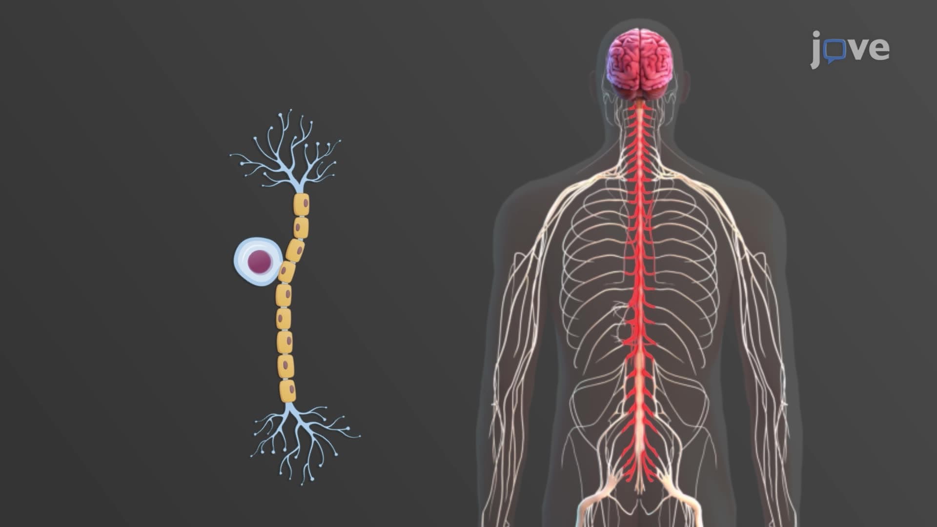
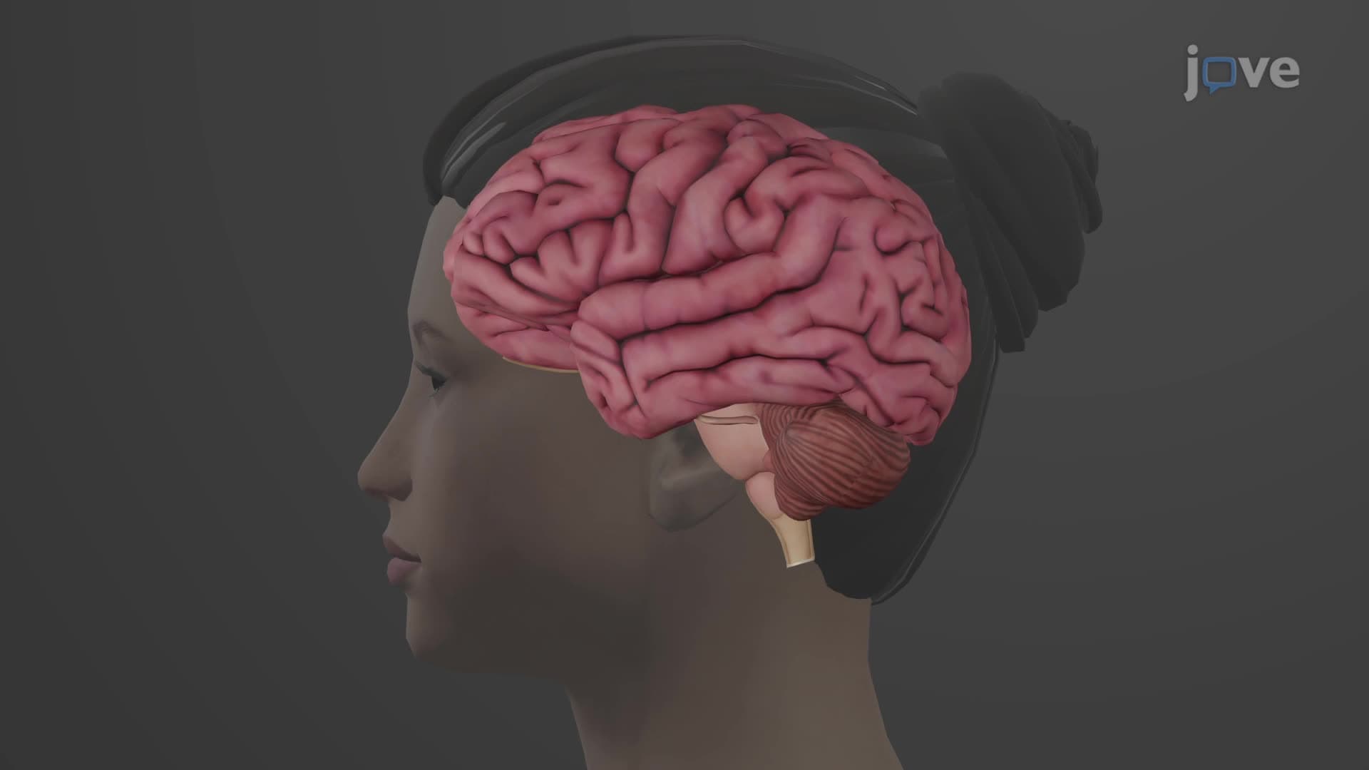
Your brain contains roughly 86 billion neurons, but surprisingly, glial cells nervous tissue types actually outnumber these famous nerve cells! While neurons grab the spotlight in AP Biology textbooks, glial cells quietly perform essential jobs like maintaining the blood-brain barrier that protects your brain during a concussion. These specialized support cells include astrocytes, oligodendrocytes, microglia, and others that keep your nervous system functioning properly. Watch the full video on JoVE Coach to master this concept with expert-led visuals and step-by-step explanations.
Neuroglia types and functions represent one of neuroscience's most underappreciated topics. While medical students preparing for the USMLE often focus intensively on neuronal action potentials, glial cells actually outnumber neurons by a 10:1 ratio in human brains. These "nerve glue" cells (from Greek "glia" meaning glue) provide structural support, immune protection, and metabolic assistance that neurons cannot survive without.
Astrocyte blood brain barrier function exemplifies how glial cells protect neural tissue. These star-shaped cells extend cytoplasmic processes that contact both neurons and blood vessels, forming tight junctions that prevent harmful substances from entering brain tissue. When someone suffers a traumatic brain injury playing high school football, astrocytes help maintain this protective barrier while also regulating potassium ion concentrations and recycling neurotransmitters like glutamate.
Oligodendrocyte CNS myelin production enables rapid nerve signal transmission essential for complex thinking and motor coordination. Each oligodendrocyte can myelinate up to 50 different axons simultaneously, creating the white matter visible in brain MRI scans. Multiple sclerosis, affecting over 400,000 Americans, occurs when the immune system attacks these myelin sheaths, demonstrating their clinical importance.
Microglia immune CNS responses protect brain tissue from infection and injury. These small, mobile cells constantly patrol neural tissue, removing cellular debris and responding to inflammation. During Alzheimer's disease progression, overactive microglia may contribute to neuronal damage, making them important therapeutic targets in current NIH-funded research.
Ependymal cells line brain ventricles and the spinal cord's central canal, producing cerebrospinal fluid that cushions neural tissue and removes metabolic waste. Their coordinated ciliary beating circulates this fluid throughout the CNS, maintaining proper intracranial pressure.
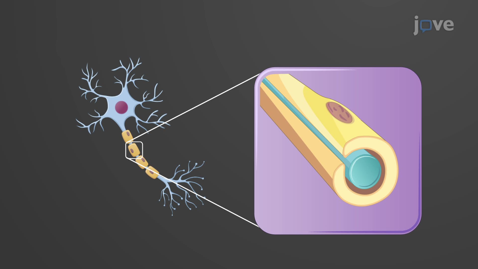
Schwann cell PNS myelin differs significantly from CNS myelination. Each Schwann cell myelinates only one axon segment, creating nodes of Ranvier that enable saltatory conduction. This one-to-one relationship facilitates peripheral nerve regeneration following injury, unlike the limited regenerative capacity of CNS neurons.
Satellite cells surround peripheral neuron cell bodies in ganglia, regulating their local environment and providing metabolic support. These cells become particularly important during diabetic neuropathy, where high blood glucose levels can damage both neurons and their supporting glial cells.
Understanding these concepts proves essential for AP Biology students tackling nervous system units and pre-med students preparing for MCAT passages about neurological disorders. College anatomy courses frequently test glial cell identification and function on practical exams using microscope slides and histological images.
Related Micro-courses