Loading subject page…


111,914 views
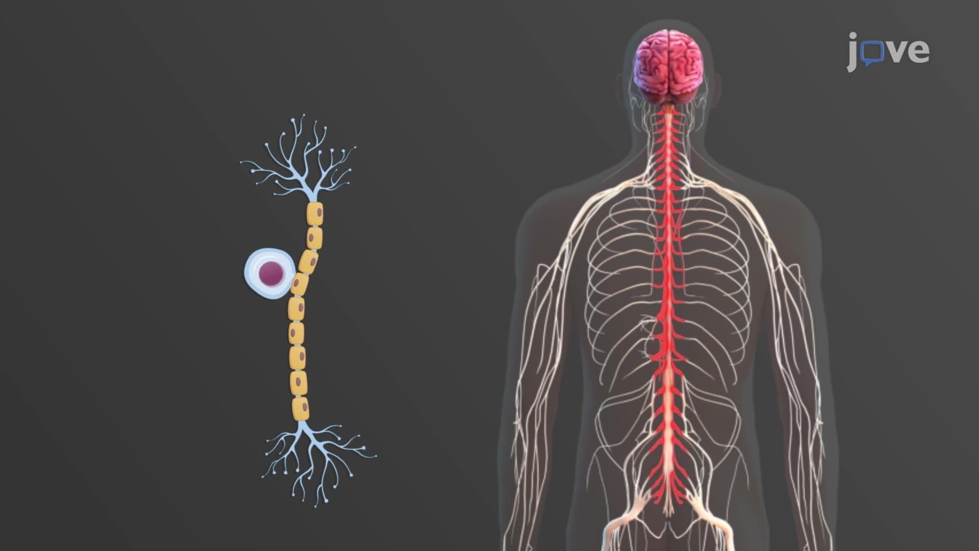

Video Summary: What are Nervous Tissue Neuron Types
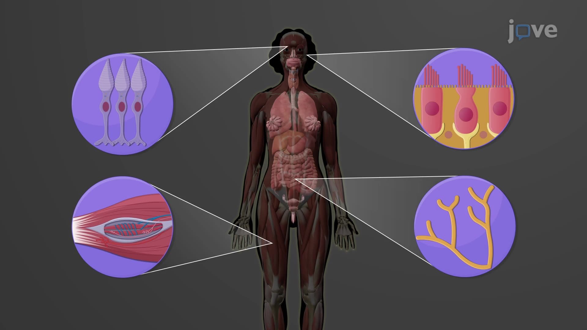
Ever wonder how your brain processes the sensation of touching a hot stove and instantly commands your hand to pull away? Neuron types nervous tissue biology reveals the fascinating cellular diversity behind this lightning-fast response. From the multipolar neurons dominating your spinal cord to the rare bipolar neurons in your retina that enable vision, understanding what are Nervous Tissue Neuron Types unlocks the secrets of neural communication. Each neuron type has evolved specific structures and functions, like how sensory neurons in your fingertips can detect pressure changes as small as a mosquito landing. Watch the full video on JoVE Coach to master this concept with expert-led visuals and step-by-step explanations.
The classification of neurons explained through both structural and functional lenses provides the cornerstone for understanding how your nervous system processes information. This dual classification system isn't just academic-it's essential for medical students preparing for the MCAT, AP Biology students tackling neural physiology, and anyone seeking to understand how 86 billion neurons coordinate seamlessly in the human brain.
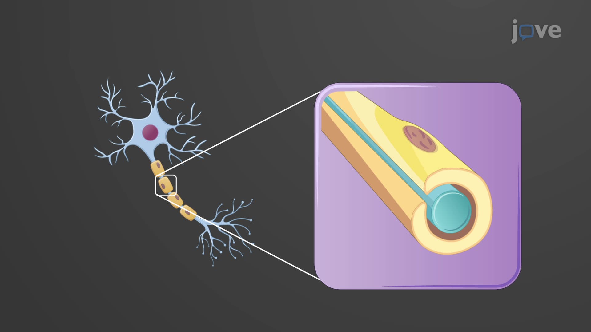
Multipolar bipolar unipolar neurons represent the three fundamental architectural designs in neural tissue. Multipolar neurons, featuring one axon and multiple dendrites, dominate your brain and spinal cord precisely because their branched structure enables complex information integration. Picture a multipolar motor neuron in your spinal cord: its numerous dendrites collect signals from hundreds of other neurons, while its single axon extends up to three feet to reach muscles in your toes.
Bipolar neurons, though rare, excel in specialized sensory roles. In your retina, bipolar neurons bridge photoreceptors and ganglion cells, creating the critical second step in vision processing. Their streamlined two-process design-one dendrite, one axon-facilitates rapid, direct signal transmission essential for real-time visual processing.
Unipolar neurons showcase evolutionary efficiency in sensory systems. These sensory afferent neurons have cell bodies tucked safely in ganglia while their combined axon-dendrite processes span remarkable distances. A single sensory neuron monitoring your big toe extends from the digit all the way to your spinal cord-potentially over three feet of continuous neural fiber.
The functional trinity of sensory afferent neuron, motor efferent neuron, and interneuron association neuron creates the input-processing-output framework underlying all neural activity. Sensory neurons serve as your body's information gathering network, converting stimuli from touch, temperature, and proprioception into electrical signals your brain can interpret.
Motor neurons translate neural commands into physical action, controlling everything from voluntary movements to involuntary reflexes. Each motor neuron in your spinal cord may innervate hundreds of muscle fibers, forming motor units that enable precise force control during activities like writing or lifting weights.
Interneurons, comprising roughly 99% of all neurons, perform the computational heavy lifting. In reflex circuits studied in college neurobiology courses, interneurons can inhibit motor neurons to opposing muscle groups while exciting target muscles, creating coordinated movement patterns that happen faster than conscious thought.
Understanding neuron type function proves crucial for interpreting neurological conditions. Multiple sclerosis primarily affects myelinated axons of motor neurons, while diabetic neuropathy typically damages the long processes of sensory neurons first. These patterns make sense when you understand the structural vulnerabilities of different neuron types-knowledge frequently tested on USMLE Step 1 examinations and featured prominently in undergraduate neuroscience curricula across major US universities.
Related Micro-courses