Loading video…


 8 Concepts
 61.3K views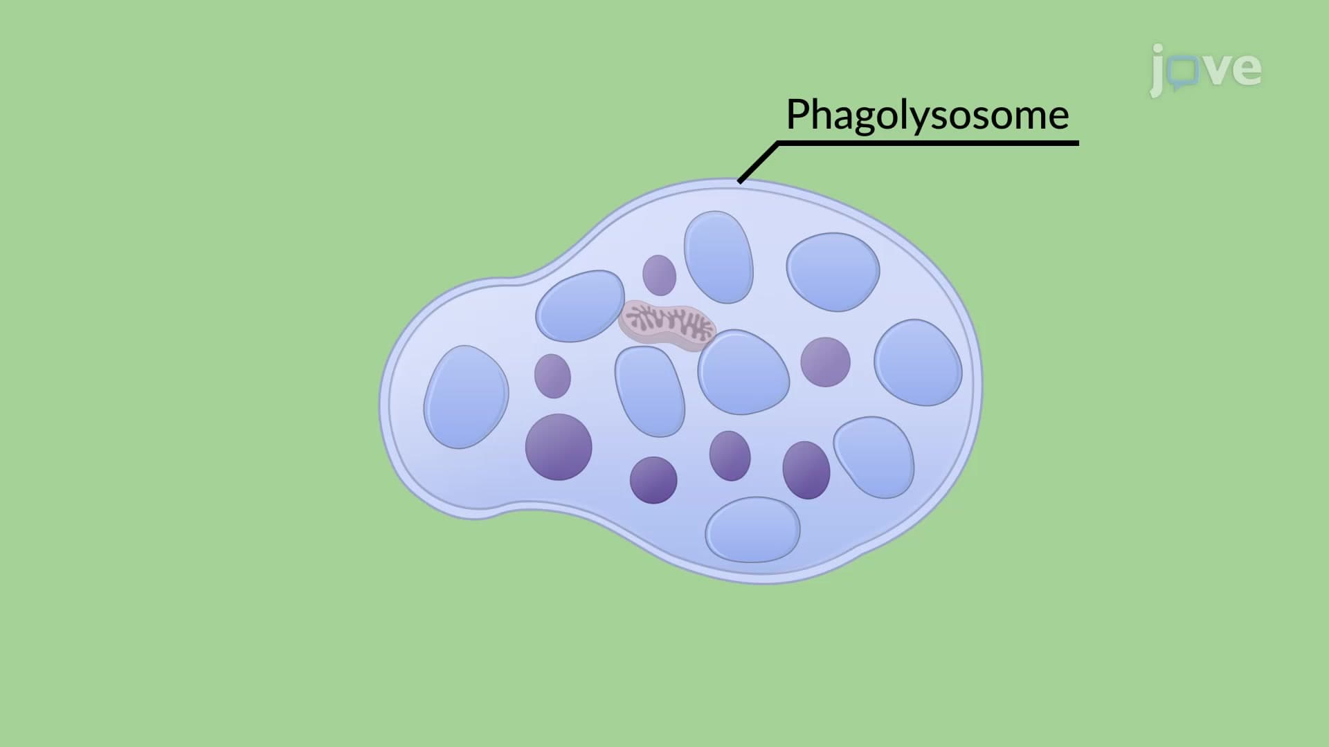

Video Summary: What are Phagocytosis of Apoptotic Cells
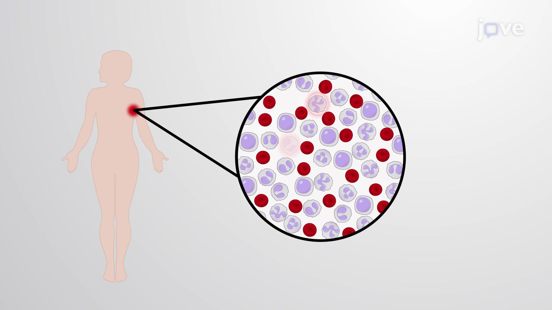
Every day, billions of cells in your body die naturally, yet you never develop massive inflammation from this cellular turnover. The phagocytosis of apoptotic cells is the critical cleanup process where specialized immune cells called phagocytes engulf and digest dying cells before they can cause harmful inflammatory responses. In the United States, researchers at Johns Hopkins University have shown this process prevents autoimmune diseases like lupus. Watch the full video on JoVE Coach to master this concept with expert-led visuals and step-by-step explanations.
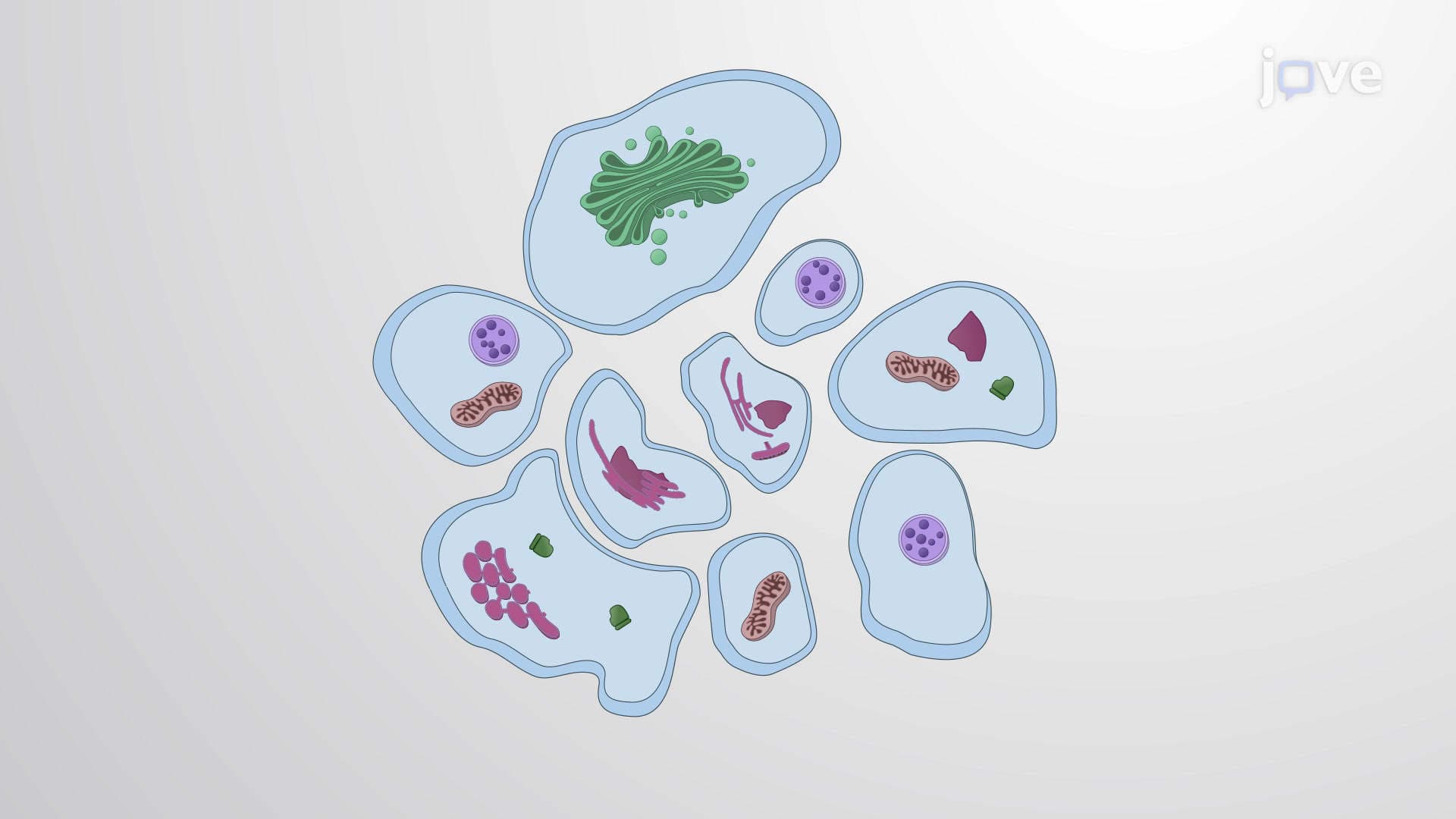
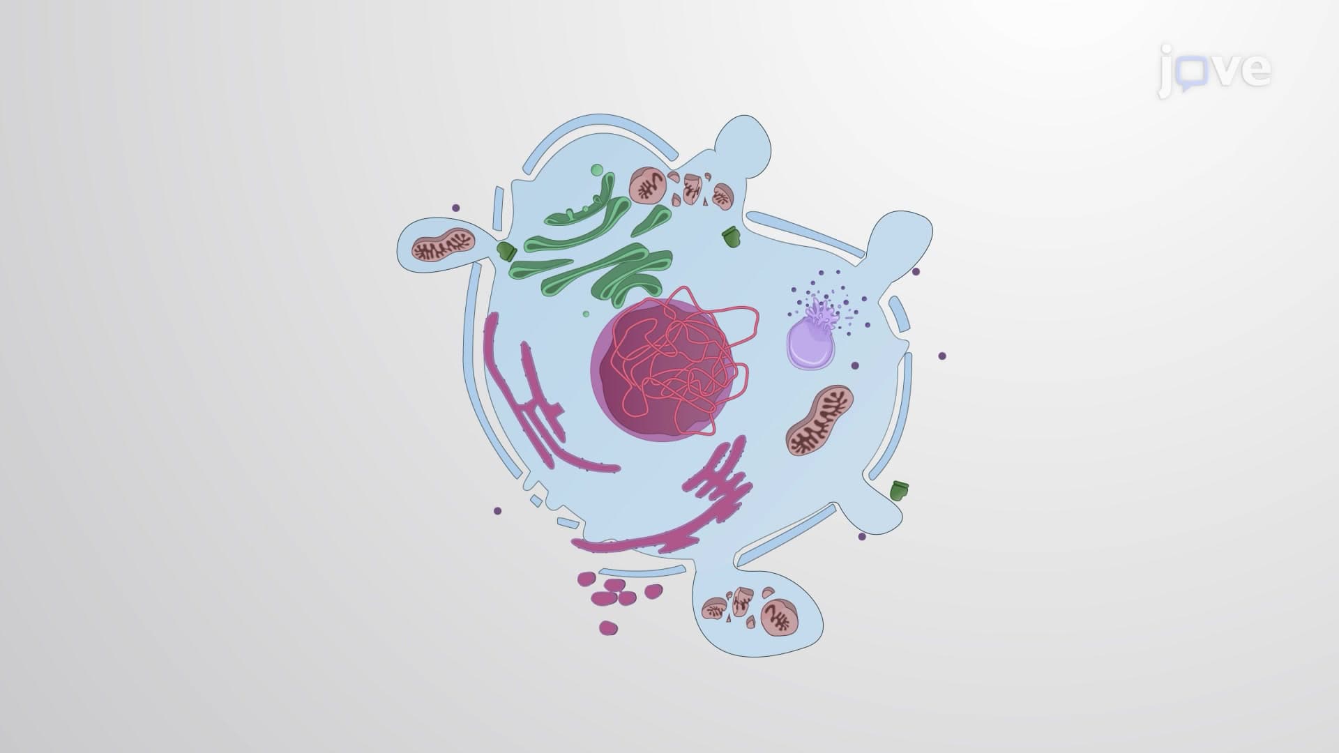
Phagocytosis of apoptotic cells represents one of the most elegant quality control mechanisms in human biology. When cells undergo programmed cell death (apoptosis), they must be rapidly removed to prevent the release of toxic intracellular contents that could trigger widespread inflammation and tissue damage.
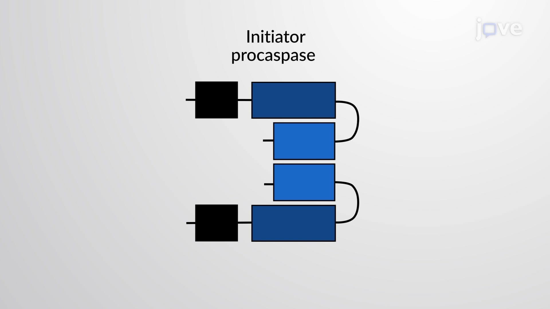
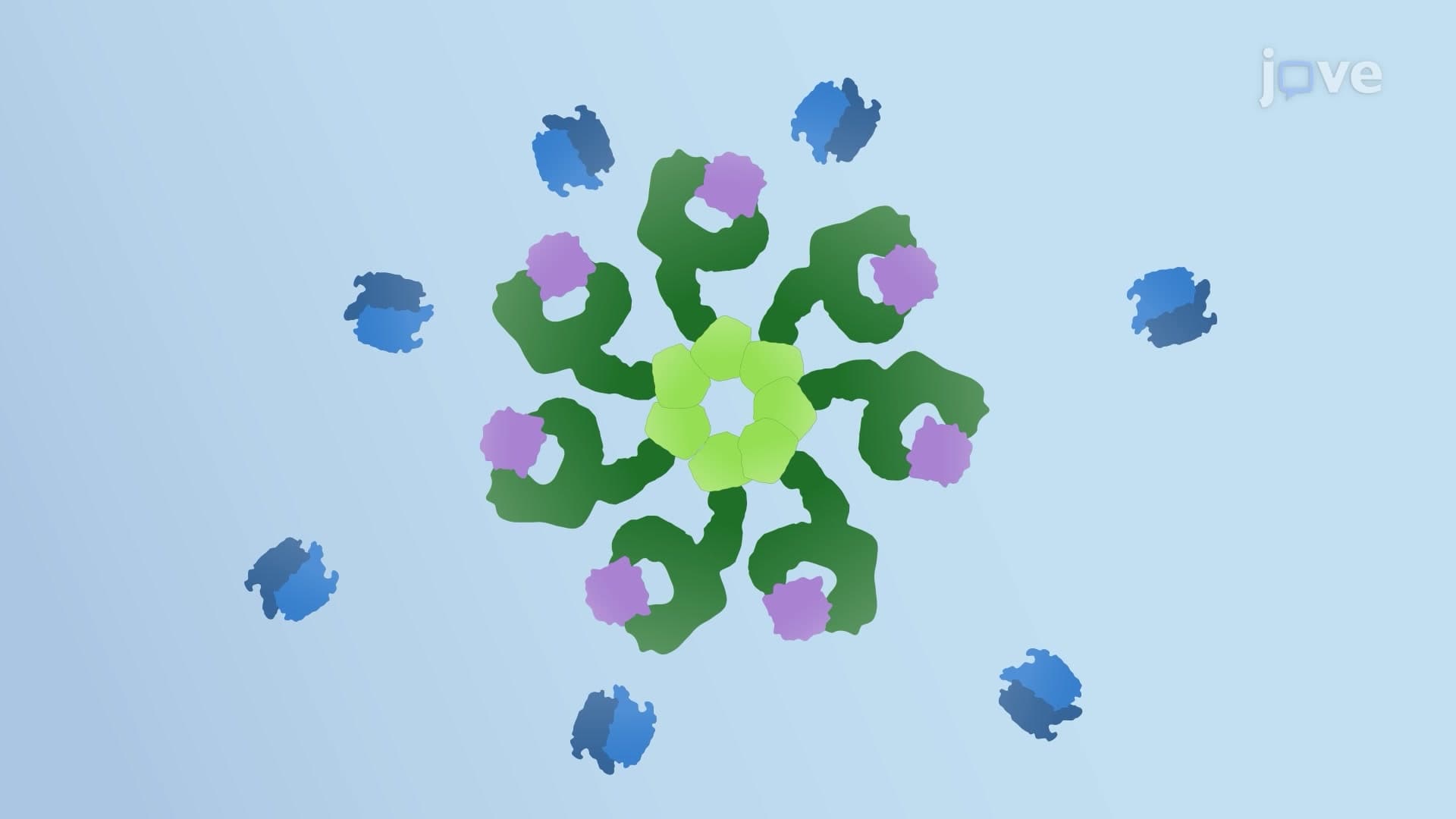
The process begins with dramatic changes in the dying cell's membrane composition. In healthy cells, an enzyme called flippase actively maintains phosphatidylserine (PS) on the inner leaflet of the plasma membrane. However, when executioner caspases activate during apoptosis, they inactivate flippase while simultaneously activating another enzyme called scramblase. This biochemical switch causes PS to flip to the outer membrane surface, creating a universal "eat-me" signal that phagocytes recognize across all cell types.
Beyond PS exposure, apoptotic cells secrete bridging proteins and release chemical attractants (chemoattractants) that function like molecular beacons, guiding nearby phagocytes to their location. These signals ensure rapid clearance-typically within hours of cell death initiation.
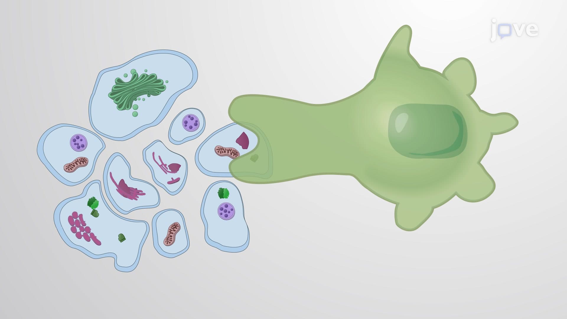
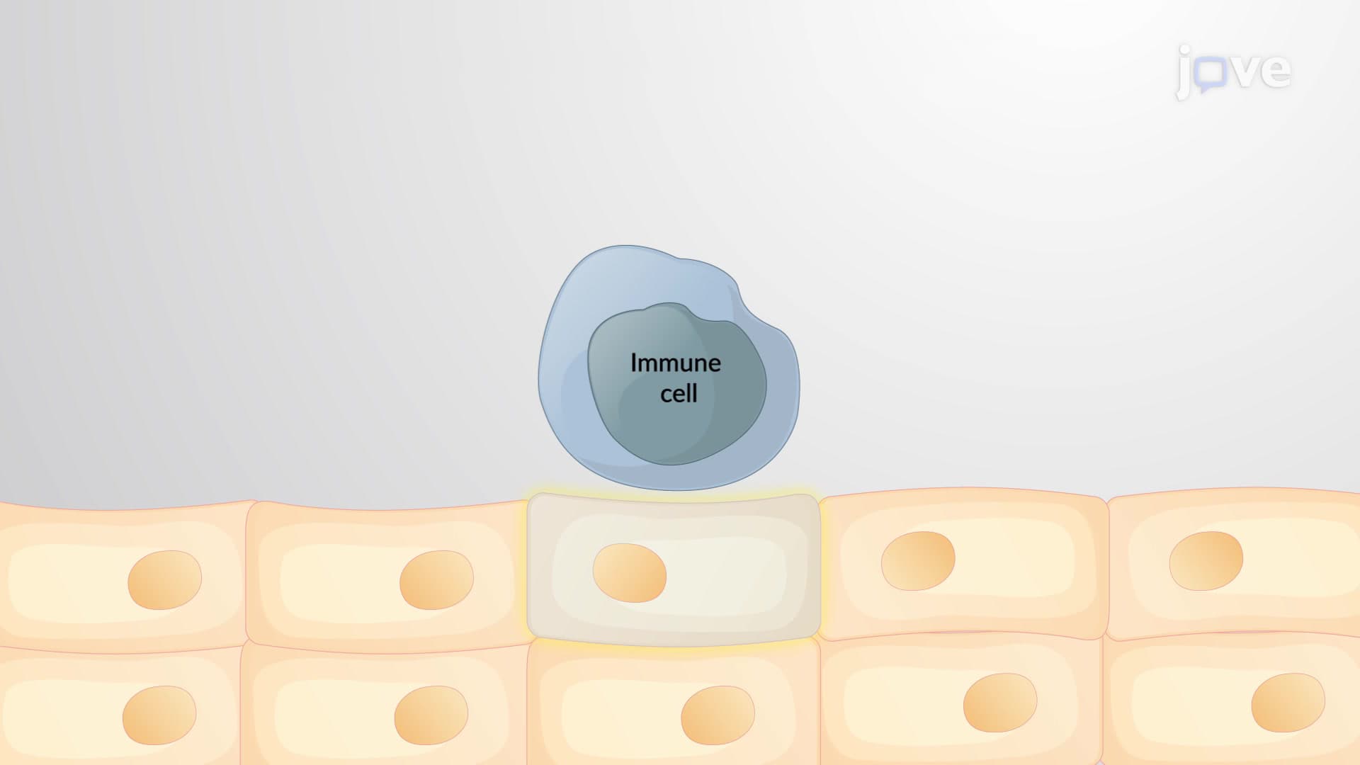
Professional phagocytes, particularly macrophages and dendritic cells, possess specialized receptors that bind either directly to exposed PS or indirectly through bridging proteins. This recognition triggers cytoskeletal rearrangements that allow the phagocyte to extend pseudopodia around the apoptotic cell, ultimately engulfing it completely.
Once internalized, the apoptotic cell becomes encased in a membrane-bound compartment called a phagosome. This structure then fuses with lysosomes-cellular organelles packed with digestive enzymes-to form a phagolysosome. Within this acidic environment, lysosomal enzymes systematically break down proteins, lipids, nucleic acids, and other cellular components for recycling.
Understanding phagocytosis of apoptotic cells proves crucial for students preparing for advanced coursework and standardized exams. On the MCAT, this concept frequently appears in passages about immune system function and cellular homeostasis. AP Biology students encounter this process when studying programmed cell death and immune responses.
In clinical settings, defective apoptotic cell clearance contributes to autoimmune diseases like systemic lupus erythematosus, where uncleared cellular debris triggers inappropriate immune responses against self-tissues. Research institutions across the United States, including Harvard Medical School and the National Institutes of Health, actively investigate how enhancing this process might treat inflammatory diseases and improve tissue repair after injury.
Related Micro-courses