Loading subject page…


61,302 views

Video Summary: What Is the Intrinsic Apoptotic Pathway
When cancer researchers at Johns Hopkins discovered that some tumor cells resist chemotherapy by blocking programmed cell death, they unlocked a crucial mystery about the intrinsic apoptotic pathway. This cellular suicide mechanism allows damaged cells to self-destruct before becoming cancerous, making it essential for preventing diseases like leukemia and solid tumors. Understanding what is the intrinsic apoptotic pathway reveals how mitochondria act as cellular executioners, releasing death signals when cells detect internal damage. Watch the full video on JoVE Coach to master this concept with expert-led visuals and step-by-step explanations.
The intrinsic apoptotic pathway represents one of biology's most sophisticated quality control mechanisms, allowing cells to eliminate themselves when internal damage threatens the organism. Unlike the extrinsic pathway triggered by external death signals, this mitochondrial-centered process responds to intracellular stress signals including DNA damage, oxidative stress, and cellular aging. Students preparing for the MCAT or AP Biology exam should recognize this pathway as a critical tumor suppressor mechanism-when it fails, cancer can develop.
The pathway's regulation centers on the Bcl-2 protein family, discovered through research on B-cell lymphomas at major US cancer centers like MD Anderson and Memorial Sloan Kettering. These proteins function as molecular switches determining cellular fate. Pro-apoptotic members include BH3-only sensors (Bad, Bim, PUMA) that detect damage signals, and effector proteins (Bax, Bak) that execute the death program. Anti-apoptotic guardians (Bcl-2, Bcl-XL, Mcl-1) prevent unnecessary cell death by restraining effector proteins. Understanding the intrinsic apoptotic pathway basics requires mastering these protein interactions, frequently tested on college biochemistry exams.
When death signals activate BH3-only proteins, they either directly activate Bax/Bak or neutralize anti-apoptotic proteins. This releases Bax and Bak to oligomerize on mitochondrial outer membranes, forming pores that release cytochrome c into the cytoplasm. This intrinsic apoptotic pathway concept transforms mitochondria from cellular powerhouses into executioners. Students should visualize this as opening floodgates-once cytochrome c escapes, there's no turning back.
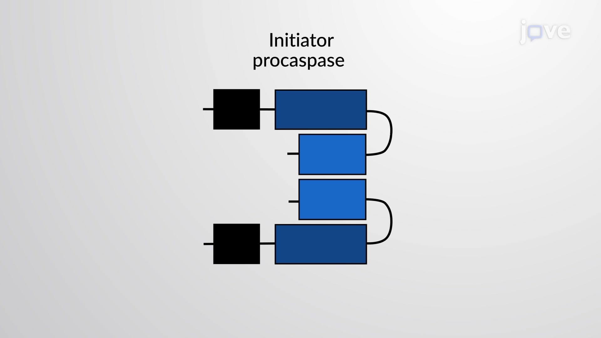
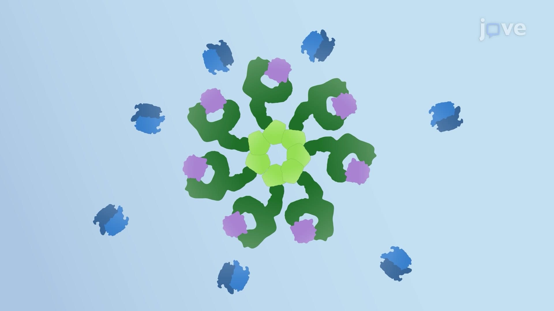
Released cytochrome c binds Apaf-1 (apoptotic protease activating factor-1), causing seven Apaf-1 molecules to assemble into the wheel-shaped apoptosome complex. This recruits procaspase-9, which undergoes proximity-induced activation to become caspase-9. The activated initiator caspase then cleaves and activates executioner caspases (caspase-3, -6, -7), which systematically dismantle cellular components. This intrinsic apoptotic pathway study guide sequence-from cytochrome c release through caspase activation-represents a common focus area for USMLE Step 1 questions testing biochemical pathways.
Related Micro-courses