Loading subject page…

- Cell Biology
- Cell Polarization and Migration
- Cancer Cell Migration through Invadopodia
Micro-course: Cell Polarization and Migration
40,212 views
Learn key concepts and earn your certificate.
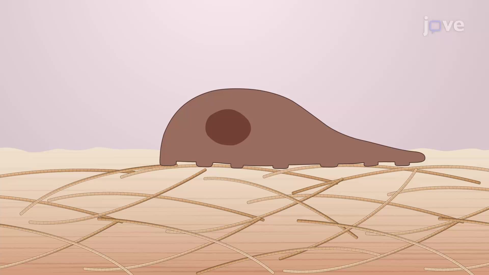

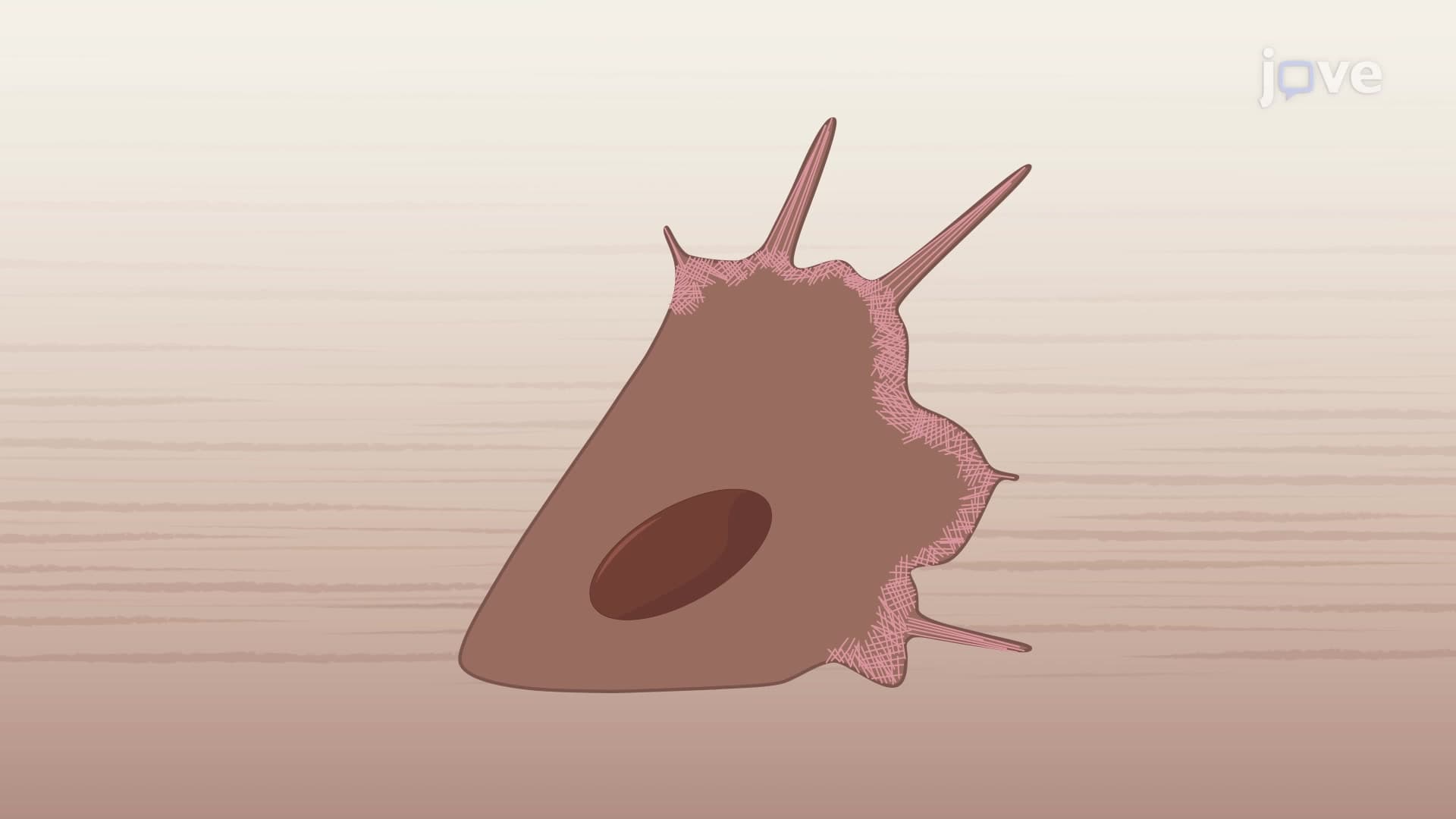
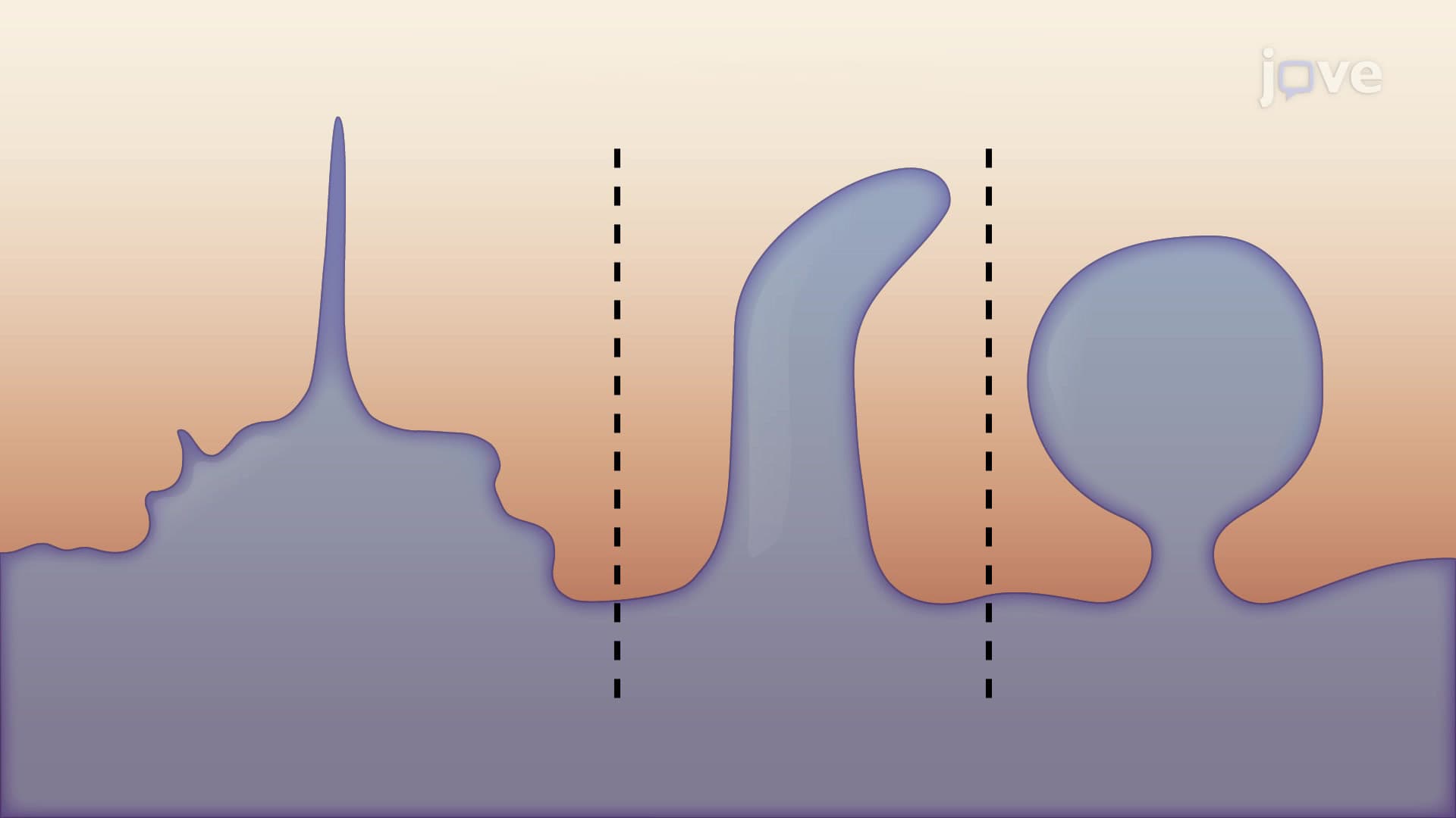
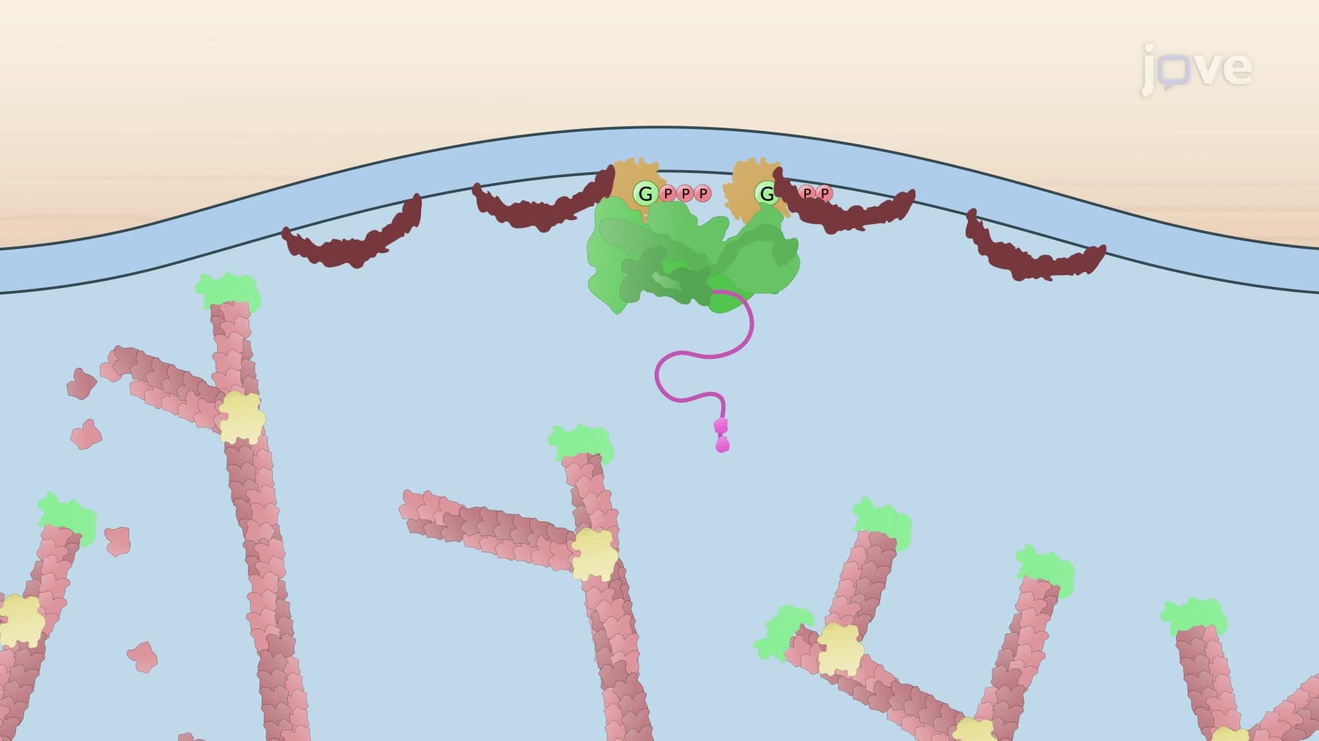
Cancer Cell Migration through Invadopodia

00:00
01:35
Video Summary: Cancer Cell Migration Through Invadopodia Explained
Did you know that cancer cells essentially grow tiny "fingers" to push through healthy tissue barriers? Cancer cell migration through invadopodia represents one of the most sophisticated cellular invasion mechanisms, where malignant cells create specialized membrane protrusions to break down surrounding tissue. Similar to how Johns Hopkins researchers study melanoma cells invading skin tissue, these invadopodia act like molecular crowbars, allowing cancer cells to squeeze through blood vessel walls and establish deadly metastases throughout the body. Watch the full video on JoVE Coach to master this concept with expert-led visuals and step-by-step explanations.
- Understand the fundamental mechanism of cancer cell migration through invadopodia and how these specialized structures enable metastasis
- Identify the key molecular components involved in invadopodia formation, including cortactin, cofilin, WASP, and Arp2/3 complexes
- Analyze the sequential steps of invadopodia development from initial membrane protrusion to ECM degradation
- Compare the roles of F-actin and fascin in creating the structural framework that drives invadopodia extension
- Evaluate how proteases delivered via microtubule networks enable cancer cells to break through tissue barriers
- Apply knowledge of invadopodia assembly and disassembly to understand both intravasation and extravasation processes
- Recognize the clinical significance of invadopodia in cancer progression and potential therapeutic targeting strategies
Understanding Cancer Cell Migration Through Invadopodia Mechanics
Cancer cell migration through invadopodia represents a highly coordinated cellular process that enables malignant transformation from localized tumors to life-threatening metastatic disease. Unlike normal cell movement, invadopodia creation involves sophisticated molecular machinery that allows cancer cells to literally drill through tissue barriers that would otherwise contain tumor growth.
Molecular Architecture and Formation Sequence
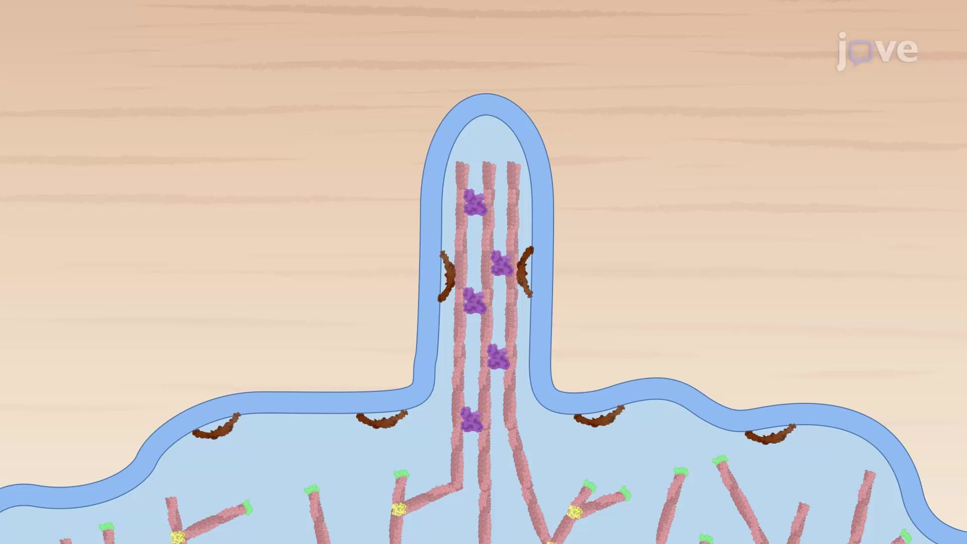
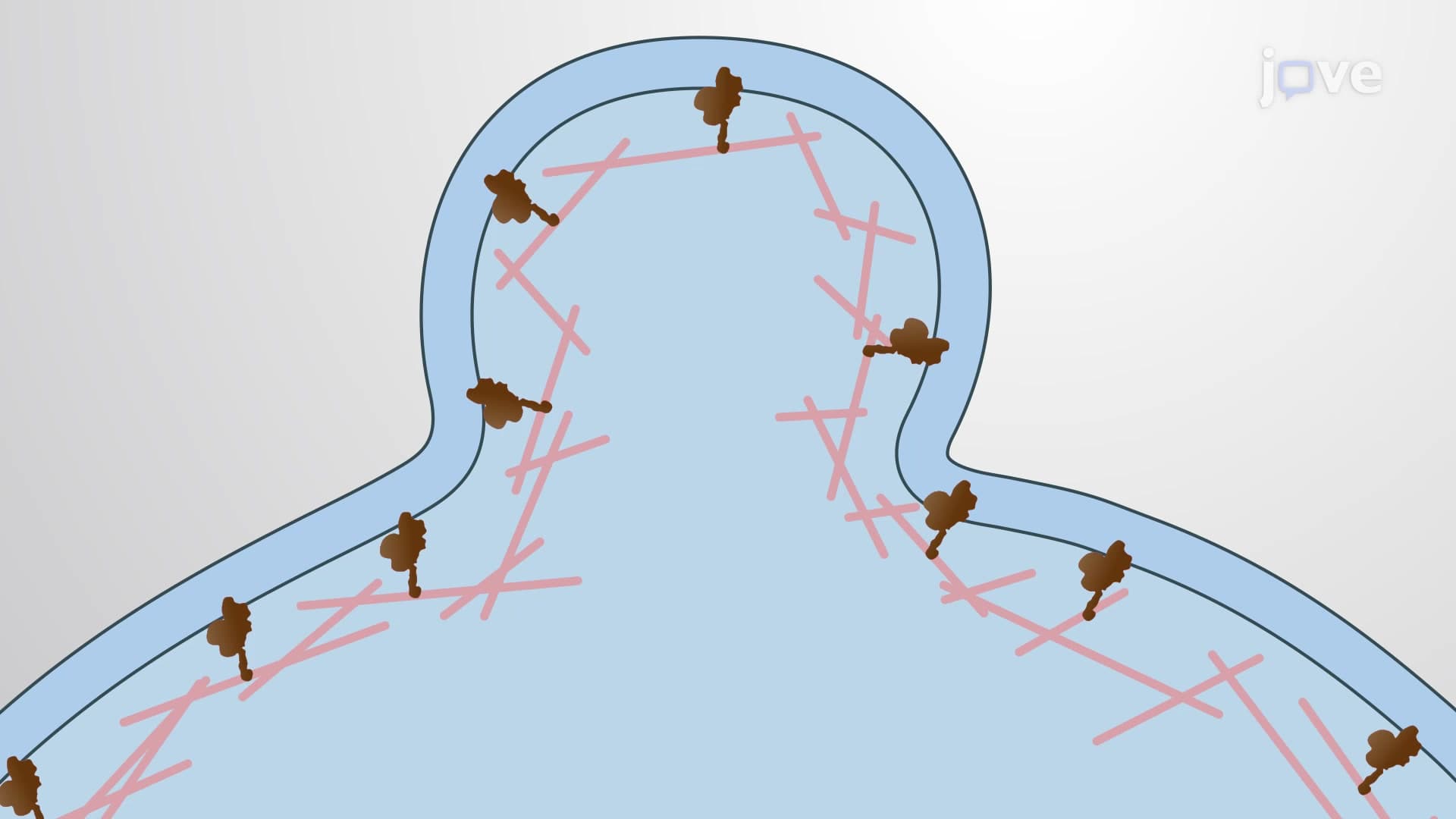
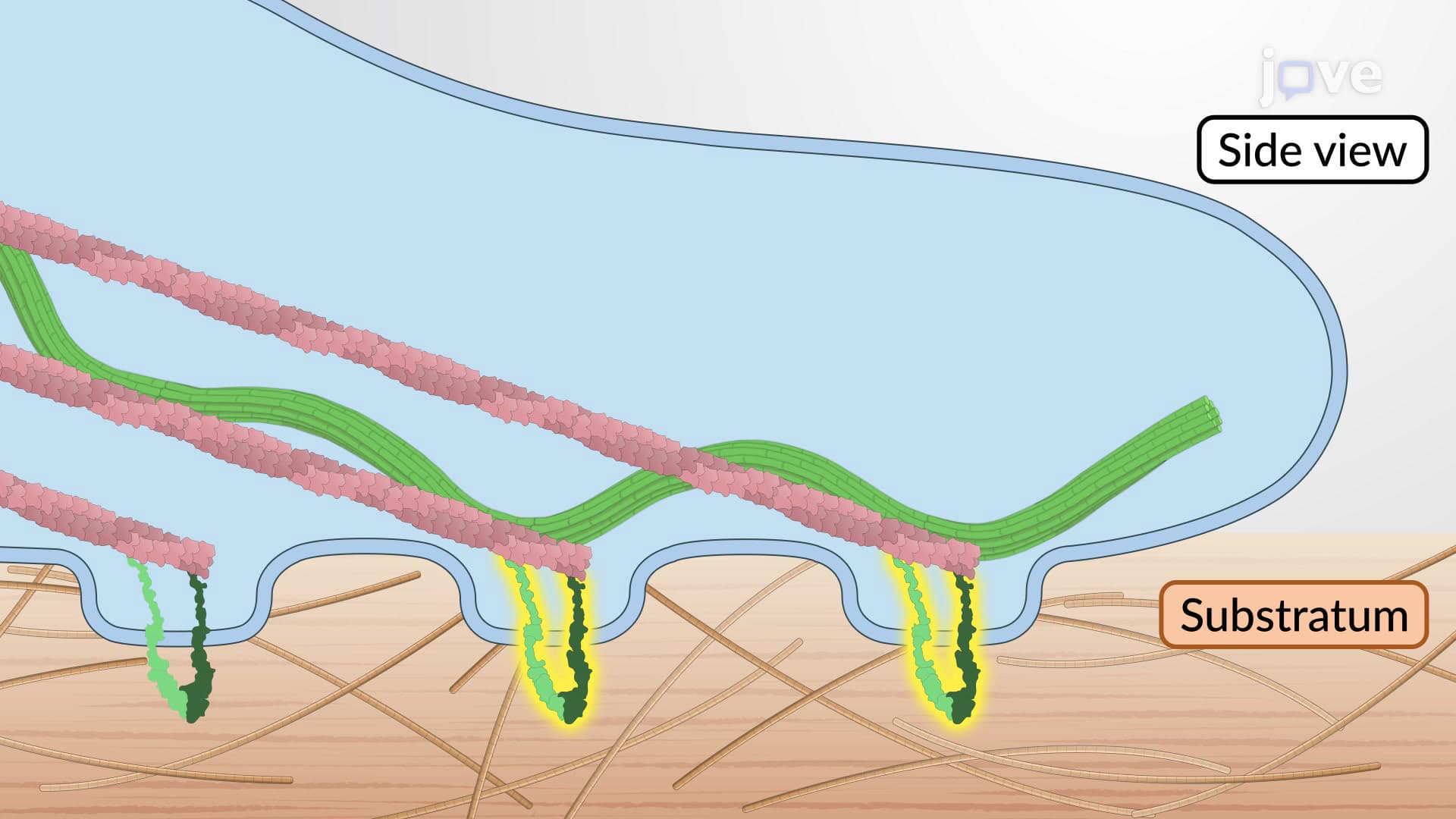
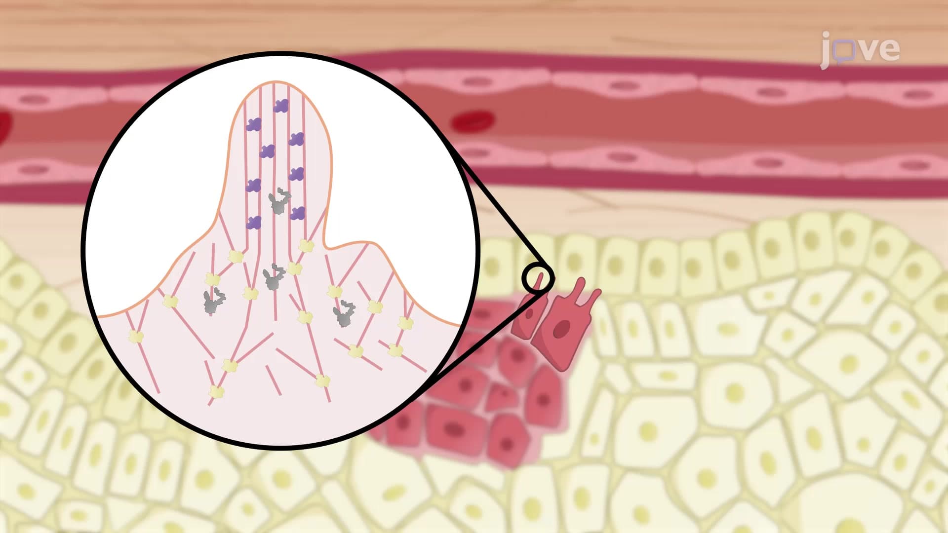
The invadopodia formation process begins with cortactin activation, a nucleation-promoting factor that serves as the cellular equivalent of a construction foreman. Cortactin orchestrates the recruitment of key players including cofilin, WASP (Wiskott-Aldrich Syndrome Protein), and the Arp2/3 complex near the cell membrane. This initial assembly creates the foundation for what becomes a powerful invasive structure.
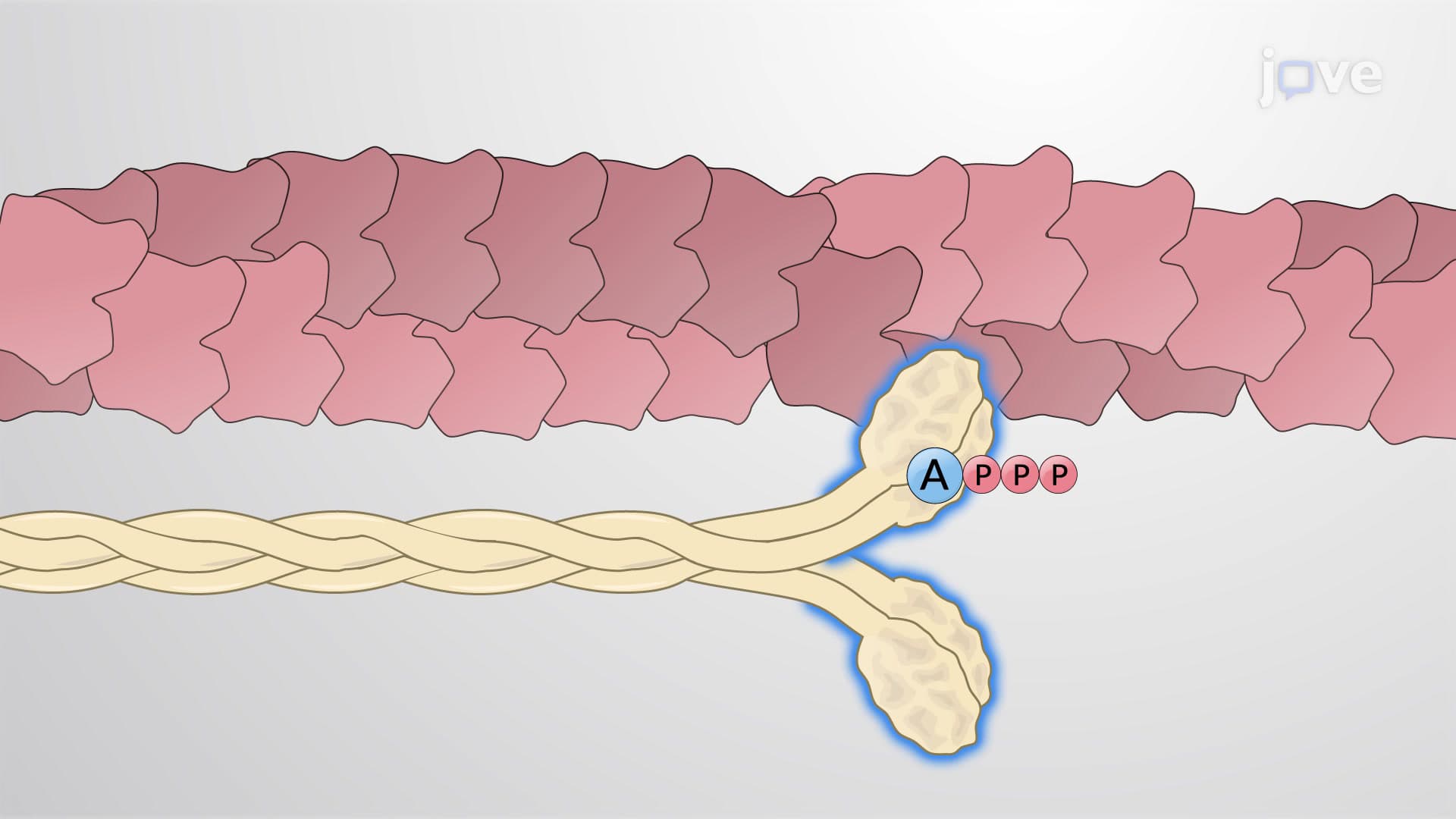
The dynamics shift dramatically when cofilin dissociates from cortactin and begins severing existing actin filaments. This process generates free barbed ends that serve as nucleation sites for rapid actin polymerization. Simultaneously, WASP and Arp2/3 proteins initiate branched actin networks that physically push the cell membrane outward, creating the characteristic finger-like protrusion. Students preparing for the MCAT should note how this branched actin arrangement differs from the parallel bundles found in cellular structures like microvilli.
Maturation and ECM Degradation
As invadopodia mature, fascin cross-links actin filaments into parallel bundles, creating a rigid core structure capable of penetrating dense extracellular matrix (ECM). The University of California San Francisco has extensively documented how this fascin-mediated bundling provides the mechanical strength necessary for sustained tissue invasion.
The final phase involves kinesin motor proteins utilizing colocalized microtubule networks to transport vesicles containing matrix metalloproteinases (MMPs) and other ECM-degrading enzymes to the invadopodia tip. These proteases create localized tissue breakdown, allowing cancer cells to intravasate into blood vessels. Understanding this mechanism proves crucial for AP Biology students studying cancer biology, as it explains how localized tumors become systemic diseases.
Clinical Relevance and Therapeutic Implications
The invadopodia disassembly process, triggered by cortactin phosphorylation, enables circulating cancer cells to extravasate and establish secondary tumors. Memorial Sloan Kettering researchers have identified this cycle as a critical therapeutic target, since disrupting invadopodia formation could potentially prevent metastatic spread while leaving primary tumors treatable through conventional methods.
Frequently Asked Questions
Cancer cell migration through invadopodia is the process by which malignant cells create specialized membrane protrusions to invade surrounding tissues and blood vessels. This mechanism is crucial for understanding metastasis because it explains how cancer cells escape their original location and spread throughout the body. The invadopodia act like molecular drills, allowing cancer cells to break through tissue barriers that would normally contain tumor growth.
The MCAT frequently tests cellular biology concepts including cytoskeleton dynamics and cancer biology mechanisms. Understanding invadopodia formation demonstrates knowledge of actin polymerization, protein-protein interactions, and disease pathophysiology. This topic commonly appears in biological sciences passages discussing cancer progression, making it essential for achieving competitive scores in medical school admissions.
AP Biology examines cell structure and function, including cytoskeleton organization and cell communication mechanisms. Invadopodia formation involves multiple AP-tested concepts: protein regulation, membrane dynamics, and cellular signaling pathways. Students should focus on understanding how cortactin, WASP, and Arp2/3 interactions demonstrate enzyme regulation and cellular response mechanisms that frequently appear on free-response questions.
Leading institutions like MD Anderson Cancer Center and Johns Hopkins use advanced imaging techniques to observe invadopodia formation in patient tumor samples. Researchers analyze tissue biopsies to identify invadopodia markers, helping predict metastatic potential and guide treatment decisions. This research directly influences cancer staging and therapeutic strategies used throughout the American healthcare system.
While invadopodia involves complex molecular interactions, the basic concept builds on fundamental biology principles taught in standard curricula. Students familiar with cell membrane structure, protein function, and basic cancer biology can grasp invadopodia formation. The key is focusing on the overall process rather than memorizing every molecular detail, making it accessible for motivated high school students.
Create a step-by-step flowchart showing cortactin activation through final ECM degradation, focusing on cause-and-effect relationships between each molecular player. Practice drawing the process from memory and explaining how disrupting any single step would prevent successful invasion. This active recall method proves especially effective for cell biology exams requiring detailed process explanations.
Advanced students should explore related topics including epithelial-mesenchymal transition (EMT), tumor microenvironment interactions, and therapeutic targeting strategies. Understanding how invadopodia relate to cancer stem cell behavior and drug resistance mechanisms provides deeper insight into modern oncology research and potential career paths in cancer biology.
Pharmaceutical companies are developing drugs that specifically target invadopodia formation proteins like cortactin and fascin to prevent metastasis. Clinical trials at institutions like the National Cancer Institute are testing whether blocking invadopodia assembly can improve patient outcomes when combined with traditional chemotherapy. This represents a promising approach to preventing cancer spread rather than just treating existing tumors.
Related Micro-courses

7 Concepts
20 Concepts

17 Concepts