Loading subject page…

- Cell Biology
- Cell Polarization and Migration
- Cell Polarization by Rho Proteins
Micro-course: Cell Polarization and Migration
40,212 views
Learn key concepts and earn your certificate.


Cell Polarization by Rho Proteins

00:00
01:21
Video Summary: Cell Polarization by Rho Proteins Explained
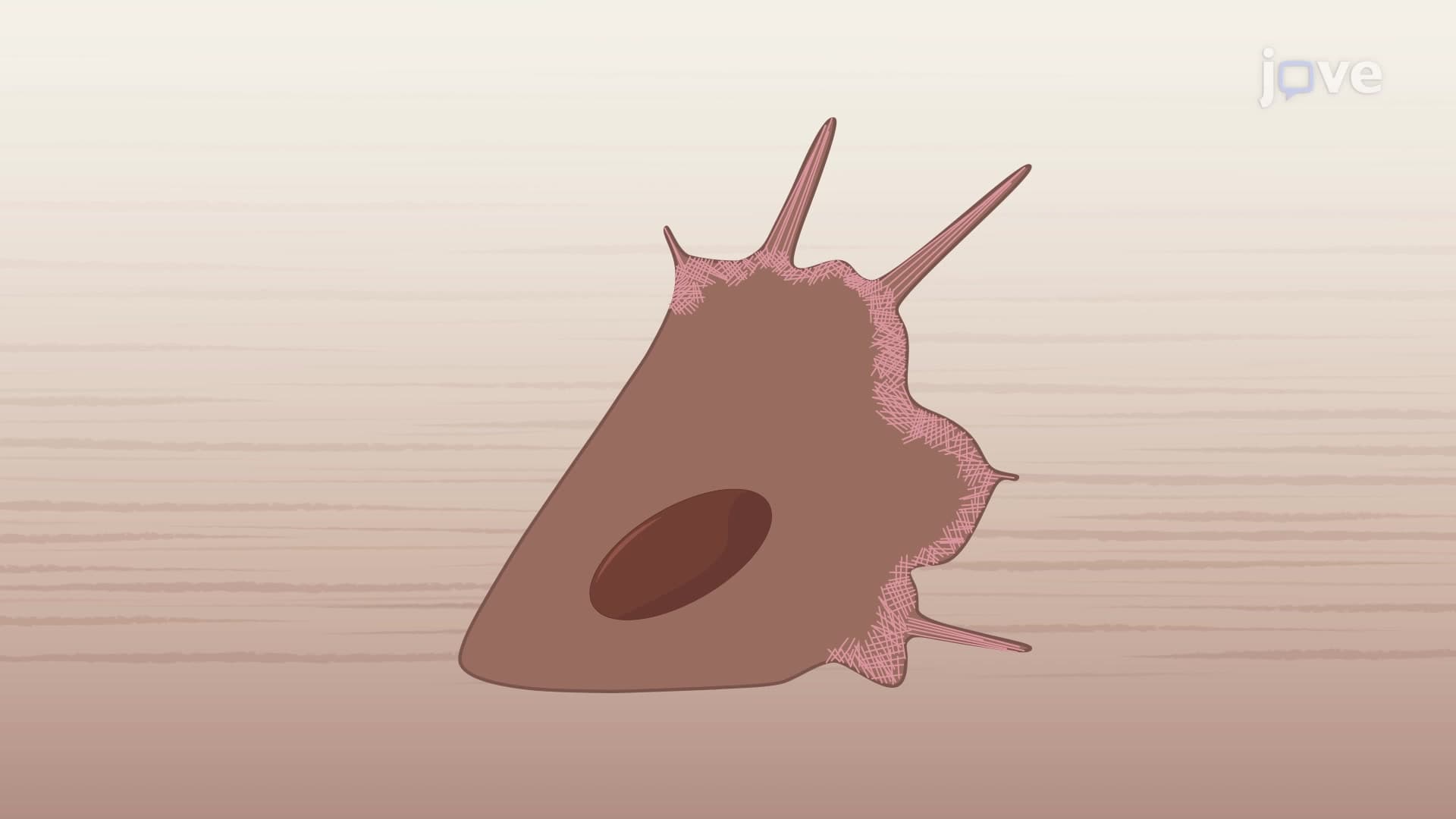
How does a single cell know which direction to move when responding to injury or infection? Cell polarization by Rho proteins is the sophisticated molecular mechanism that enables cells to establish distinct front and rear regions during migration. Consider immune cells rushing to a wound site in your skin, they rely on three key Rho family proteins (Rac, Cdc42, and Rho) to coordinate their directional movement and cellular organization. Watch the full video on JoVE Coach to master this concept with expert-led visuals and step-by-step explanations.
- Understand how cell polarization by Rho proteins establishes distinct cellular regions with specialized functions during migration
- Identify the three primary Rho family proteins (Rac, Cdc42, and Rho) and their specific roles in cell polarity
- Analyze the molecular mechanisms by which Rac and Cdc42 proteins create the leading edge through actin cytoskeleton modifications
- Compare the opposing functions of Rac/Cdc42 at the cell front versus Rho protein activity at the trailing edge
- Evaluate how chemical gradients and receptor activation initiate the polarization cascade in migrating cells
- Apply knowledge of actin branching, polymerization, and myosin contraction to explain cellular movement patterns
- Recognize the inhibitory relationship between Rac and Rho that maintains stable cell polarity
- Explore real-world applications in wound healing, immune responses, and cancer cell metastasis
Understanding Cell Polarization By Rho Proteins: The Foundation of Directed Movement
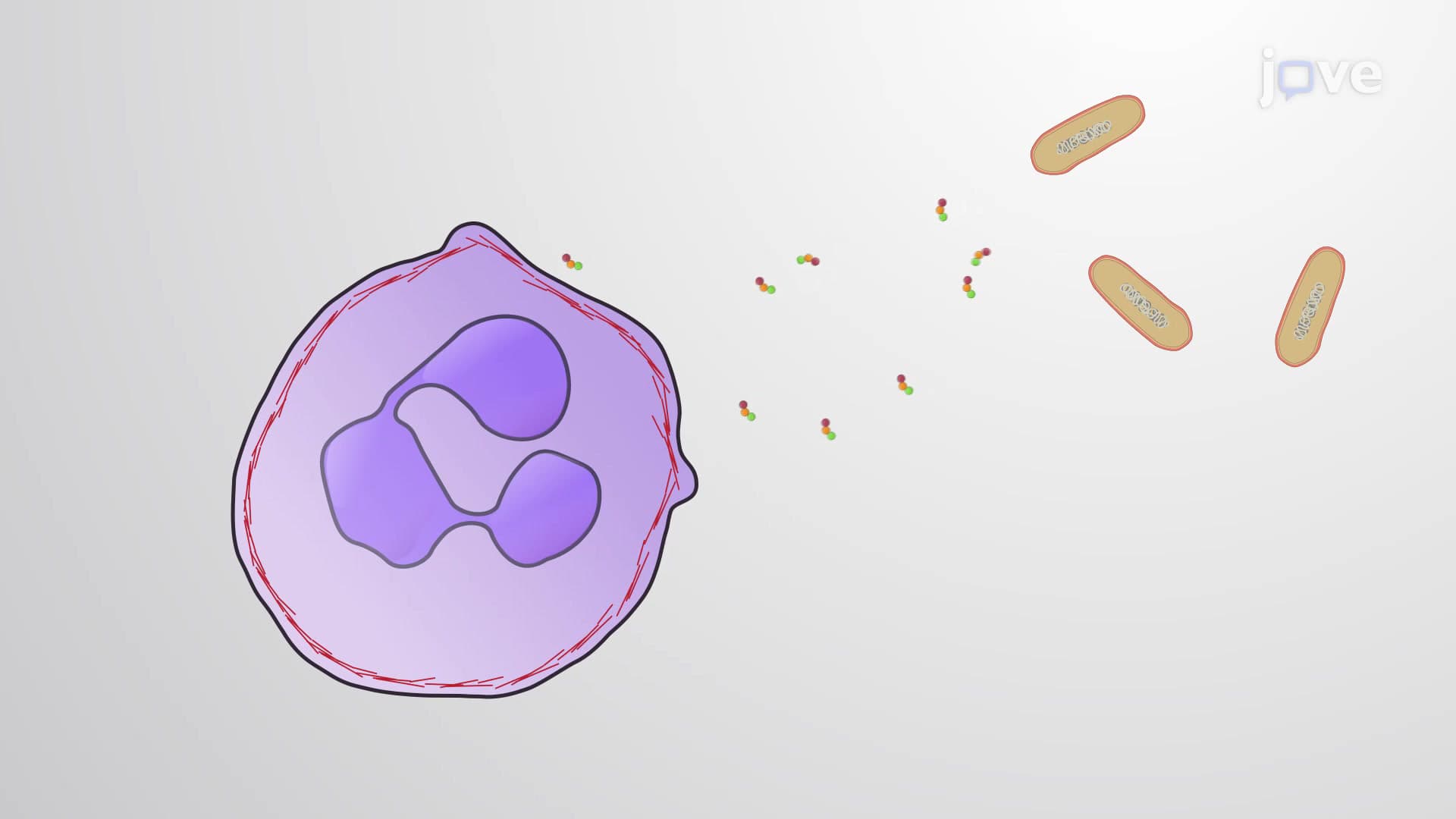
Cell polarization represents one of biology's most elegant solutions to the challenge of directional movement. When cells receive chemical signals, whether from damaged tissue releasing inflammatory molecules or pathogens triggering immune responses, they must rapidly reorganize their internal architecture to move effectively toward the stimulus source.
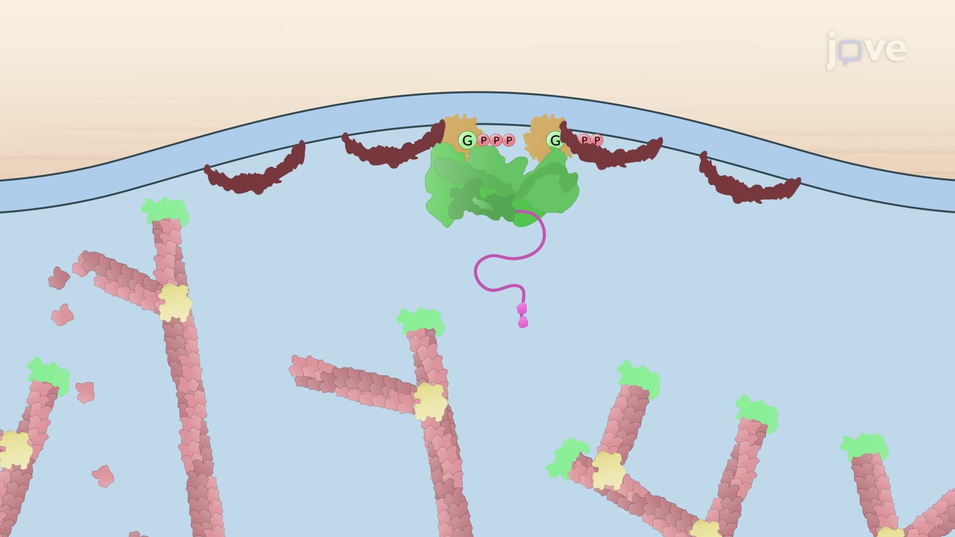
The Rho family of proteins serves as the master regulators of this polarization process. These small GTPases act like molecular switches, becoming activated in response to upstream signals and subsequently orchestrating dramatic changes in cell structure and function.
The Leading Edge: Rac and Cdc42 Protein Functions
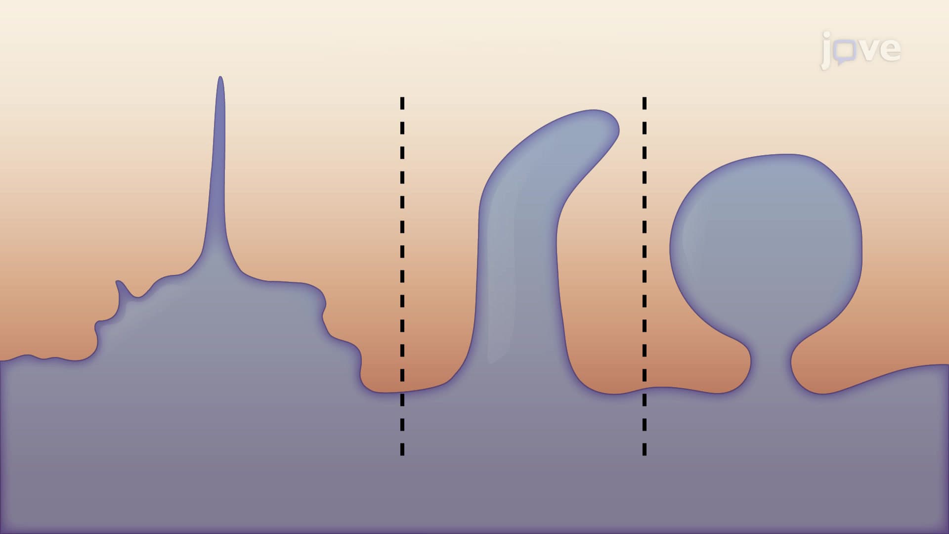
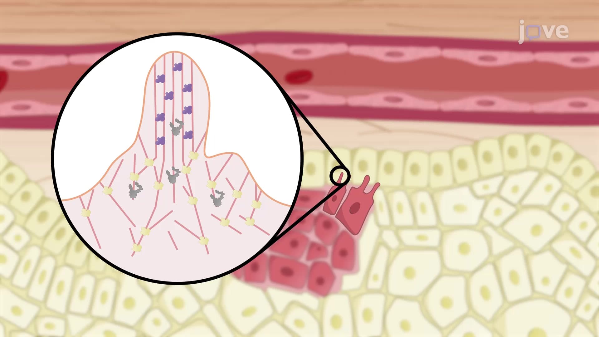
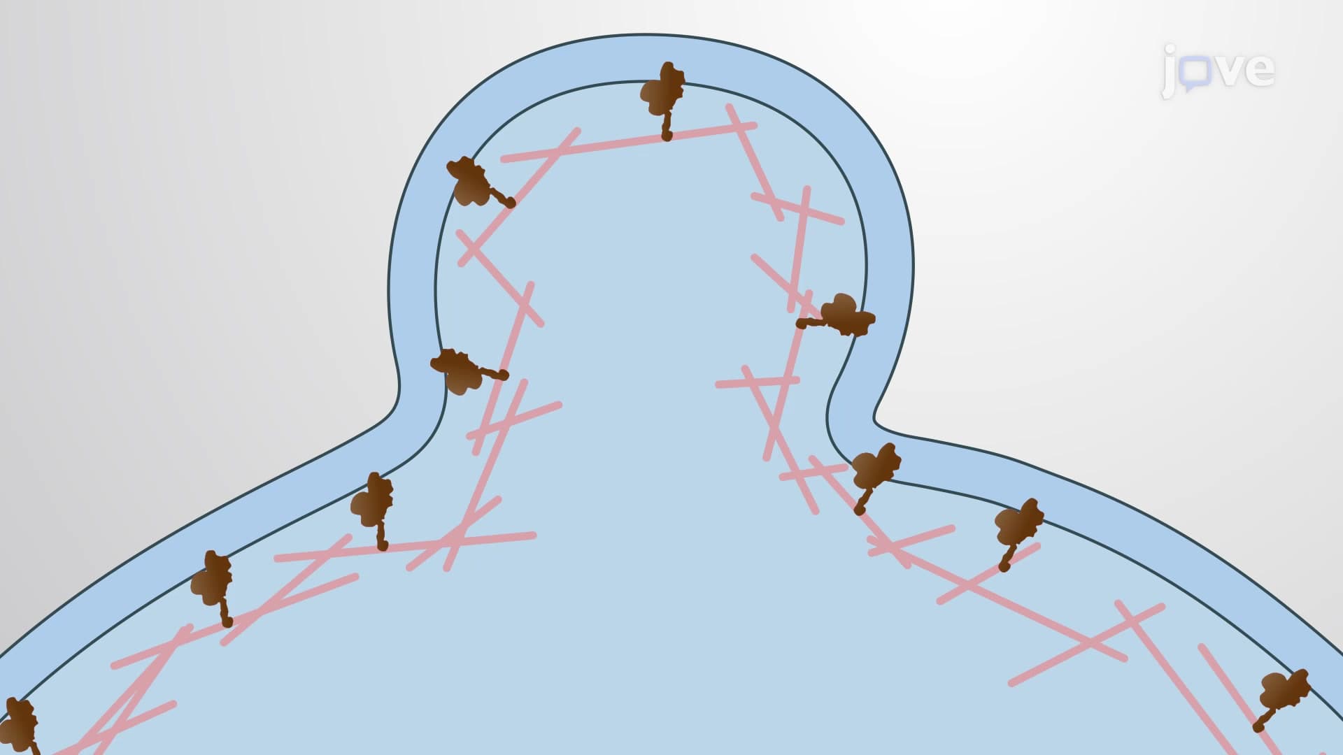
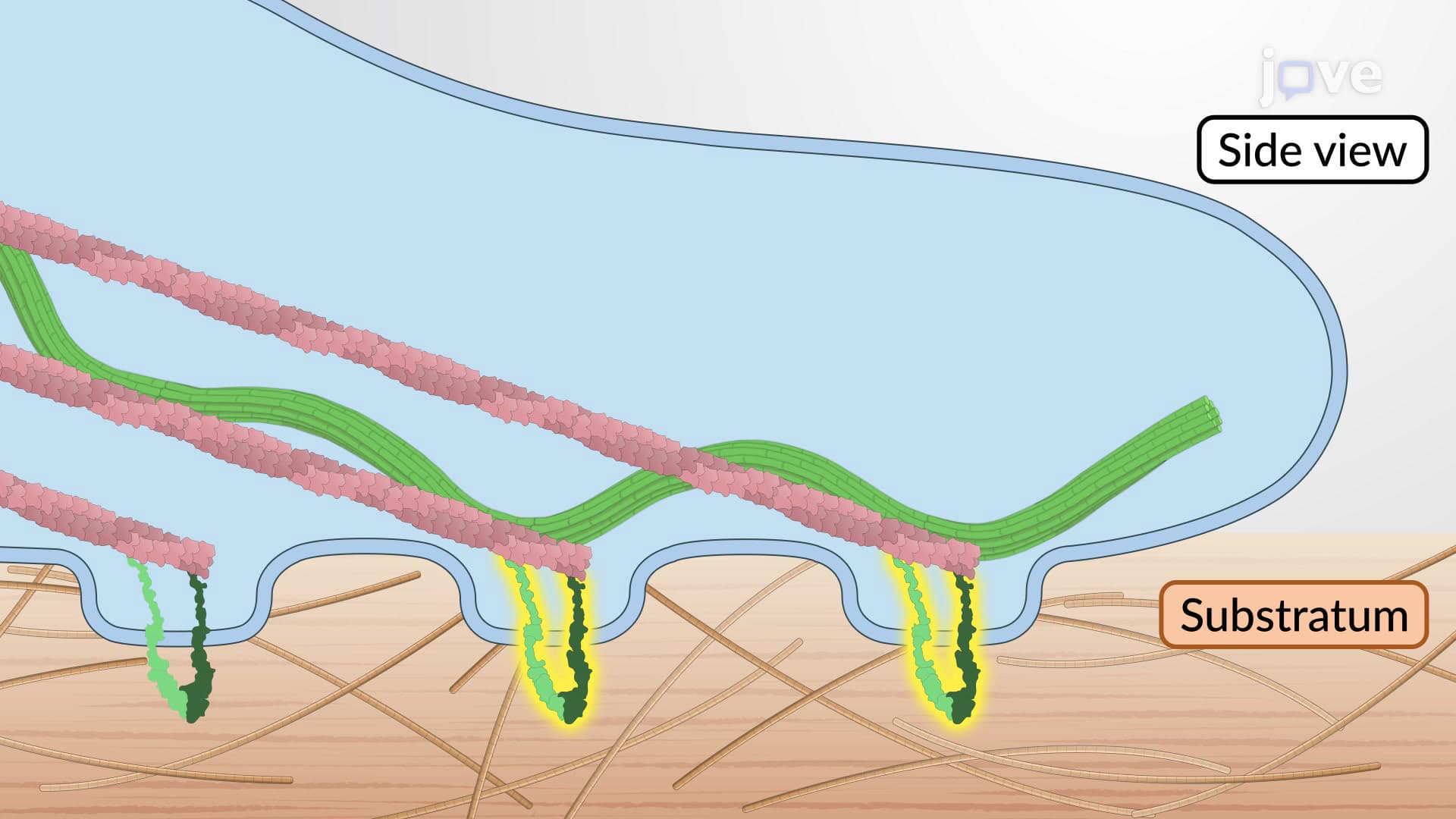
At the cell's leading edge, two Rho proteins work in concert to drive forward movement. Rac protein activation triggers extensive actin branching through the Arp2/3 complex, creating broad, sheet-like membrane protrusions called lamellipodia. These structures act like the cell's "bulldozer blade," pushing the membrane forward and providing the primary driving force for migration.
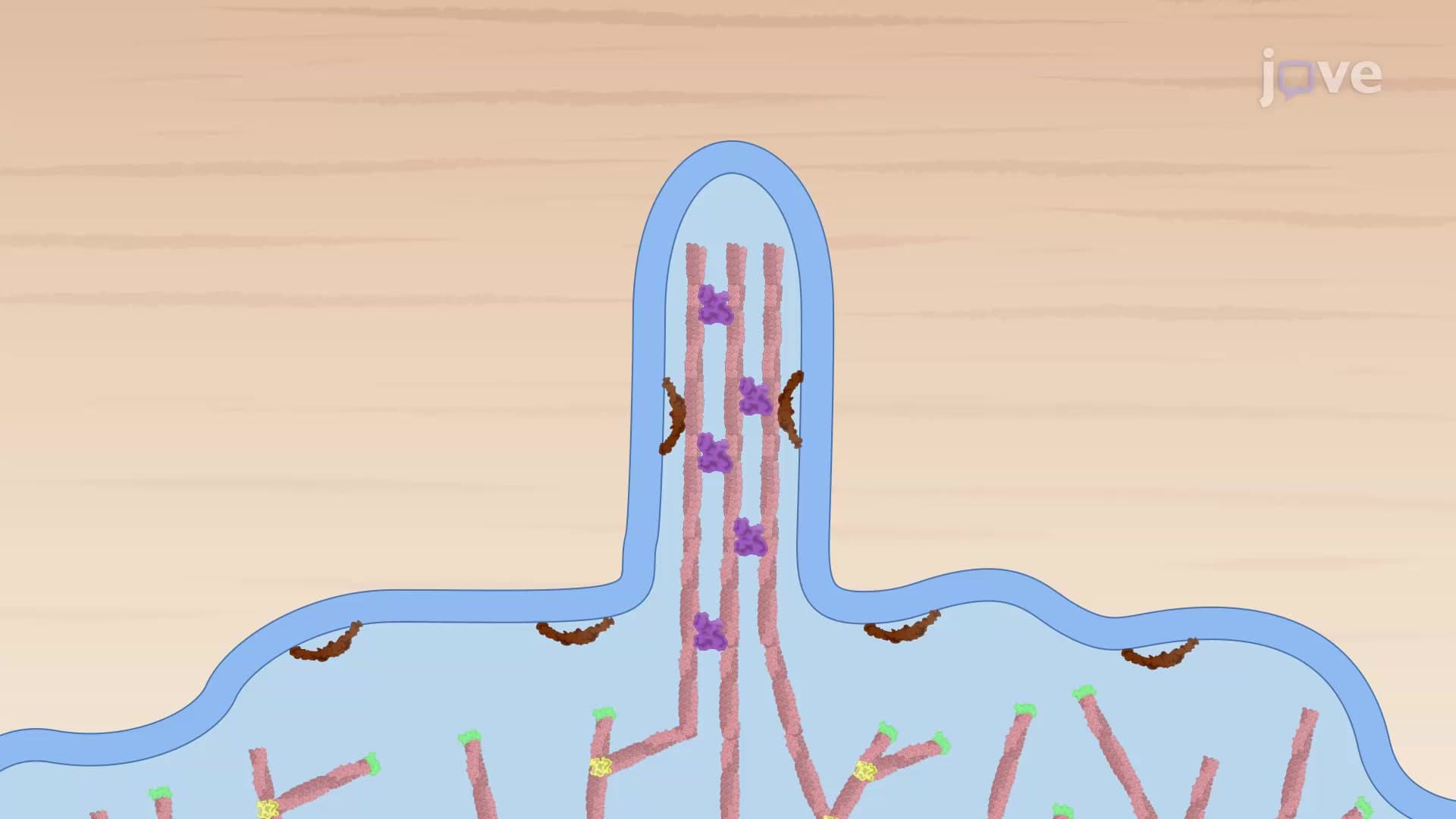
Simultaneously, Cdc42 protein promotes the formation of finger-like projections called filopodia. Unlike the broad lamellipodia, filopodia contain bundled actin filaments that extend outward like cellular antennae. These structures help the cell sense its environment and establish initial contact points with surrounding surfaces.
Students preparing for AP Biology or college-level cell biology courses should recognize that this spatial organization isn't random, the precise localization of Rac and Cdc42 to the cell front requires sophisticated molecular machinery that responds to external chemical gradients.
The Trailing Edge: Rho Protein-Mediated Contraction
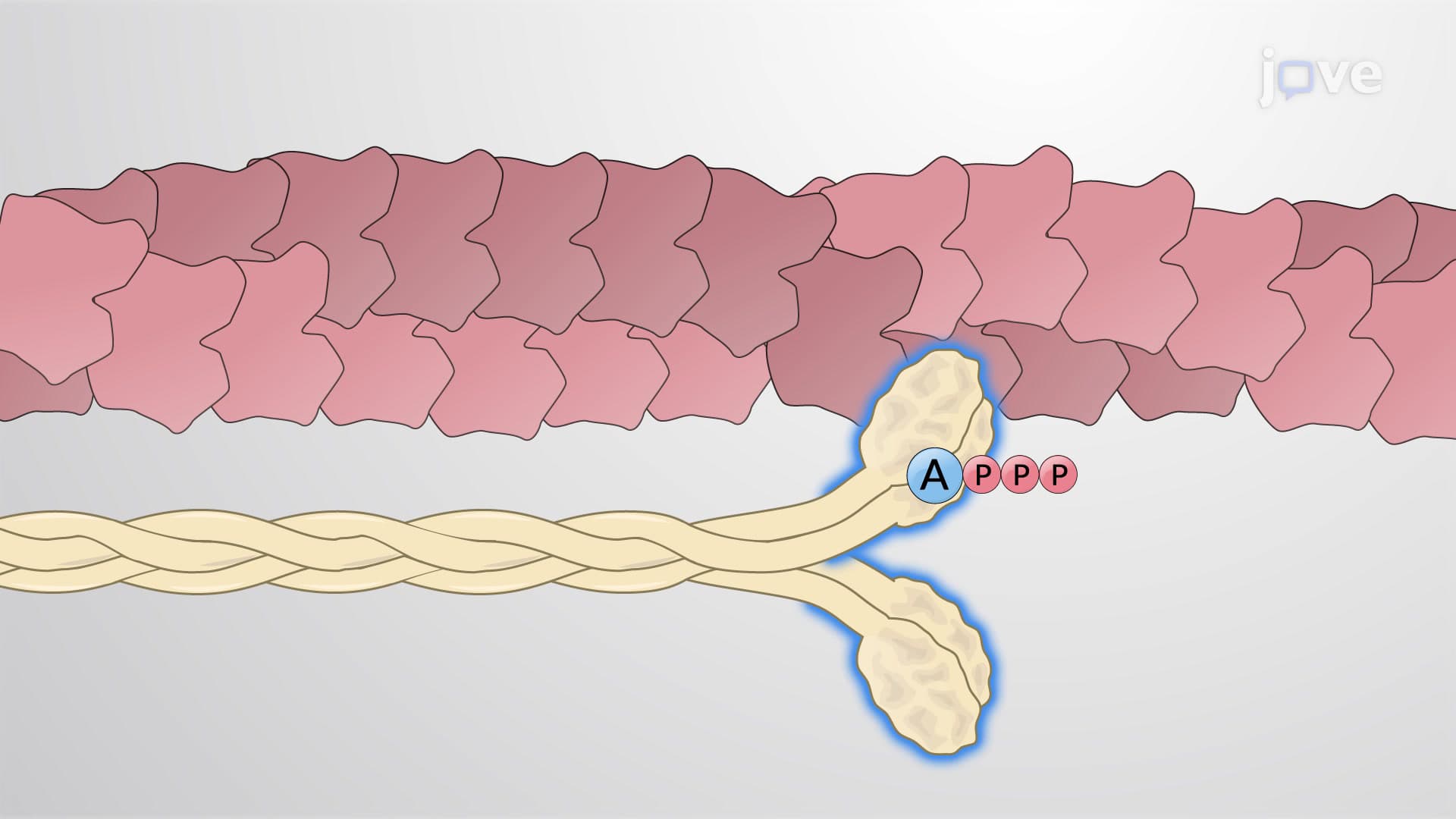
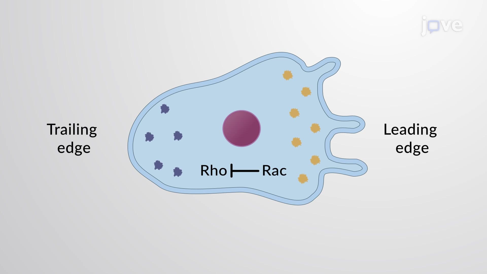
While Rac and Cdc42 establish the cell front, Rho protein dominates the trailing edge with an entirely different function. Rho activation increases myosin II motor protein activity, generating powerful contractile forces that literally squeeze the cell's rear forward. This process, called rear retraction, is essential for efficient migration, without it, cells would simply extend forward without achieving net displacement.
The molecular mechanism involves Rho-mediated activation of Rho kinase (ROCK), which phosphorylates myosin light chain and increases actomyosin contractility. For MCAT preparation, understanding this pathway proves crucial for questions involving cell motility and cytoskeletal regulation.
Maintaining Polarity: The Rac-Rho Inhibitory Circuit
Perhaps the most sophisticated aspect of cellular polarization involves the mutual inhibition between Rac and Rho proteins. Active Rac at the leading edge directly inhibits Rho activation in the same cellular region, ensuring that contractile forces don't interfere with forward protrusion. This creates a stable, self-reinforcing polarity where the cell front remains "front" and the rear remains "rear" throughout migration.
This regulatory circuit explains why cells maintain consistent directional movement rather than randomly changing direction, a concept frequently tested in college-level cell biology midterms and comprehensive exams.
Frequently Asked Questions
Cell polarization by Rho proteins is the process where cells establish distinct front and rear regions using three key signaling molecules: Rac, Cdc42, and Rho. This mechanism is crucial for directed cell migration during wound healing, immune responses, and embryonic development. The spatial organization allows cells to move efficiently toward chemical signals rather than wandering randomly through tissues.
MCAT questions often focus on the molecular mechanisms linking Rho protein activation to cytoskeletal changes, particularly actin dynamics and myosin contractility. AP Biology exams emphasize the functional relationships between cell structure and migration, including how chemical gradients initiate polarization. Students should understand both the individual protein functions and their coordinated regulation during directed movement.
Create visual diagrams showing the spatial distribution of each Rho protein and their effects on the cytoskeleton. Practice drawing the inhibitory relationship between Rac and Rho, and memorize the specific actin structures each protein creates (lamellipodia vs. filopodia vs. contractile bundles). Focus on connecting protein names to their cellular locations and primary functions rather than just memorizing isolated facts.
No advanced prerequisites are required beyond basic cell biology concepts. Understanding requires familiarity with protein structure, enzyme activation, and cytoskeletal components (actin, myosin), all covered in introductory biology courses. The key is visualizing how these molecular players work together spatially rather than memorizing complex biochemical pathways.
Consider diabetic wound healing complications seen in US hospitals, impaired Rho protein signaling prevents immune cells and fibroblasts from migrating effectively to injury sites. Cancer metastasis research at institutions like MD Anderson focuses on how tumor cells hijack normal polarization mechanisms to invade surrounding tissues. Understanding these pathways helps explain both normal physiology and disease processes.
Explore related cell migration mechanisms including integrin-mediated adhesion, chemotaxis signaling pathways, and epithelial-mesenchymal transition. Advanced topics include Rho GTPase regulation by guanine nucleotide exchange factors (GEFs) and GTPase-activating proteins (GAPs). These concepts build naturally on the foundational polarization principles and appear frequently in upper-level cell biology and biochemistry courses.
This topic sits at intermediate difficulty level, more complex than basic organelle functions but less challenging than detailed metabolic pathways. The main difficulty involves visualizing three-dimensional spatial relationships and understanding how multiple proteins coordinate simultaneously. With consistent practice drawing cellular diagrams and connecting structure to function, most students master these concepts within 2-3 study sessions.
Related Micro-courses

7 Concepts
20 Concepts

17 Concepts