Loading subject page…

- Cell Biology
- Cell Polarization and Migration
- Cytoskeletal Coordination in Cell Migration
Micro-course: Cell Polarization and Migration
40,212 views
Learn key concepts and earn your certificate.

Cytoskeletal Coordination in Cell Migration

00:00
01:32
Video Summary: What Is Cytoskeletal Coordination
Ever wondered how a white blood cell chases down bacteria through your bloodstream, changing direction with precision? Cytoskeletal coordination makes this cellular navigation possible by orchestrating three key protein networks-actin filaments, microtubules, and intermediate filaments-to work as a unified team. Consider how immune cells in COVID-19 patients must rapidly migrate to infected lung tissue. Watch the full video on JoVE Coach to master this concept with expert-led visuals and step-by-step explanations.
- Understand what is cytoskeletal coordination and how it enables precise cellular movement through synchronized protein networks
- Identify the three main cytoskeletal components (actin filaments, microtubules, intermediate filaments) and their specific roles in cell migration
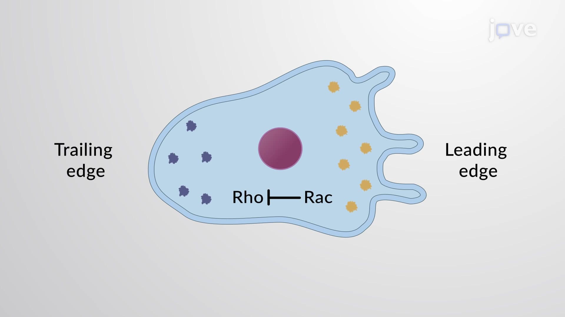
- Analyze how centrosome positioning creates cellular polarity by establishing distinct leading and trailing edges
- Recognize the function of motor proteins (kinesin and dynein) in cargo transport along microtubule highways
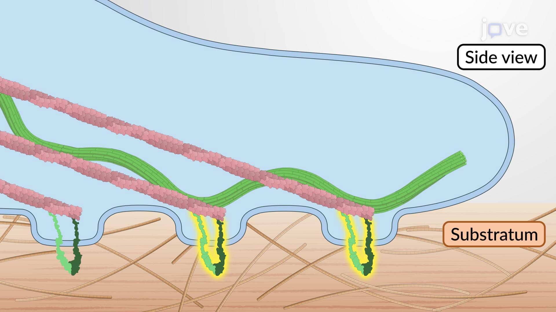
- Evaluate how focal adhesions form and disassemble to create traction points during cell movement
- Apply knowledge of integrin recycling pathways to understand how cells maintain adhesion while migrating
- Explore cross-linking mechanisms that synchronize different cytoskeletal networks into a cohesive migration machine
- Compare the coordinated roles of organelles and protein complexes in maintaining directional cell movement
Understanding Cytoskeletal Coordination Fundamentals
Cytoskeletal coordination represents one of cell biology's most sophisticated orchestrations-the synchronized interaction of three distinct protein networks that enable cells to move with purpose and precision. Unlike a simple mechanical system, this biological coordination involves dynamic communication between actin filaments, microtubules, and intermediate filaments, each contributing unique properties to cellular migration.
This concept frequently appears on AP Biology exams and college cell biology midterms, where students must demonstrate understanding of how multiple cellular systems work together. The coordination process resembles a well-rehearsed dance, where each component knows its role and timing.
Establishing Cellular Polarity Through Cytoskeletal Organization
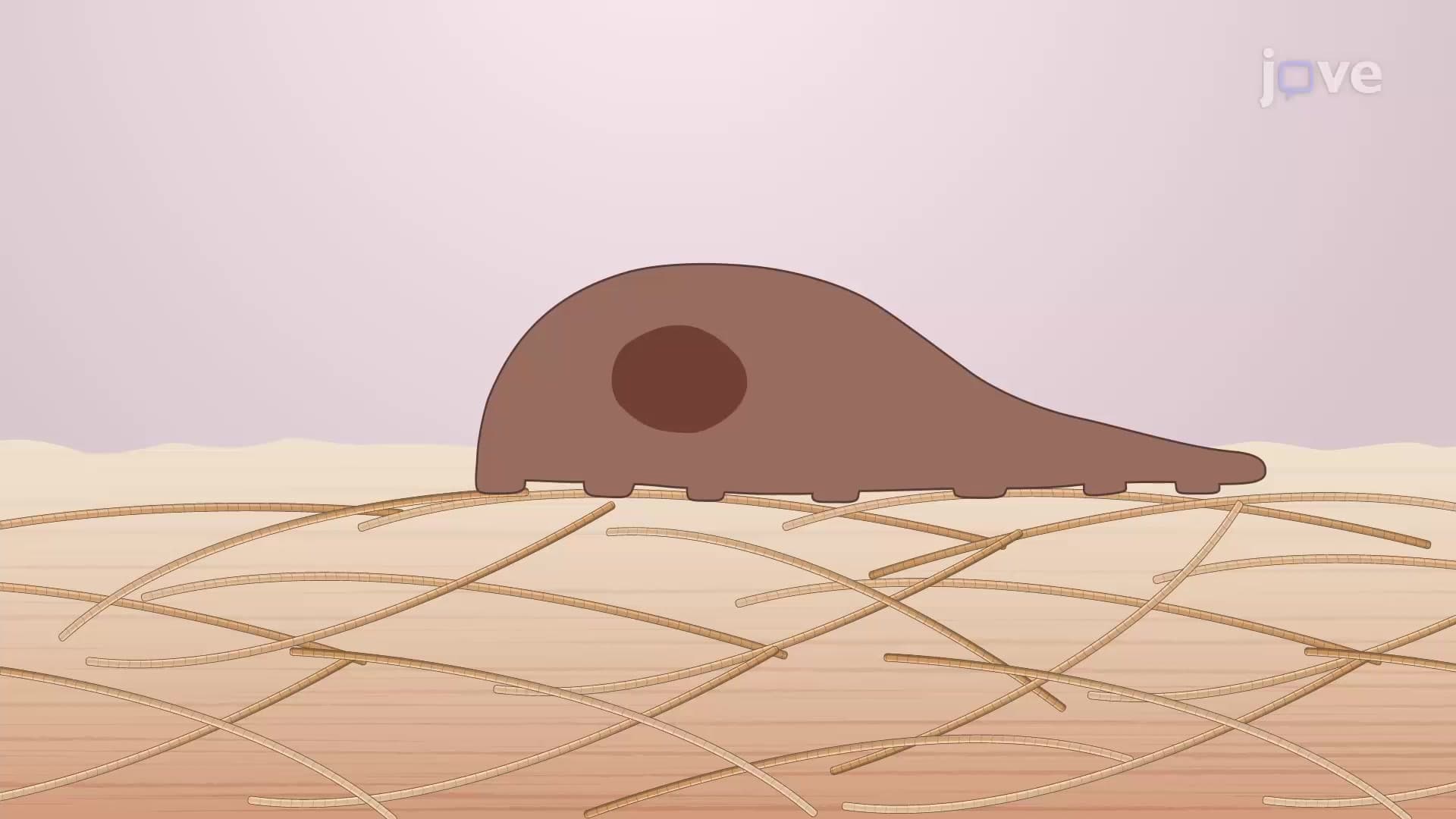
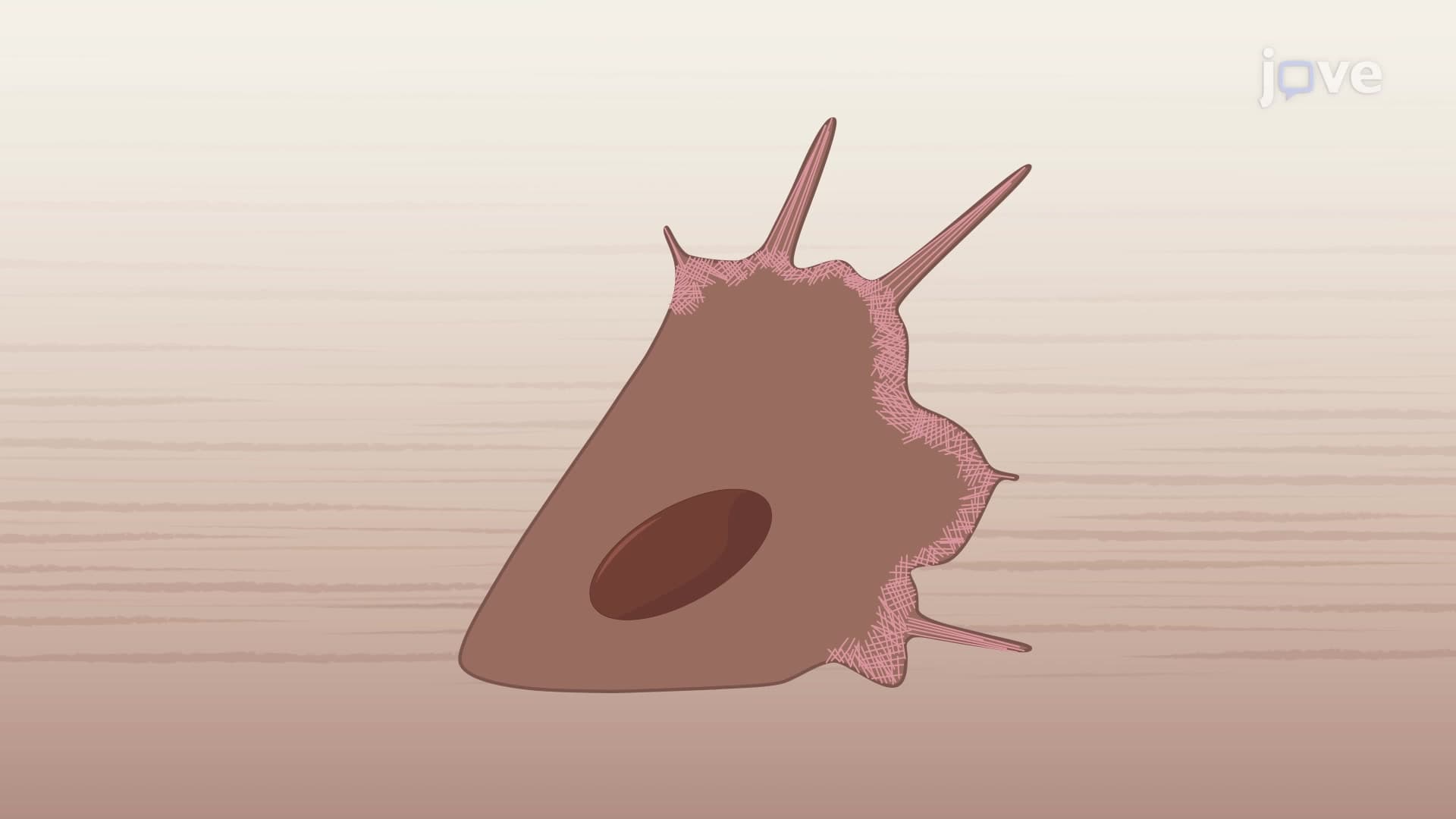
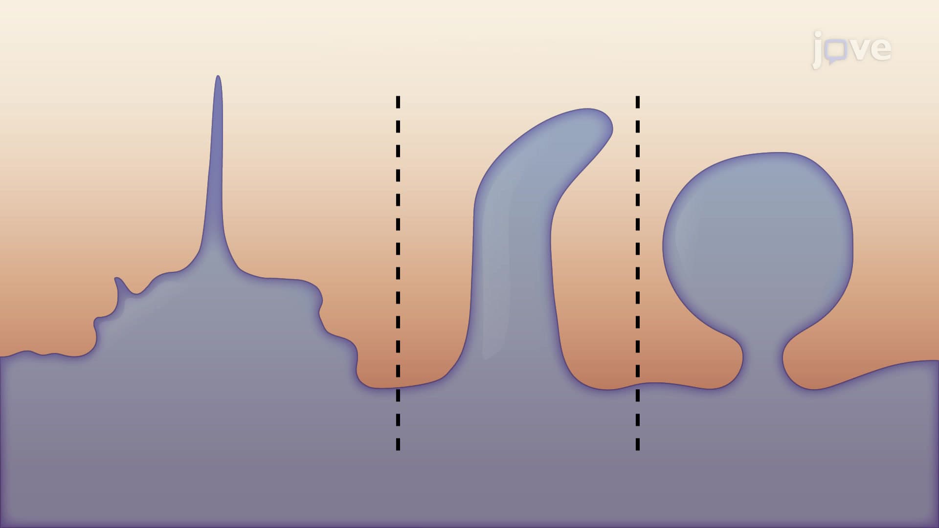
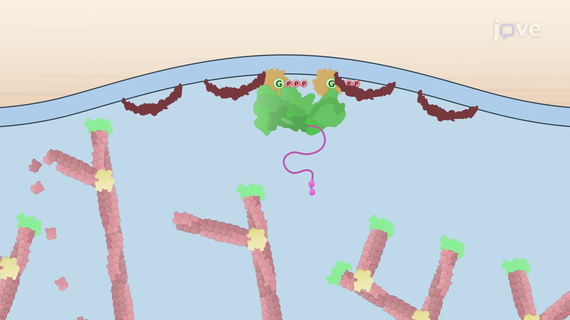
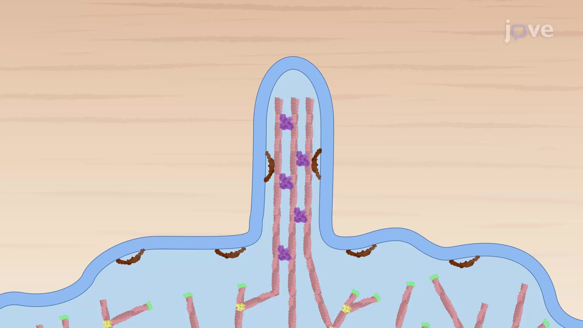
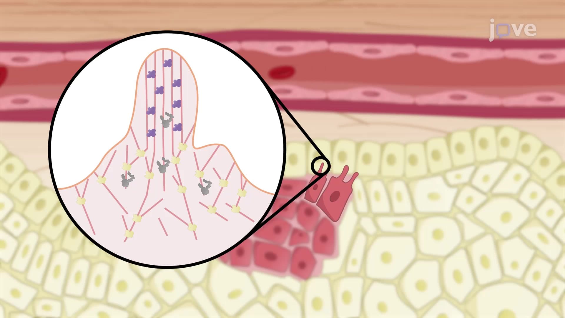
The migration process begins with establishing cellular polarity-creating distinct front and back regions. Actin filaments concentrate at the cell's leading edge, forming dynamic structures called lamellipodia that probe the environment like cellular antennae. Meanwhile, the centrosome, serving as mission control for microtubules, repositions itself between the nucleus and leading edge.
This polarization is crucial for directed movement. Cancer researchers at Johns Hopkins University have shown that disrupting this polarity prevents metastatic cells from invading surrounding tissues, highlighting the clinical importance of cytoskeletal coordination.
Motor Protein Highways and Cargo Transport Systems
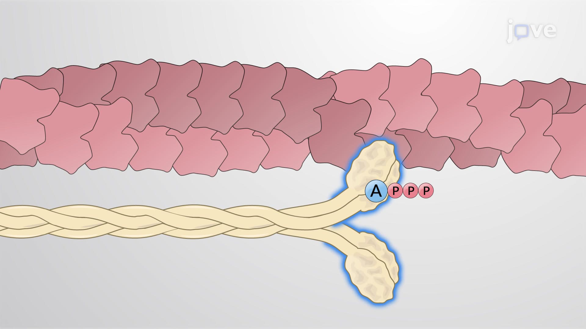
Microtubules function as cellular highways, with motor proteins acting as sophisticated transport vehicles. Kinesin motors carry essential cargo-including integrin proteins, membrane components, and actin-remodeling factors-toward the cell's leading edge. This transport system ensures that the advancing cell front receives necessary materials for continued migration.
Simultaneously, dynein motors operate the recycling system, collecting used integrins from disassembling focal adhesions at the trailing edge and transporting them forward for reuse. This recycling mechanism appears frequently in MCAT cell biology passages, where test-takers must trace molecular pathways through cellular compartments.
Cross-Linking Networks and Coordinated Movement
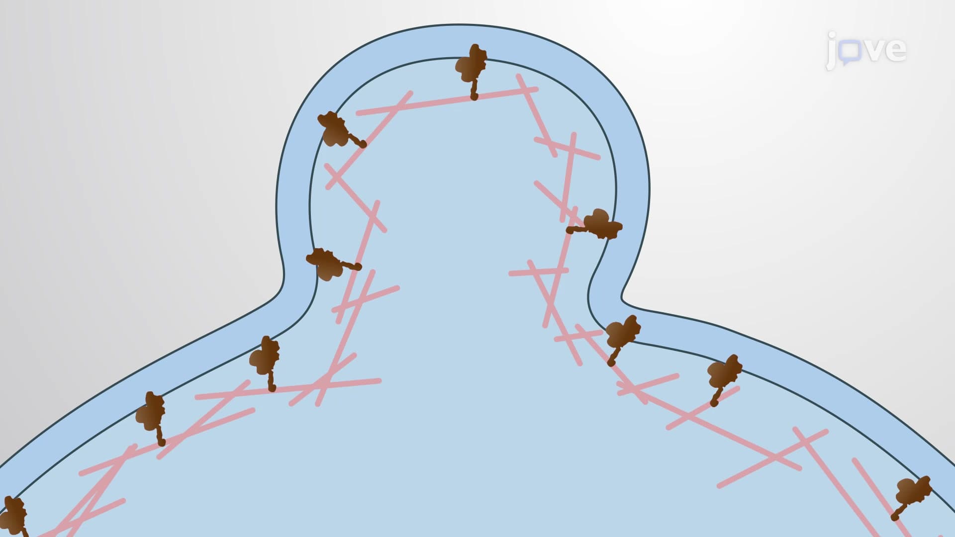
The true elegance of cytoskeletal coordination lies in cross-linking proteins that connect different cytoskeletal networks. These molecular bridges ensure that when actin filaments reorganize at the leading edge, microtubules and intermediate filaments respond appropriately. Intermediate filaments provide structural stability to newly formed focal adhesions, while cross-linked networks synchronize the entire cellular migration machinery.
Understanding these interconnections helps explain how cells achieve such precise control during migration-knowledge that proves essential for students pursuing careers in biomedical research or clinical medicine.
Frequently Asked Questions
Cytoskeletal coordination is the synchronized interaction between actin filaments, microtubules, and intermediate filaments that enables precise cellular movement and organization. It's essential because cells need to migrate during development, immune responses, and wound healing. Without proper coordination, cells would move randomly or remain stationary, preventing crucial biological processes like fighting infections or repairing tissue damage.
This topic commonly appears in cell biology units focusing on cellular movement and organization. Expect questions about identifying cytoskeletal components, explaining motor protein functions, or analyzing experimental data showing migration defects. The College Board often includes diagrams where you must trace the path of proteins during cell migration or explain how disrupting one component affects the entire system.
The MCAT frequently tests cytoskeletal coordination within cell biology and physiology passages. You'll encounter it in contexts involving muscle contraction, neuronal transport, immune cell migration, and cancer metastasis. Understanding motor proteins, cellular polarity, and membrane dynamics becomes crucial for passages describing cellular movement disorders or experimental manipulations of migration pathways.
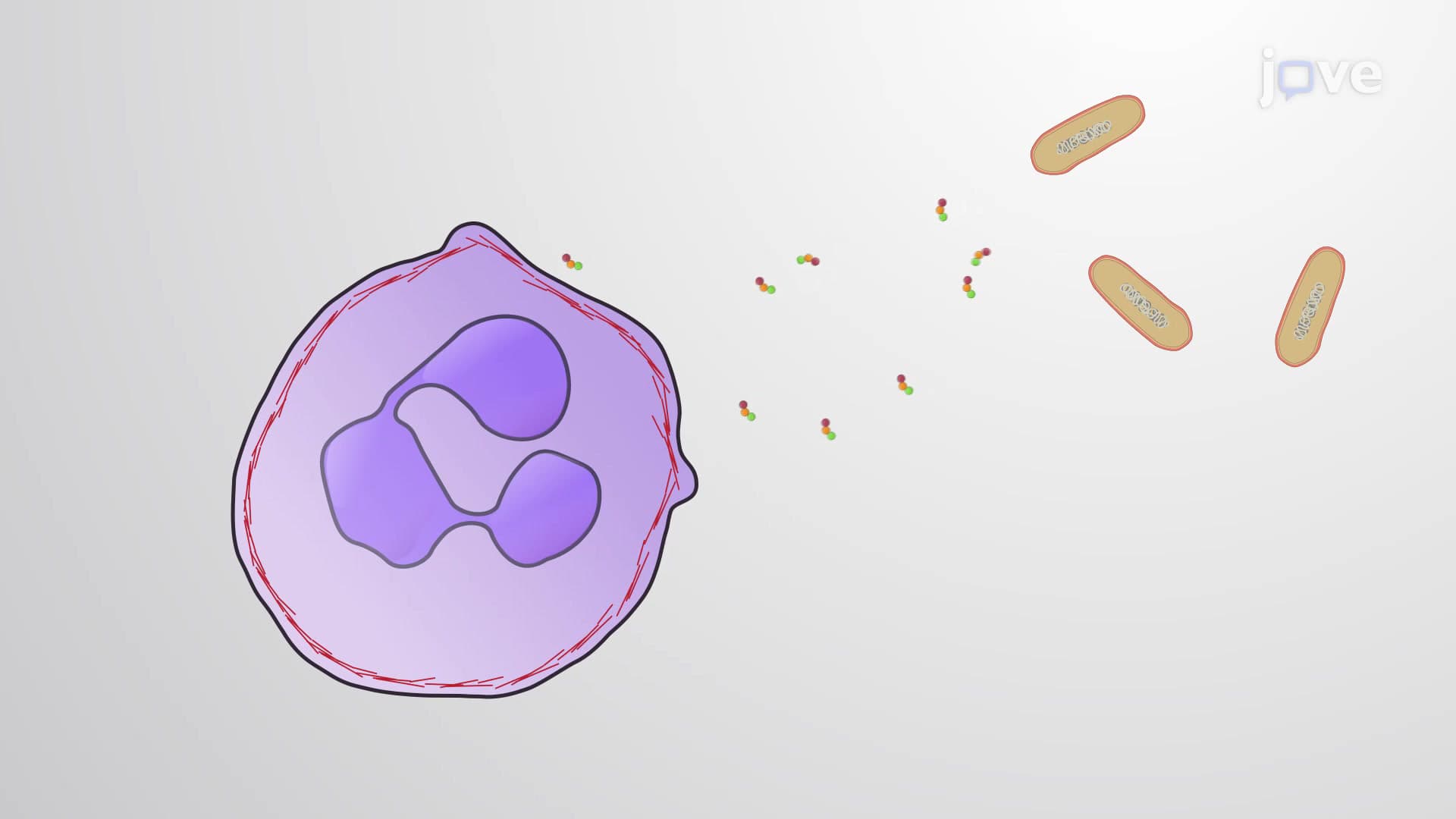
During bacterial infections, neutrophils demonstrate remarkable cytoskeletal coordination as they navigate blood vessel walls and migrate toward infection sites. These cells rapidly reorganize their actin networks to squeeze through tissue spaces while maintaining directional movement toward chemical signals. This process, studied extensively at the CDC, represents cytoskeletal coordination in action during real immune responses.
Not at all-cytoskeletal coordination builds naturally from basic cell structure concepts you already know. If you understand that cells have internal scaffolding (cytoskeleton) and can move, you're ready to learn how these systems coordinate. Think of it like understanding how different parts of a car work together; once you grasp the individual components, their coordination becomes logical.
Create concept maps connecting the three cytoskeletal components to their specific functions during migration. Practice drawing cell diagrams showing leading/trailing edges with appropriate protein locations. Use active recall by explaining the process aloud, focusing on cause-and-effect relationships between molecular events and cellular movements.
This foundational concept becomes essential for understanding muscle physiology, neuron function, developmental biology, and cancer biology in upper-level courses. Students who master cytoskeletal coordination find these advanced topics more accessible because they understand the underlying molecular mechanisms that drive cellular behavior.
Several genetic disorders affect cytoskeletal coordination, including certain forms of muscular dystrophy and neurological conditions. Cancer metastasis also involves dysregulated cytoskeletal coordination, allowing tumor cells to invade surrounding tissues. Research at institutions like MD Anderson Cancer Center focuses on targeting these coordination mechanisms as potential therapeutic strategies.
Build on this foundation by exploring muscle contraction mechanisms, neuronal axonal transport, or cell division processes. Each topic applies cytoskeletal coordination principles in specialized contexts, deepening your understanding of how cells achieve complex behaviors through coordinated molecular interactions.
Related Micro-courses
7 Concepts
20 Concepts

17 Concepts